
Framework Developmental processes are driven by differential gene

Framework • Developmental processes are driven by differential gene expression • Gene expression programs are induced by signals between neighboring tissues of the developing embryo • Cell movements during embryogenesis place particular tissues into juxtaposition
“Official glossary” (from Wolpert) • Morphogen: Any substance active in pattern formation whose spatial concentration varies and to which cells respond differently at different levels • Morphogenesis: The process involved in bringing about changes in form in the developing embryo • Pattern formation: Process by which cells in a developing embryo acquire identities that lead to a well-ordered spatial pattern of activities
What development accomplishes • Differentiation of all of the required celltypes from a single fertilized egg (oocyte) • Morphogenesis: Precise arrangement of these cells into tissues and organs • Pattern formation: Precise arrangement of tissues and organs to achieve a reproducibly working organism capable of reproduction • Epigenesis: the de novo formation of an organism from “disordered” egg cytoplasm
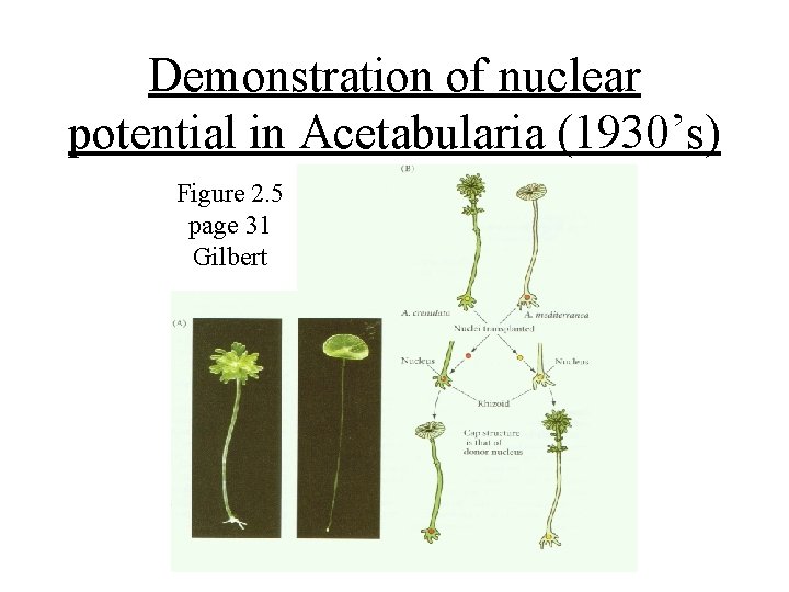
Demonstration of nuclear potential in Acetabularia (1930’s) Figure 2. 5 page 31 Gilbert
The nucleus and Epigenesis • The nucleus contains the instructions that drive epigenesis/development • Chromatin is the instructional unit (DNA plus proteins). The state of the chromatin is set by “epigenetic control” mechanisms • Covert “epigenetic” changes occur during the early cleavages of the fertilized egg into the blastomeres of the embryo
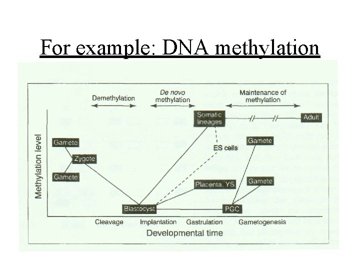
For example: DNA methylation
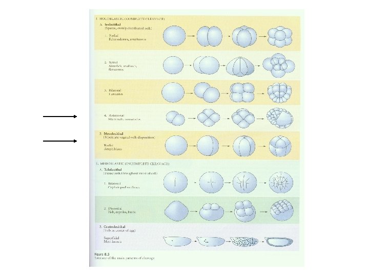
Embryonic cell division is not the same in all kinds of organisms Next slide So there is a sense that these zygotes “know what they are” when the begin to divide. The yolk and its position obviously plays a role in determining the cleavage pattern. But, there are sometimes other products stored in the egg by the mother that act as morphogenetic determinants.
Major morphogenetic strategies • Autonomous specification – Morphogenetic determinants deposited in the egg become segregated by cell division – Determination of cell fate is early • Conditional specification – Cell fate determination is later and depends on the position of the blastomere in the embryo – Removal of cells is compensated for by others – Each cell has the potential to give rise to more cells than it normally does
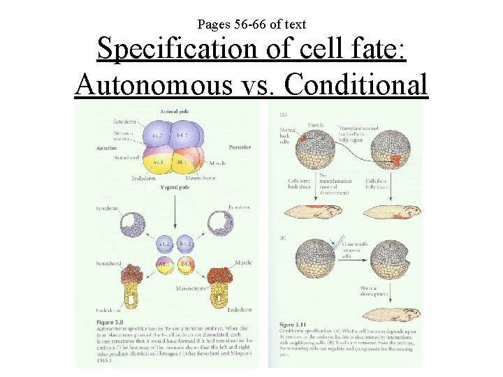
Pages 56 -66 of text Specification of cell fate: Autonomous vs. Conditional
Syncitial specification • Mainly seen in insects (Drosophila) • Gradients form, over time, of morphogens deposited in the egg by the mother • These morphogens become segregated by cell membranes which grow into the egg • Drosophila lectures: End of February
GASTRULATION • The process that puts cells into position to have their fates determined • Gastrulation involves a particular repertoire of cell movements which can be classified • Gastrulation will result in the formation of three “germ layers” from which the organs of the embryo will arise
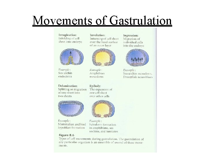
Movements of Gastrulation
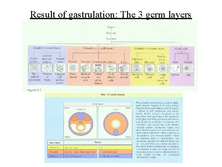
Result of gastrulation: The 3 germ layers
Gastrulation and Conditional Specification • Newly positioned tissues (germ layers) interact with one another to “induce” organ formation – Cell-cell interactions via receptors (juxtacrine) – Soluble signaling molecules (paracrine) • Morphogen gradients link position to cell fate • Signal transduction activates gene expression which leads to specification and lineage commitment
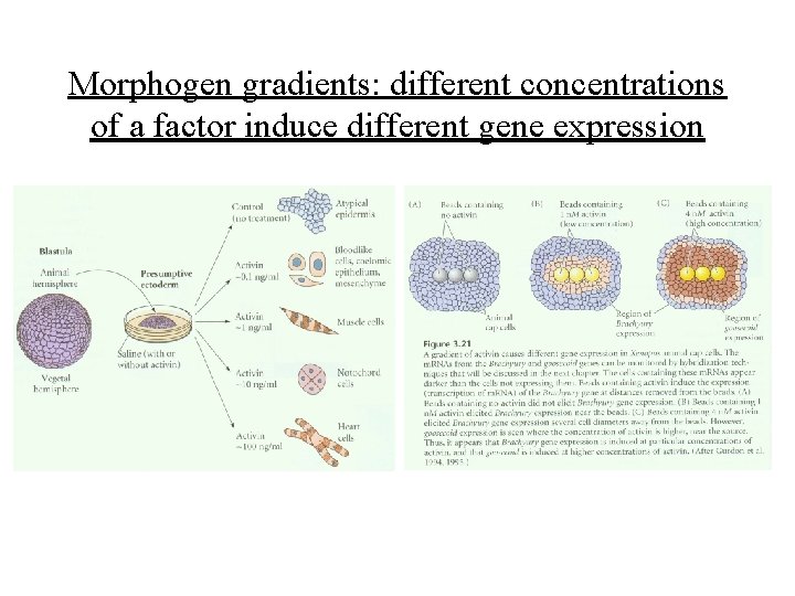
Morphogen gradients: different concentrations of a factor induce different gene expression
Stages of commitment to cell fate • Specification – Changes in gene expression which are labile and changeable. The gene expression “allows” that cell to differentiate along a pathway but does not irreversibly commit the cell • Determination – Further changes in gene expression which seal the lineage fate of the cell and eliminate alternative choices
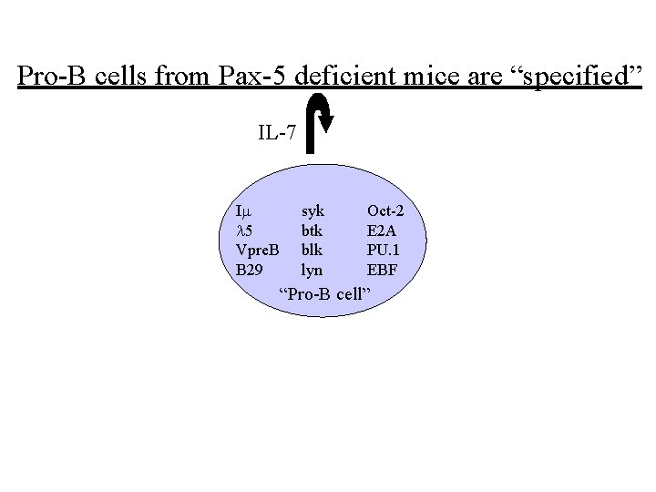
Pro-B cells from Pax-5 deficient mice are “specified” IL-7 Im l 5 Vpre. B B 29 syk btk blk lyn Oct-2 E 2 A PU. 1 EBF “Pro-B cell”
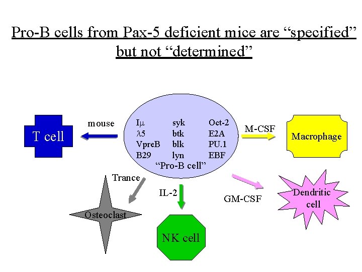
Pro-B cells from Pax-5 deficient mice are “specified” but not “determined” mouse T cell Im l 5 Vpre. B B 29 syk btk blk lyn “Pro-B cell” Oct-2 E 2 A PU. 1 EBF M-CSF Macrophage Trance IL-2 Osteoclast NK cell GM-CSF Dendritic cell
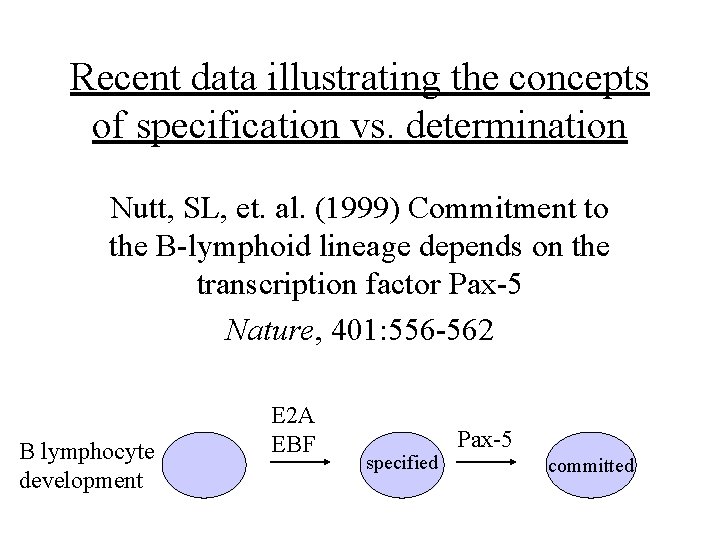
Recent data illustrating the concepts of specification vs. determination Nutt, SL, et. al. (1999) Commitment to the B-lymphoid lineage depends on the transcription factor Pax-5 Nature, 401: 556 -562 B lymphocyte development E 2 A EBF specified Pax-5 committed
Forming a solid organ • How do cells “stick together” to form tissue? • Coordination of tissues from multiple germ layers to form a single functional organ – Tissue layers – Organ polarity • Cell adhesion molecules: Cadherins (p 66 -74)
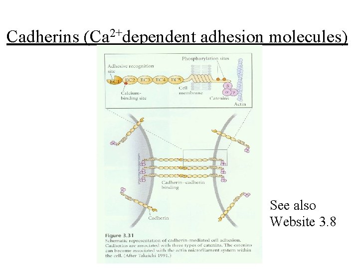
Cadherins (Ca 2+dependent adhesion molecules) See also Website 3. 8
Types of cadherins • E- cadherin: expressed on early embryonic cells in mammals. Later becomes restricted to embryonic and adult epithelial tissue • P-cadherin: Trophoblast cells (placental) • N-cadherin: First mesodermal, later CNS • EP-cadherin: frog blastomere adhesion • Protocadherins: not connected to catenin
Mechanisms of cadherin based cell “sorting” into tissues • Differential expression of cadherin type – Neural vs epidermal cells (N vs. E cadherin) • Different levels of cadherin expression – Oocyte positioning in follicle • Loss or switch of cadherin expression – Neural crest emigration from the neural tube – Protocadherin switching in frog gastrulation • See pages 311 -312 (Chapter 10 of Gilbert)
- Slides: 24