Fracture of shaft of femur Introduction It is


 • Skeletal traction: • Hip spica")


- Slides: 26
Fracture of shaft of femur
Introduction. - • It is a fracture of femoral diaphysis occurring between 5 cm distal to lesser trochanter and 5 cm proximal to adductor tubercle. • Usually occur in young men after high energy trauma and elderly women (even after a low energy fall).
Fig : -
Anatomy
Fig :
Fig
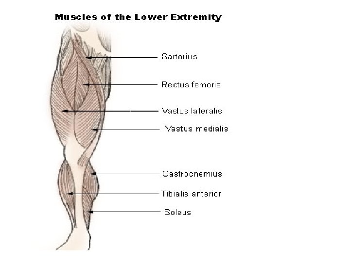
Anatomy • Femur is the largest tubular bone. • Surrounded by large muscle mass. • Major deforming muscle forces– Abductors: Gluteus medius and minimus. – Iliopsoas ; - flexion and external rotation. – Adductors -pectineus, adductor brevis , adductor longus , gracilis and adductor magnus.
Thigh muscles • Anterior compartment: quadriceps femoris, iliopsoas, sartorius. • Medial compartment: gracilis, adductor longus, brevis, magnus, obturator externus. • Posterior compartment: biceps femoris, semitendinosus, semimembranosus. • Because of the large volume, compartment syndrome is much less common.
Vascular supply • Mainly from profunda femoris.
Mechanism of injury • Almost always due to high energy trauma: RTA, gunshot injury, fall from height. • Pathologic fractures occur at the metaphyseal/diaphyseal junction. • If degree of trauma inconsistent with fracture, rule out pathological #.
Classification: Winquist and Hansen’s comminution
Clinical features -Pain, - swelling, - deformity, - shortening of the lower limb and complete external rotation deformity. - severe blood loss ( up to 1500 m. L ) - shock features : - unconsciousness , pallor , cold nose , tachycardia , cold and clammy skin , hypotension etc.
Associated injuries • Ligament and meniscus injuries of ipsilateral knee. • Spinal injuries. • Injury to the Pelvis.
Radiographic evaluation • AP and lateral views of the femur, hip and knee. • AP view of the pelvis. • Fracture pattern, comminution, shortening should be evaluated.
Treatment (Non operative) • Skeletal traction: • Hip spica
• Skin traction: Gallow’s traction – For children upto 2 yrs. – Legs of the child are tied to an overhead beam. – Hips are raised about 2 inches from the bed so that weight of the body provides counter traction. – For 4 to 6 weeks.
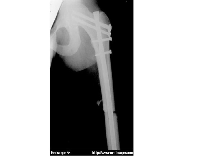
Operative methods • Standard treatment for most femoral shaft fractures. • Early surgery is recommended.
Closed Intramedullary nailing Advantages • Inside the medullary cavity, so more stable than plate, less exposure required. • Fracture hematoma is maintained. • Early use of limb, restoration of length and alignment, rapid union and low re-fracture rates are the advantages.
Interlocking nailing • Nail is introduced in the medullary cavity. • The screws are passed from the cortex through the holes in the nail.
Kuntscher’s cloverleaf Intramedullary nail. • Open reduction required. • For # at the junction of upper and middle 1/3. • Not suitable for comminuted fractures, fractures in the distal shaft and in open fractures.
Plate fixation • Advantage: no additional trauma. • Disadvantage: – more risk of infection, more bleeding, soft tissue injury. – Higher rate of implant failure as it is load bearing. – Decreased vascularization beneath the plate.
• Indications of plating – Extremely narrow medullary canal where IM nailing is difficult. – Fractures that occur through previously malunited fracture. – Fractures that have extended to the trochanters or condyles. – For comminuted fractures.
Complications – Shock. – Fat embolism. – Femoral artery injury. – Sciatic nerve injury. – Infection.
• Late – Delayed union. – Non union. – Malunion. – Knee stiffness.