Foundations in Biology Block 1 A Cell structure

Foundations in Biology Block 1 A - Cell structure 2. 1. 1 Using the magnification formula

Spec

Starter Which of these specimens could be viewed by a light or electron microscope or both? Justify your answers. 1. Nucleus 2. Cell wall 3. Mitochondria 4. White blood cell ingesting a bacteria 5. Chloroplast 6. Embryo dividing 7. Sperm cell swimming 8. Ribosomes 9. Detailed structure of the cytoplasm 10. Fertilisation taking place
Nucleus – visible under both microscopes 2)Cell wall – visible under both microscopes")
Answers 1)Nucleus – visible under both microscopes 2)Cell wall – visible under both microscopes 3)Mitochondria – mainly visible by electron microscope; too small for optical 4)White blood cell ingesting a bacteria – only optical as the cells must be alive 5)Chloroplast – electron 6)Embryo dividing – only optical as cells must be alive 7)Sperm cell swimming – only optical as cell must be alive 8)Ribosomes – electron microscope 9)Detailed structure of the cytoplasm – electron microscope. Fertilisation taking place – only optical as cells must be alive •

Objectives and Success Criteria Objectives Success criteria • Use the magnification formula • Recall the magnification formula (Grade E) • Describe how to use an eye piece graticule (Grade C) • Manipulate the magnification formula (Grade A)

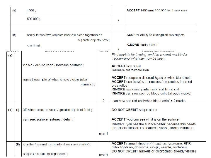
Task • Complete exam questions
 in a millimetre")
Practicing magnification maths • There are: • 1 000 nanometres (nm) in a millimetre (mm) • 1000 micrometres (mm) in a millimetre • 1000 millimetres in a metre (m) • 1 000 micrometres in a metre • 1 000 000 nanometres in a metre.

How small is a cell?

key terms

Light microscope • Actual size = image size magnification • Magnification = image size actual size
 15 000/2. 6 =")
Worked Example Image size is 15 mm (15 000 mm) 15 000/2. 6 = 5769 times

Task You may have to work out the magnification of an image 1. A micrograph of a plant cell is 150 mm long. The plant cell measures 120 μm long. Calculate the magnification. step 1 – convert μm into mm (1000 μm = 1 mm) so 120/1000 = 0. 12 mm step 2 – Use equation = 150/0. 12 = x 1250 2. A cell is 50 mm across on a page, the magnification is x 1250. What is its actual (real size) in μm? Actual = image/magnification 50 000/1250 = 40 μm

Magnification used The magnified image is produced by 2 lens, and eye piece and an objective lens, these can be used to work out the total magnification • Magnification used= Magnification of eyepiece × Magnification of objective lens Magnification of eyepiece = 10 Magnification of objective lens = 100 Magnification used = 10 × 100 = x 1000

Eye Piece Graticule • Microscopes can be fitted with an EPG • Ruler etched on it • A specimen can be measured in eyepiece units (an arbitrary measurement) the image changes size depending on the magnification, but the graticule stays the same size for each magnification. • To measure the size of objects in the field of view, the graticule needs to be calibrated)
 Place a stage graticule on the stage 2)")
Calibration - Using Stage Micrometer 1) Place a stage graticule on the stage 2) The ruler is 1 mm long and split into 100 divisions • Each division = 10µm (0. 01 mm) • 1µm is equal to 1 millionth of a metre
 Align the eyepiece graticule with the stage micrometer")
Calibration - Using Stage Micrometer 3) Align the eyepiece graticule with the stage micrometer 4) Find the value of one eyepiece division In (a) where mag = x 40, The stage graticule is equal to 40 eyepiece divisions. Each eyepiece division = 1000 mm/40 = 25 mm In (b) where mag = x 100 The stage graticule is equal to 100 epd Remember the stage graticule is 1 mm or 1000 mm) Each epd = 1000 mm/100 = 10 mm Total magnification = mag of eyepiece x mag of objective lens
, the nucleus is 3. 2 epd With")
Calculating size • In the image (a), the nucleus is 3. 2 epd With x 100 mag. 1 epd = 10 mm So: the nucleus is 3. 2 x 10 = 32 mm You are observing a specimen of squamous tissue under high power. Each individual cell has an average diameter of 60 mm & the diameter of the field of view is 2 mm. • Calculate the maximum number of cells that are visible in the field of view.

TASK • Complete worksheet organelle sizes

Plenary Question 1. If a nucleus measures 100 mm on a diagram, with a magnification of x 10000, what is the actual size of the nucleus? Actual = Image size/Magnification 100000/10000 = 10 micrometers

Objectives and Success Criteria Objectives Success criteria • Use the magnification formula • Recall the magnification formula (Grade E) • Describe how to use an eye piece graticule (Grade C) • Manipulate the magnification formula (Grade A)
- Slides: 21