Forsterite Crystal Myanmar Olivine structure Two cation sites


- Slides: 19

Forsterite Crystal Myanmar

Olivine structure Two cation sites: c M 2 M 1 b M 1 M 2

Columns of M 1 in the olivine structure b The M 1 sites form columns parallel to the c-axis a

Compare M 1 and M 2 sites M 1 M 2 Distorted 6 -coordination <M-O> = 2. 16 Å <M-O> = 2. 19 Å Smaller site Larger site

All atoms

Olivine structure

Top layer of oxygens

Almost hexagonal close packing

Problem of site occupancy (I) Into what site do cations go? Does site occupancy make a difference?

Problem of site occupancy (II) Ni can be directed into either the M 1 or the M 2 site by appropriate substitutions in the other site. Ni 2+ occupancy M 1 site M 2 site Li. Sc. Si. O 4 M 1 site blocked by Sc 3+

Optical spectra of olivine Optical absorption spectrum of a 1. 0 mm thick olivine from San Carlos, AZ. The three spectra are taken with linearly polarized light vibrating parallel to the three orthorhombic axes. The intense band at 1040 nm is from Fe 2+ in the M(2) site.

Distortion changes the energetics The M(2) site is more distorted than the M(1) site Therefore, the t 2 g and eg orbitals will split more in the M(2) site eg E t 2 g octahedral distorted

Conclusion from optical studies MIT says: Fe is dominantly in the M(2) site

X-Ray studies At VPI and University of Chicago a precision X-ray diffraction study to determine the structure. (called a 3 -D refinement of the structure)

Conclusion from X-ray study No evidence of M(1) – M(2) disorder M(1) = M(2)

Mössbauer spectra 57 Fe(excited state) 57 Fe (ground state) With the emission of a 14. 4 ke. V gamma ray. 57 Co Gamma source sample detector

Mössbauer spectrum of Fo 26 Mössbauer results

Conclusion from Mössbauer Slight preference for Fe in the M 1 site

Summary of studies MIT Chicago/VPI Carnegie M 2 >> M 1 = M 2 M 1 > M 2
 Msdp myanmar
 Solvus temperature
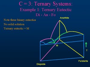
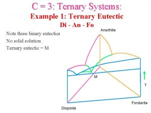
 Diopside-anorthite-forsterite ternary system
 Formule chimique du plagioclase
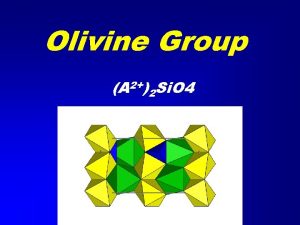
 Olivine group of minerals
 Homogeneous inorganic substances
 Oceaness
 What two sites did the narrator go back to see at devon
 Cscl structure
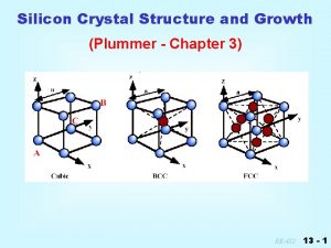
 Silicon crystal structure
 Titanium crystal structure
 Electrostatic attraction
 Volume of bcc unit cell
 Piezoelectric crystal atomic structure
 Basis in crystal structure
 Crystal structure of ceramics
 Ideal crystal
 Crystalline solid
 Atomic packing factor for bcc
 Motif in material science

