Forensics Chapter 11 Trace Evidence Hair and Fibers

Forensics Chapter 11: Trace Evidence Hair and Fibers

Introduction • Hair is encountered as physical evidence in a wide variety of crimes. • It is useful evidence because it does not easily break down. • Although it is not yet possible to individualize a human hair to any single head or body through its morphology, it still has value as physical evidence. Sometimes, DNA can be extracted from a hair to individualize it as evidence. • When properly collected and submitted to the laboratory accompanied by an adequate number of standard/reference samples, hair can provide strong corroborative evidence for placing an individual at a crime scene.

Morphology of Hair • Hair is an appendage of the skin that grows out of an organ known as the hair follicle. • The length of a hair extends from its root or bulb embedded in the follicle, continues into a shaft, and terminates at a tip end. • Hair on mammals helps to regulate body temperature, decrease friction, and protect against sunlight. • The skin is a complex organ whose tissues serve many functions.
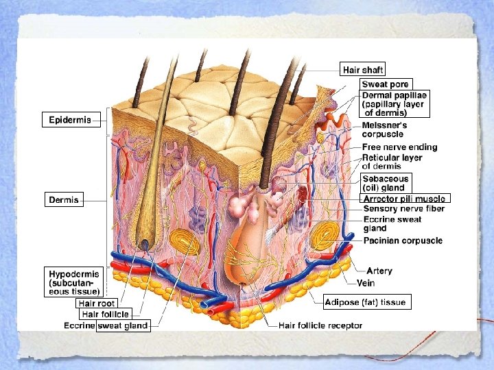

The skin has 3 layers: • The epidermis, which consists mostly of dead cells that serve as a waterproof covering for the body. • The dermis, which is made primarily of connective tissue that gives skin its strength and flexibility. This layer contains numerous blood vessels and nerve endings. • The hypodermis (also called the subcutaneous layer), which contains a layer of adipose (fat) tissue that provides insulation and serves as a shock absorber. At the border between the epidermis and dermis lies the dermal papillae whose bumpy surface gives us the ridges and grooves we call fingerprints. Along each ridge is a line of pores leading from the sweat glands. It is through these pores that sweat is discharged to cool the body.

• The hair follicles extend from the epidermis down into the dermis. The follicle is lined with both a layer of epidermal cells (found inside, next to the hair) and dermal cells (found outside the epidermal cells). • Deep in the base of the follicle is the hair root which is rich in blood supply. This is where the new hair is formed. Because of its blood supply, chemicals from the blood (including drugs) may be incorporated into the hair. • The hair shaft protrudes from the follicle above the epidermis. • Most hair follicles have sebaceous gland around the follicle. These glands produce oils that help to keep the skin from drying out. • Each follicle also has a tiny muscle known as the arrector pili attached. It is this muscle that pulls the hair upright to cause “goosebumps”.

The hair shaft is made up of a protein called keratin. Keratin makes the hair both strong and flexible. It is the shaft, which is composed of three layers— the cuticle, cortex, and medulla — that is subjected to the most intense examination by the forensic scientist.

Cuticle • The cuticle is the scale structure made up of dead cells covering the exterior of the hair. – The scales always point towards the tip of the hair. – The scale pattern is useful in species identification. • Three basic patterns of cuticles are observed: Human hair always has an imbricate cuticle. Cuticles in other animals may be any of the three types

Cortex • The cortex is the main body of the hair shaft. • Its major forensic importance is the fact that it is embedded with the pigment granules that impart hair with color. • The color, shape, and distribution of these granules provide the criminalist with important points of comparison among the hairs of different individuals. • Melanin is the chief pigment imbedded in the cortex. • Hair containing a high concentration of melanin (eumelanin) will appear brown or black whereas hair with a low concentration of melanin (pheomelanin) will appear blond or red. Hair that lacks melanin will appear white or gray.

Medulla • The medulla is a row of cells like a canal running through the center of the hair. • The medulla may be continuous, interrupted, fragmented, solid, or absent. • The presence of the medulla vary from individual to individual and even among hairs of a given individual.

While in humans the medulla is not patterned, the medulla of some animals show specific patterns:

• The medullary index is the ratio of the diameter of the medulla relative to the diameter of the hair shaft. • For humans, the medulla generally occupies less than one-third the diameter of the shaft, while for animals it is generally one-half or greater. Dog Human

• Which of these would most likely be human? • How could you tell? • What medulla patterns do you see?
 (multisereal) Mouse (Medullary Index > 1/3) (unisereal) Human (Medullary")
Muskrat (Medullary Index > 1/3) (multisereal) Mouse (Medullary Index > 1/3) (unisereal) Human (Medullary Index < 1/3) (not patterned)

• Human or animal? • How could you tell? • What is the medulla pattern?

Medulla diameter Hair shaft diameter This is deer hair. Notice that the medullary index is much greater than 1/2. Also notice the lattice pattern of the medulla.

Root • The root and other surrounding cells in the hair follicle provide the tools necessary to produce hair and continue its growth. • When pulled from the head, some translucent tissue surrounding the hair’s shaft near the root may be found. This is called a follicular tag. • By using DNA analysis on the follicular tag, the hair may be individualized.

The Life Cycle of Hair proceeds through 3 stages as it develops: • During the long anagen stage, hair actively grows. The cells around the follicle rapidly divide and deposit materials in the hair. • In the catagen stage, the hair grows and changes. • Hair is in the telogen stage when the follicle becomes dormant. During this stage, hairs easily can be lost.

Comparing Strands • The comparison microscope is an indispensable tool for comparing the morphological characteristics of hair. • When comparing strands of human hair, the criminalist is particularly interested in matching the color, length, curliness, and diameter. • A careful microscopic examination of hair will reveal morphological features that can distinguish human hair from the hair of animals. • Scale structure, medullary index, and medullary shape are particularly important in animal hair identification.

• Other important features for comparing human hair are: – the presence or absence of a medulla. – the distribution, shape, and color intensity of the pigment granules present in the cortex. • The most common request is to determine whether or not hair recovered at the crime scene compares to hair removed from the suspect. • However, microscopic hair examinations tend to be subjective and highly dependant on the skills and integrity of the analyst.

Treated Hair Forensic investigators sometimes can link hair from a location with an individual. • Bleaching disturbs the scales on the cuticle and removes pigment leaving hair brittle and a yellowish color. • Dyeing colors the cuticle and the cortex of the hair shaft. Because of this and because hair grows daily, a person’s treated hairs will have specific characteristics in common with her or his lost hairs.

Hair and DNA • Recent major breakthroughs in DNA profiling have extended this technology to the individualization of human hair. • The probability of detecting DNA in hair roots is more likely for hair being examined in its anagen as opposed to its catagen or telogen phases. • Often, when hair is forcibly removed a follicular tag, a translucent piece of tissue surrounding the hair’s shaft near the root may be present. • This has proven to be a rich source of nuclear DNA associated with hair.

Hair and Mitochondrial DNA • Mitochondrial DNA can be extracted from the hair shaft. • Mitochondrial DNA is found in cellular material located outside of the nucleus and it is transmitted only from the mother to child. • As a rule, all positive microscopical hair comparisons must be confirmed by DNA analysis.

Collection and Preservation • As a general rule, forensic hair comparisons involve either head hair or pubic hair. • The collection of 50 full-length hairs from all areas of the scalp will normally ensure a representative sampling of head hair. • A minimum collection of two dozen full-length pubic hairs should cover the range of characteristics present in pubic hair. • Hair samples are also collected from the victim of suspicious deaths during an autopsy.

Types of Fibers • Natural fibers are derived in whole from animal or plant sources. – Examples: wool, silk, mohair, cashmere, furs from animals and cotton, hemp, linen, and jute from plants. • Man-made fibers are manufactured. – Regenerated fibers are manufactured from natural raw materials and include rayon, acetate, and triacetate. – Synthetic fibers are produced solely from synthetic chemicals and include nylons, polyesters, and acrylics.

Fiber Evidence • The quality of the fiber evidence depends on the ability of the criminalist to identify the origin of the fiber or at least be able to narrow the possibilities to a limited number of sources. • Obviously, if the examiner is presented with fabrics that can be exactly fitted together at their torn edges, it is a virtual certainty that the fabrics were of common origin.

• When examining fiber evidence, characteristics such as fiber type, shape, diameter, color, variation of color in the fiber, length of fiber, and lengthwise striations on the fiber's surface are noted. • Another useful test is called the burn test. Different types of fibers react differently to fire. Some burn readily and continue to burn even when the flame is removed. Others stop burning when the flame is removed or tend to melt more than burn. This analysis can be helpful in determining the type of fiber, but they are destructive of the evidence. They are only useful if there is a large sample of the fiber evidence available. • If a whole section of fabric is available microscopic observation can reveal the type of weave and the thread count (number of threads per inch).

• The visible light microspectrophotometer is a convenient way for analysts to compare the colors of fibers through spectral patterns. • A more detailed analysis of the fiber’s dye composition can be obtained through a chromatographic separation. • Infrared spectrophotometry is a rapid and reliable method for identifying the generic class of fibers, as does the polarizing microscope. • Depending on the class of fiber, each polarized plane of light will have a characteristic index of refraction.

Collection and Preservation • The investigator’s task of looking for minute strands of fibers often becomes one of identifying and preserving potential “carriers” of fiber evidence. • Relevant articles of clothing should be packaged carefully in separate paper bags. • If it is necessary to remove a fiber from an object, the investigator must use clean forceps, place it in a small sheet of paper, fold and label the paper, and place the paper packet inside another container.
- Slides: 29