Forensic DNA Fingerprinting Using Restriction Enzymes DNA Fingerprinting


 DNA at a")


 electrode")


 Distance (mm) 23, 000 11. 0 9, 400 13.")

- Slides: 20
Forensic DNA Fingerprinting: Using Restriction Enzymes
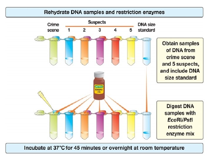
DNA Fingerprinting Procedure Overview
DNA is Tightly Packaged into Chromosomes Which Reside in the Nucleus
Model of DNA is Comprised of Four Base Pairs
DNA structure
DNA restriction enzymes Evolved by bacteria to protect against viral DNA infection Endonucleases = cleave within DNA strands Over 3, 000 known enzymes
Enzyme Site Recognition Restriction site Palindrome • Each enzyme digests (cuts) DNA at a specific sequence = restriction site • Enzymes recognize 4 - or 6 - base pair, palindromic sequences (eg GAATTC) Fragment 1 Fragment 2
5 vs 3 Prime Overhang Enzyme cuts • Generates 5 prime overhang
Common Restriction Enzymes Eco. RI – Eschericha coli – 5 prime overhang Pstl – Providencia stuartii – 3 prime overhang
The DNA Digestion Reaction Restriction Buffer provides optimal conditions • Na. CI provides the correct ionic strength • Tris-HCI provides the proper p. H • Mg is an enzyme co-factor 2+
Why incubate at 37°C? DNA Digestion Temperature • Body temperature is optimal for these and most other enzymes What happens if the temperature is too hot or cool? • Too hot = enzyme may be denatured (killed) • Too cool = enzyme activity lowered, requiring longer digestion time
Restriction Fragment Length Polymorphism RFLP Allele 1 1 Allele 2 Pst. I Eco. RI CTGCAG GAGCTC GAATTC GTTAAC 2 CGGCAG GCGCTC Different Base Pairs No restriction site GAATTC GTTAAC Fragment 1+2 M Electrophoresis of restriction fragments M: Marker A-1: Allele 1 Fragments A-2: Allele 2 Fragments + A-1 A-2 3 3
Agarose Electrophoresis Loading Electrical current carries negativelycharged DNA through gel towards positive (red) electrode Buffer Dyes Agarose gel Power Supply
Agarose Electrophoresis Running • Agarose gel sieves DNA fragments according to size • Small fragments move farther than large fragments Gel running Power Supply
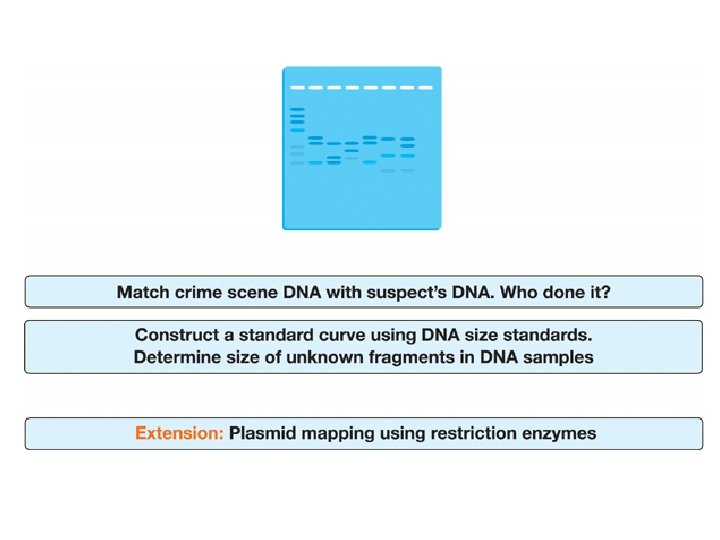
Analysis of Stained Gel Determine restriction fragment sizes • Create standard curve using DNA marker • Measure distance traveled by restriction fragments • Determine size of DNA fragments Identify the related samples
Molecular Weight Determination Size (bp) Distance (mm) 23, 000 11. 0 9, 400 13. 0 6, 500 15. 0 4, 400 18. 0 2, 300 23. 0 2, 000 24. 0 Fingerprinting Standard Curve: Semi-log
Electrophoresis Equipment