FOREARM Deep fascia of forearm Antebrachial fascia It

FOREARM
: -It is attached to olecranon and")
• Deep fascia of forearm (Antebrachial fascia): -It is attached to olecranon and posterior border of ulna -From its deep surface many intermuscular septa pass inwards to separate superficial from deep muscles. - It is thickened in front and behind wrist to form flexor and extensor retinacula - Fascial sheath with interosseous membrane and septa divide forearm into several compartments

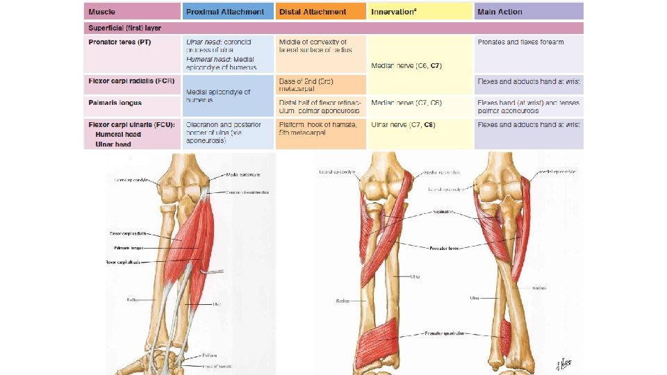
Superficial muscles of front of forearm Pronator teres Flexor carpi radialis Palmaris longus Flexor digitorum superficialis Flexor carpi ulnaris
 head: -Common flexor origin (front of medial epicondyle)")
Pronator Teres • Origin: -Superficial (humeral) head: -Common flexor origin (front of medial epicondyle) -Lower part of medial supracondylar ridge -Deep (ulnar) head: -From medial side of coronoid process of ulna • Insertion: - The two heads join to be inserted into middle of lateral surface of shaft of radius at maximum convexity

• Nerve supply: -Median nerve in cubital fossa • Action: -Pronation of forearm -Helps flexion of elbow joint

• Relations: -Median nerve passes between its 2 heads - Deep head separates median nerve from ulnar artery - Its lateral border forms the medial boundary of cubital fossa -Its insertion is crossed by superficial radial nerve and radial artery
 •")
Flexor carpi radialis • Origin: -From common flexor origin (front of medial epicondyle) • Insertion: -Into palmar surface of bases of 2 nd and 3 rd metacarpal bones • Nerve supply: -Median nerve in cubital fossa • Action: -Flexion of wrist joint -Helps in abduction of the hand

• Relations: -In the forearm, its tendon descends vertically -At wrist: -Its tendon pierces flexor rtinaculum occupying the groove in front of trapezium. -Radial artery lies lateral to its tendon and median nerve is medial to it.
 •")
• Origin: Palmaris Longus From common flexor origin (front of medial epicondyle) • Insertion: -It has a long slender tendon descends superficial to flexor retinaculum with its deep fibres attached to it -It is inserted into apex of palmer aponeurosis • Nerve supply: -Median nerve in cubital fossa • Action: -Tension of palmer aponeurosis - Flexion of wrist joint

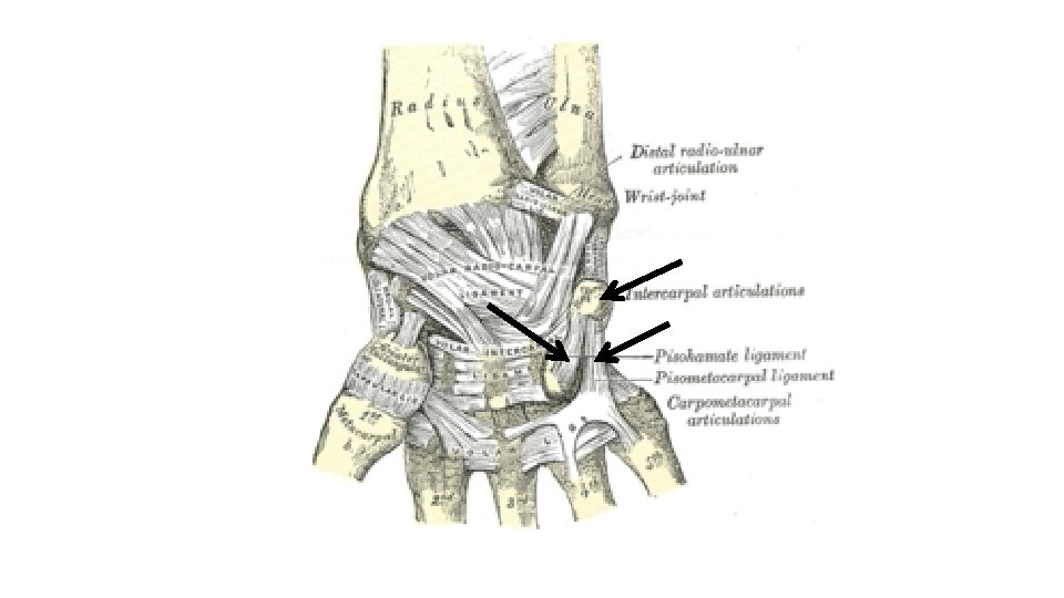
Flexor Carpi Ulnaris • Origin: -Humeral head From common flexor origin (front of medial epicondyle) -Ulnar head: medial border of olecranon -By aponeurosis from upper 2/3 of posterior border of ulna • Insertion: -Into pisiform bone - This insertion is prolonged by 2 ligaments: 1 - piso-hamate ligament to hook of hamate 2 Piso-metacarpal ligament to base of 5 th metacarpal bone

• Nerve supply: Ulnar nerve • Action: -Flexion of wrist joint -Adduction of hand • Relations: -At its origin: ulnar nerve passes between its 2 heads -Along its course: it overlies flexor digitorum profundus with ulnar nerve in between -At its insertion: ulnar nerve and artery lie just lateral to its insertion

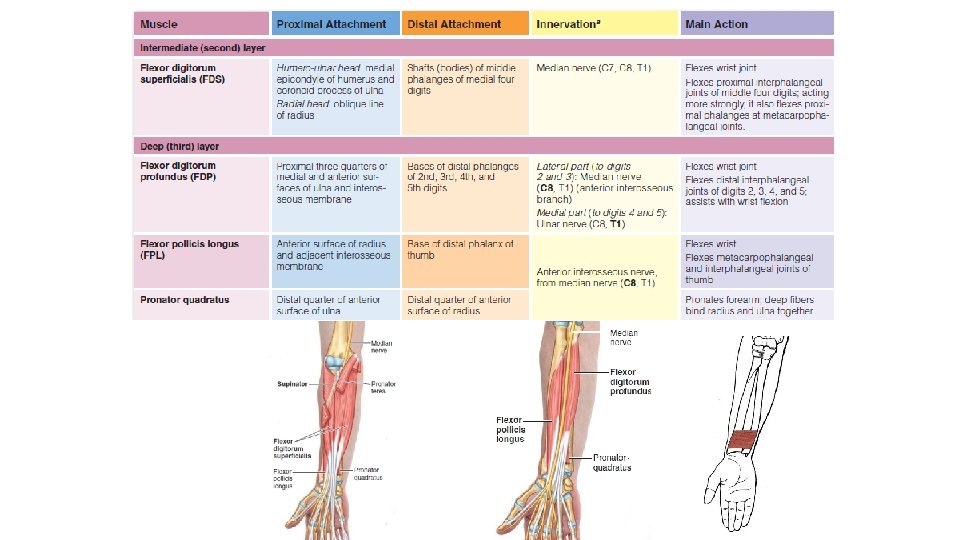
Flexor Digitorum Superficialis • Origin: -Humero-ulnar head: - From common flexor origin (front of medial epicondyle) -Medial border of coronoid process -Radial head: -From anterior oblique line of radius and its anterior border down to pronator teres insertion • Insertion: - At middle of forearm the muscle divides into 4 parallel tendons - At wrist tendons pass through carpal tunnel where those of middle and ring fingers are superficial to those of index and little fingers - On palmer surface of proximal phalanx, each tendon splits into 2 slips for passage of flexor digitorum profundus - The 2 slips are inserted into margins of palmer surface of middle phalanx

Wrist joint • Nerve Supply: Median Nerve • Action: -Flexion of proximal interphalangeal joints of medial 4 fingers -Flexion of metacarpo-phalangeal joints of medial 4 fingers -Assists in flexion of wrist joint

• Relations: -Median nerve: -In the forearm it is adherent to deep surface of the muscle -In the carpal tunnel, it is lateral its tendons -Radial artery & superficial radial nerve Descend superficial to its radial head -In hand tendons runs in midpalmar space deep to: - Palmer aponeurosis - Digital branches of median and ulnar nerves - Superficial palmar arch and its branches

Deep muscles of front of forearm Flexor Digitorum Profundus Flexor Pollicis Longus Pronator Quadratus

Flexor Digitorum Profundus • Origin: -Upper ¾ of anterior and medial surface of ulna -Posterior border of ulna (by aponeurosis with flexor& extensor carpi ulnaris) -Medial side of coronoid process -Adjoining part of interosseous membrane

• Course and insertion: -Above the wrist: The muscle gives rise to 4 tendons to medial 4 fingers -At wrist: Tendons pass through carpal tunnel deep to those of flexor digitorum superficialis

• In the palm: The tendons diverge to reach the fingers. Each tendon gives rise to a lumbdical muscle. • In the fingers: -Tendons enter fibrous flexor sheathes of the medial four fingers. -Opposite proximal phalanges, each tendon passes through the 2 slips of the corresponding flexor digitorum superficialis tendon. - Each tendon is inserted into palmar surface of the base of terminal phalanx

• Nerve Supply: -Its medial part: by ulnar nerve lateral part: by anterior interosseous branch of median nerve. • Action: -Flexion of all joints of medial 4 fingers. - Assists in flexion of the wrist joint.

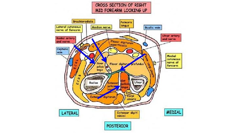
• Relations: -On its anterior surface: Median nerve between it passes and flexor digitorum superficialis. -Along its medial border: Ulnar nerve and ulnar artery descend between it and flexor carpi ulnaris -Along its lateral border: Anterior interosseous nerve and artery descend between it and flexor pollicis longus muscle.

Flexor Pollicis longus • Origin: -From middle of anterior surface of radius below anterior oblique line - From adjoining part of interosseous membrane. • Course insertion: and -The tendon passes through lateral part of carpal tunnel. -Then, it passes along medial side of thenar eminence -It is inserted into palmer surface of base of distal phalanx of thumb

• Nerve Supply: Anterior interosseous nerve • Action: -Flexion of all joints of thumb -Helps in flexion of wrist joint • Relations: -Along its medial border: Anterior interosseous nerve and artery descend between it and flexor digitorum profundus. -Above wrist: The tendon lies deep to tendon of flexor carpi radialis. -At carpal tunnel: The tendon enters along its lateral part.

Pronator Quadratus • Origin: From oblique ridge on lower ¼ of anterior surface of ulna • Insertion: Into lower ¼ of anterior surface of radius • Nerve supply: Anterior interosseous nerve • Action: Pronation

Superficial muscles of back of forearm Brachioradialis Extensor carpi radialis longus Extensor carpi radialis brevis Extensor digitorum Extensor digiti minimi Extensor carpi ulnaris

Brachioradialis • Origin: -Upper 2/3 of lateral supracondylar ridge of humerus - Front of lateral intermuscular septum • insertion: Into the lateral side of lower end of radius above styloid process

• Nerve supply: Radial nerve (by branch arises at lateral side of arm. • Action: -Flexion of elbow especially in midprone position (muscle of military salute). -Initiation of supination or pronation. • Relations: -It forms lateral boundary of cubital fossa. -Its upper part is separated from brachialis by a groove lodges radial nerve -Radial artery descends undercover of its upper part.

Extensor carpi radialis longus • Origin: -lower 1/3 of lateral supracondylar ridge of humerus -Front of lateral intermuscular septum. • Insertion: Into dorsum of base of 2 nd metacarpal bone. • Nerve supply: Radial nerve (by branch arises at lateral side of arm. • Action: Extension and abduction of the wrist

Extensor carpi radialis brevis • It lies deep to extensor carpi radialis longus throughout its course • Origin: From common extensor origin (front of lateral epicondyle) • Insertion: Into dorsum of base of 3 rd metacarpal bone. • Nerve supply: Posterior interosseous nerve (of radial nerve) before piercing supinator. • Action: Extension and abduction of the wrist

• Origin: From common extensor origin Extensor Digitorum • Insertion: -By 4 tendons for medial 4 fingers -They pass deep to extensor retinaculum -On the back of hand they are connected by inter-tendinous connections. -Each tendon joins extensor expansion on dorsum of proximal phalanx. -Each expansion divides into 3 slips: -1 median: to the base of middle phalanx -2 collaterals: converge and unite to be attached to base of distal phalanx • Nerve supply: Posterior interosseous nerve after coming from supinator • Action: -Extension of metacarpophalangeal joints and interphalangeal joints of medial 4 fingers - Assists in extension of wrist joint

Extensor digiti minimi • it is a slip from medial side of extensor digitorum • Origin: Common extensor origin • Insertion: Its tendon joins extensor expansion on dorsum of little finger with that from extensor digitorum • Nerve supply: Posterior interosseous nerve • Action: -Extends all joints of little finger -Assists in extension of wrist joint

Extensor carpi ulnaris • Origin: -Common extensor origin -Posterior border of ulna through aponeurosis with flexor carpi ulnaris and flexor digitorum profundus. • Insertion: Into dorsal surface of base of 5 th metacarpal bone. • Nerve supply: Posterior interosseous nerve • Action: -Adduction of wrist with flexor carpi ulnaris. -Extension of wrist with extensor carpi radialis longus and brevis.

Anconeus • Origin: By a rounded tendon from back of lateral epicondyle of humerus • Insertion: -Into lateral surface of olecranon -Into upper ¼ of posterior surface of ulna • Nerve supply: Radial nerve by a long branch arises in spiral groove and descends through medial head of triceps to reach anconeus • Action: Helps in extension of elbow

The Extensor Muscles of Forearm The extensor muscles are organized anatomically into superficial and deep layers. Four superficial extensors (ECRB, extensor digitorum, EDM, and ECU) are attached proximally by a common extensor tendon to the lateral epicondyle

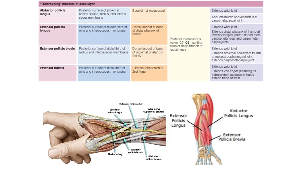
Deep muscles of back of forearm Supinator Abductor pollicis longus Extensor pollicis brevis Extensor indicis

Abductor pollicis longus • Origin: -Back of ulna just below oblique ridge and lateral to vertical ridge. -Back of interosseous membrane -Back of middle 1/3 of radius -Insertion: -Tendon of the muscle accompanies tendon of extensor pollicis brevis. -Both wind around lateral side of lower end of radius. -Both cross over tendons of extensor carpi radialis longus and brevis. -Both form lateral boundary of anatomical snuffbox. -It is inserted into lateral side of base of 1 st metacarpal bone.

• Nerve supply: Posterior interosseous nerve • Action: Abduction of the thumb

Extensor pollicis brevis • Origin: -Posterior surface of radius below origin of abductor pollicis longus - Adjoining part of interosseous membrane. • Insertion: -It becomes adherent to medial side of abductor pollicis longus. -It is inserted into dorsum of base of proximal phalanx of thumb. • Nerve supply: Posterior interosseous nerve. • Action: Extension of metacarpophalangeal joint of thumb

Extensor pollicis longus • Origin: - Middle 1/3 of posterior surface of ulna lateral to vertical ridge below origin of abductor pollicis longus -Adjoining part of interosseous membrane • Insertion: -The tendon psses in a narrow groove on back of lower end of radius medial to dorsal tubercle. -It forms medial boundary of anatomical snuffbox. -It is inserted into dorsum of base of terminal phalanx of thumb • Nerve supply: Posterior interosseous nerve. • Action: Extension of all joints of thumb

Extensor indicis • Origin: -Back of ulna below origin of extensor pollicis longus. Adjoining part of interosseous membrane. • Insertion: -The tendon joins extensor expansion of index finger. • Nerve supply: Posterior interosseous nerve. • Action: Extension of all joints of index finger.

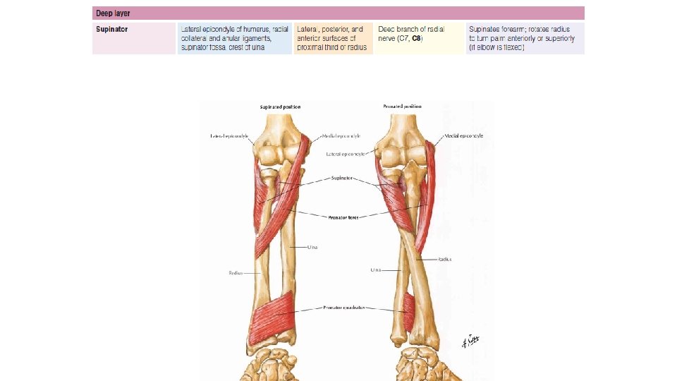
Supinator • Origin: • Superficial part: -Lateral epicondyle of humerus -Radial collateral ligament • Deep part: -Supinator crest and fossa of ulna • Insertion: Into upper 1/3 of lateral surface of radius • Nerve supply: Posterior interosseous nerve before piercing the muscle • Action: • Supination forearm of extended

• Relations; -It forms a part of floor of cubital fossa. -Posterior interosseous nerve pierces supinator and emerges on the back of forearm - Posterior interosseous artery appears on the back of forearm between supinator and abductor pollicis longus

THANK YOU 1/7/2022 48
- Slides: 48