For FCM Training Injector Tip Fluorescence signals Focused


")


 Excitation Emisson 300 nm 400 nm Wavelength 500 nm Protein 600 nm")
 Excitation Emisson 300 nm 400 nm 500 nm Protein 600 nm 700")
 Protein 300 nm 400 nm 500 nm 632. 5 nm (He. Ne)")


 一种蛋白质,在DNA复制和核苷酸的切除修复中起 到作用 Ki-67 - proliferation related antigen Ki-S 1")

")


 80 pg/m. L")

 7")

 Lck Itk P P PLC")
 Jurkat cells were pre-incubated with")
 Jurkat cells were pre-incubated")


- Slides: 141
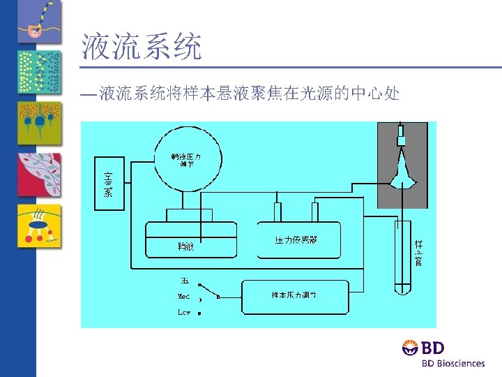
流式细胞术简介 For FCM Training
Injector Tip Fluorescence signals Focused laser beam Sheath fluid
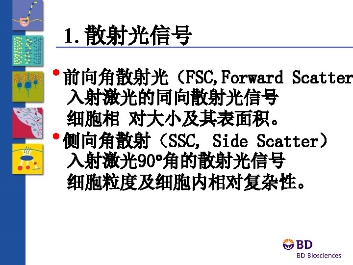
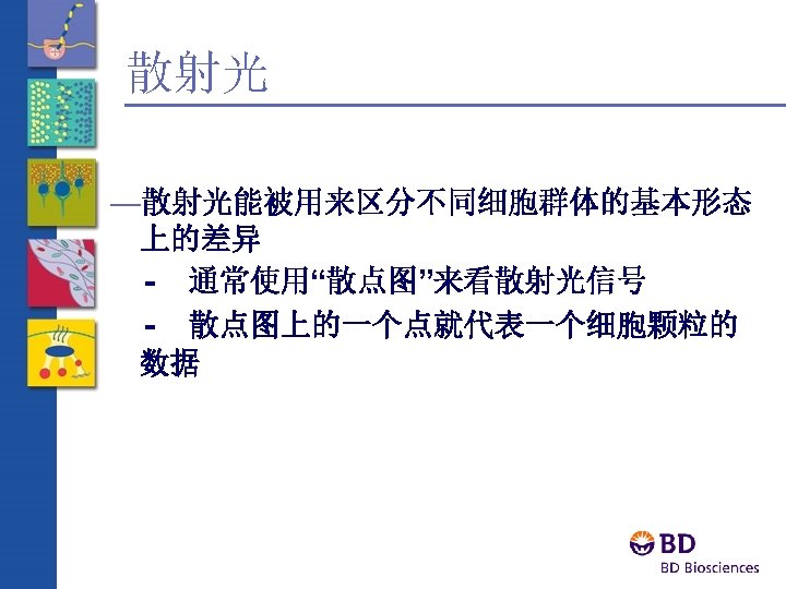
前向角散射光 ——FSC —Forward Angle Light Scatter Laser FALS Sensor
侧向角散射光——SSC Laser FALS Sensor 90 LS Sensor
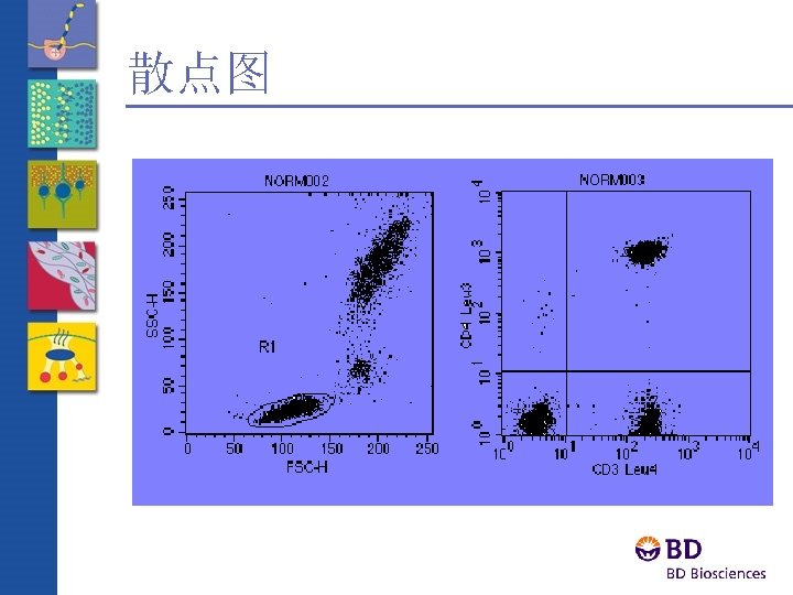
散点图——Dot Plot lysed whole blood
Review Question Dead cells are known to be smaller and to exhibit more internal complexity than live cells. Which of the populations on this plot would you expect to be dead?
荧光检测器 FALS Sensor Fluorescence detector (PMT 3, PMT 4 etc. )
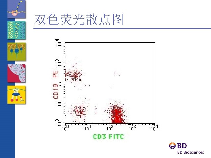
Two-Color Cell Analysis Ab B Which of the three populations has the most Ab A binding sites? Ab A
Sample Flow in Optical Cuvette Low Sample Flow Rate High Sample Flow Rate 60 m. L/min 12 m. L/min Laminar Flow Sheath Sample Sheath Sample
Review Question Which of the following would cause disturbance in the laminar flow of the optical cuvette? A. bubbles B. cellular concentration C. sample flow rate
Optics · Excitation optics consist of: - Lasers - Lenses and mirrors that route the laser light to the fluidic stream · Collection optics consist of: Filters that direct the signals to the appropriate optical detectors
Optical Filters Longpass 460 500 LP 500 540 Shortpass 460 500 SP 500 540 Bandpass 460 500 540 BP 500/50
FACSCalibur光路图
Optics Laser Fluorochromes 488 nm FITC 635 nm Detector Parameter Filter GFP FL 1 530/30 PE PI FL 2 585/42 Per. CP-CY 5. 5 FL 3 670 LP FL 4 661/16 APC
Electronics —Converts analog signals to proportional digital signals —Computes Height for each pulse —Calculates width and area —Interfaces with the computer for data transfer
Laser Voltage Creation of a Voltage Pulse - Analog Signal Laser Voltage Time
Quantification of a Voltage Pulse Height Volts — Height is a measurement for all parameters. — Width = Area/Height Pulse Area 0 Pulse Width Time
Effect of the Instrument Controls on the Data Detecto r Instrument Controls FSC detector 250 gain FSC detector 350 gain
SSC Review FL 2 FSC FL 1 FL 4 Data Processin g FL 3
Review Questions 1. Which of the following fluorochromes cannot be used with the FACSCalibur? a. DAPI (ex. 345 nm, emits 455 nm) b. Propidium Iodide (ex. 536 nm, emits 617 nm) c. Alexa Fluor 647 (ex. 650 nm, emits 668 nm) 2. What are three measurements of a particle that can be determined by FACSCalibur? 3. Briefly describe the functions of the fluidics, optics, and electronics systems.
Review Questions 4. What would happen to the population below if you increased the Red parameter value in the Instrument controls?
Review Questions 5. Which instrument components ensure that the fluorescence signal of a specific fluorochrome is only measured by a designated detector? For example, APC is only measured by the FL 4 detector.
Propidium Iodide Emisson Excitation 300 nm 400 nm 500 nm 600 nm DNA 700 nm PI
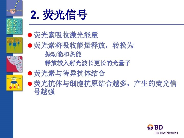
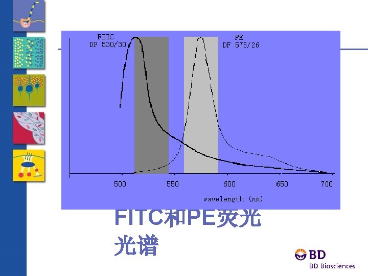
Fluorescein (FITC) Excitation Emisson 300 nm 400 nm Wavelength 500 nm Protein 600 nm 700 nm
Phycoerytherin (PE) Excitation Emisson 300 nm 400 nm 500 nm Protein 600 nm 700 nm
Allophycocyanin (APC) Protein 300 nm 400 nm 500 nm 632. 5 nm (He. Ne) 600 nm 700 nm Excitation Emisson
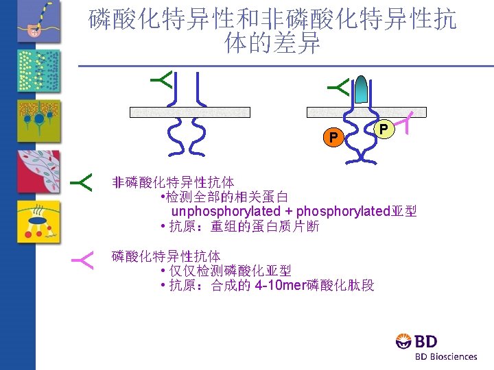
直接染色 — Fluorescent probe attached to antibody — Specific signal: weak — Nonspecific binding: low
间接染色 — Fluorescent probe attached to a 2 nd antibody — Specific signal: strong, 5 -6 2 nd Ab/each 1 st Ab; — Nonspecific binding: high
Avidin-Biotin method I biotinylated primary Ab biotin avidin biotinylated dye
Flow Cytometry in clinics and research
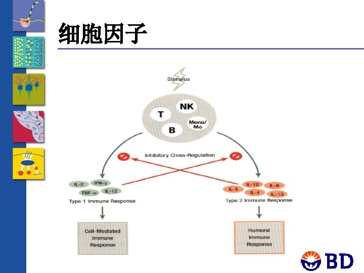
What can Flow Cytometer tell us about a cell? Cytokine/Chemokine Migration/ Production Repertoire Homing Phenotypre Cytokine/Chemokine Receptor Repertoire Cell cycle status Cell Lineage/Subset Phenotype
Nucleic acid Probes 菲啶基 Propidium Iodide Ethidium Bromide 苯甲亚胺 Hoechst 33342 抗生素 Mithramycin, Chromamycin A 3 Acridine Orange - AO Pyronyn Y
Normal Cell Cycle G 2 M G 0 DNA Analysis G 1 s G 0 G 1 C o u n t s 0 200 400 G 2 M 600 4 N 2 N DNA content 800 1000
A typical DNA Histogram # of Events G 0 -G 1 G 2 -M S Fluorescence Intensity
增殖相关基因 PCNA(Proliferating cell nuclear antigen) 一种蛋白质,在DNA复制和核苷酸的切除修复中起 到作用 Ki-67 - proliferation related antigen Ki-S 1 - proliferation related antigen Phospho-histone Cyclin D 1, E, A, B 1
Brd. Urd Incorporation
Tracing Dye(CFSE)
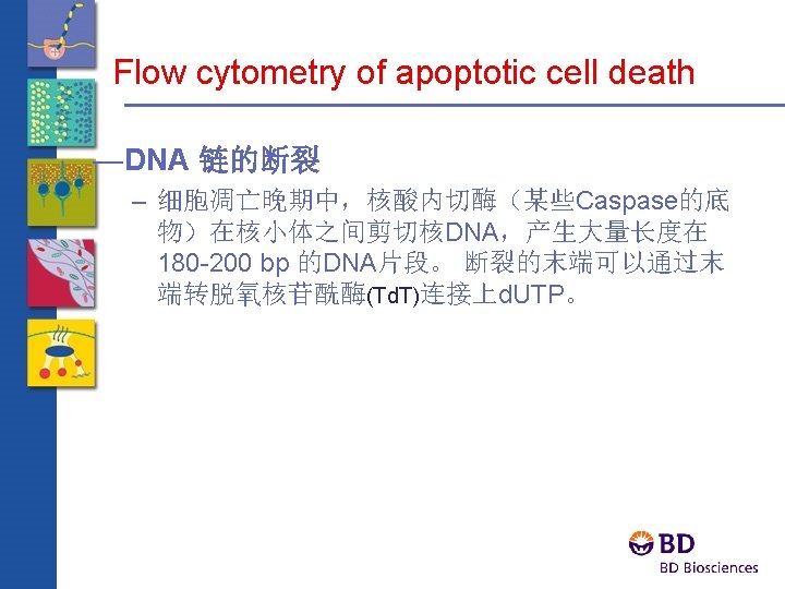
Apoptotic Cell Death -- a genetically encoded cell death program, which is morphologically, biochemically and molecularly distinct from necrosis
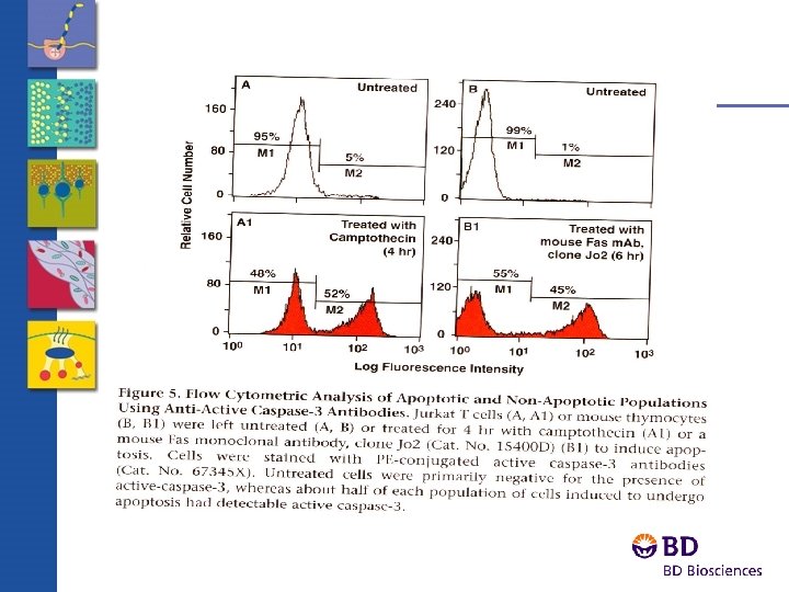
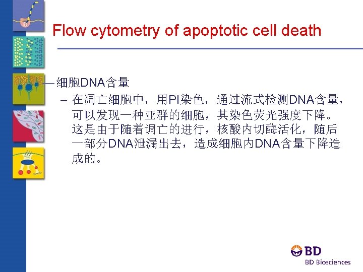
Flow cytometry of apoptotic cell death — FCM of Caspases – 通过抗体和活化的caspase-3片断相结 合来检测 – 使用特异性的caspase-3荧光底物 – 新的抗原决定基CK 18
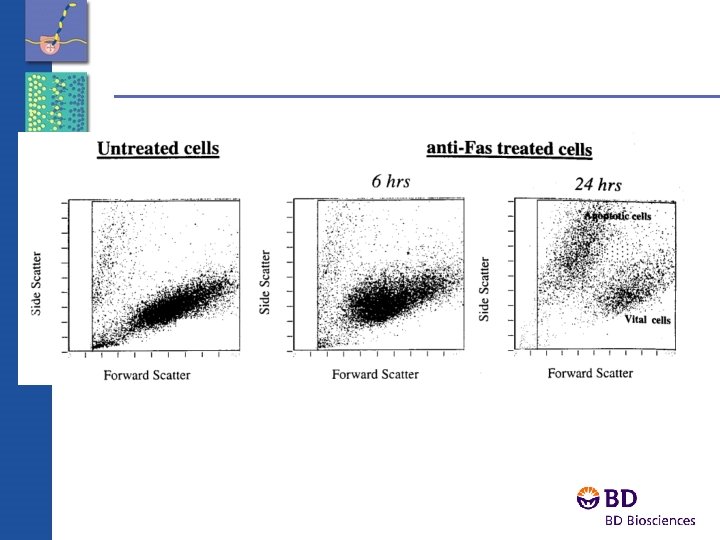
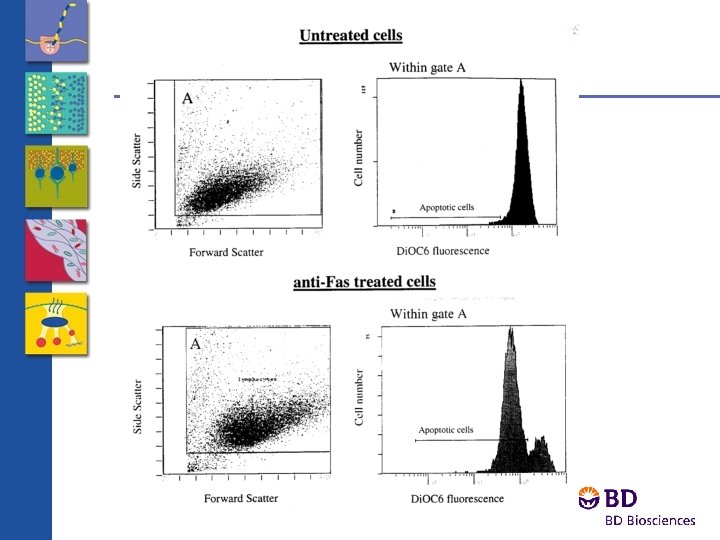
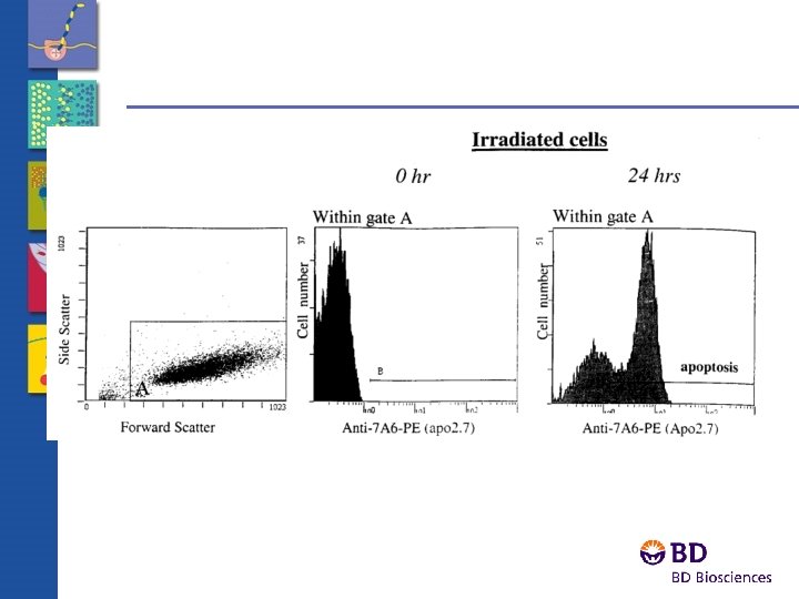
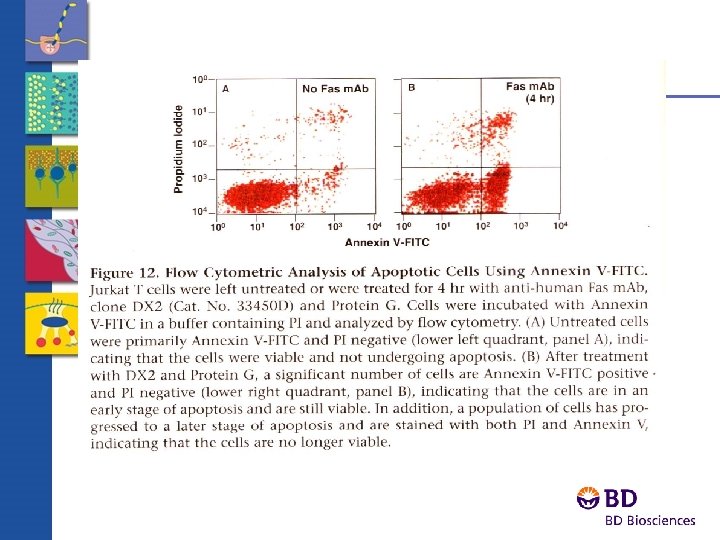
Flow cytometry of apoptotic cell death Phospholipid redistribution
Flow Cytometry of Apoptotic Cells Apoptotic cells # Events Normal G 0/G 1 cells PI - Fluorescence
An Integrated Approach to Cell Immunology
Immune Function assay – 免疫细胞亚群检测 – Antigen-peptide specific T cells 检测 – T-cells 活化 – Treg Cells – 细胞内细胞因子/趋化因子检测 – 磷酸化
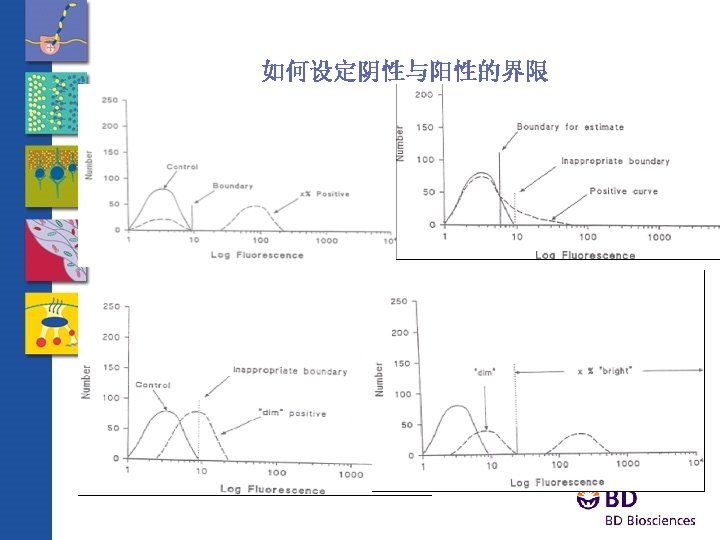
Traditional Analysis -- Percent positive Negative Cell CD 4 PE Positive cell
Absolute Counts -- Cells per m. L Absolute Count Beads Negative Cell CD 4 PE Positive Cell
MHC: Class I and Class II Gene Products —T cell受体不直接和可溶性抗原相连 • 抗原必须通过MHC由抗原呈递细胞传递给T细胞 (Macrophages, Dendritic cells, B cells) —MHC Class I Gene Products • 呈递抗原给 CD 8+ T cells • 诱导细胞毒应答 —MHC Class II Gene Products • 呈递抗原给 CD 4+ T cells • 诱导细胞因子产生,免疫球蛋白的分泌
MHC/TCR Signaling
Quantitation of antigen-specific T lymphocytes in peripheral blood Control Gag-A 2/Ig Tax-A 2/Ig HAM CD 8 HIV
IL-2 Phycoerythrin Traditional Cytokine Flow Cytometry CD 4 FITC
Biological Variation Among CMV-seropositive Donors in Response to CMV
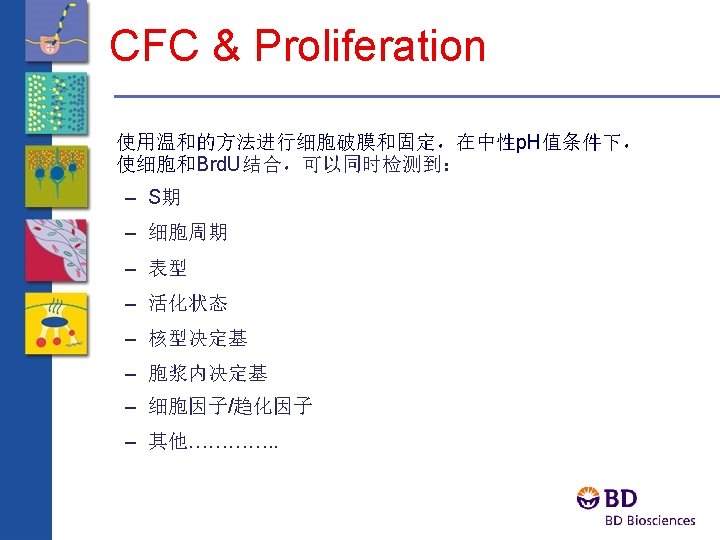
CFC & Proliferation Allows the correlation of: • Phenotype • Cytokine expression • Cell Cycle • Proliferation
BD Phos. FLOW和Western blot 的比较 A theoretical experiment comparing Western blot and flow cytometry with three samples and a protein of interest at 1, 10, or 50 copies per cell. Sample 2 and 3 look the same via Western blot, but when stained with fluorescently labeled antibodies, the differences between the samples become more relevant. (Source: P. O. Krutzik et al. / Clinical Immunology 110 (2004) 206– 221)
BD Phos. Flow not only tells you activation of a single cell for one particular phospho protein and pathway, but allows you to study multiple phosphorylation events and pathways simultaniously. Genomics & Proteomics -- Single Cells Have Big Proteomics Story to Tell.
Multi-Color Phos. Flow in CD 3/CD 28 or CD 3 crosslinked human PBMCs or Whole Blood 200 SSC-H 150 100 50 21. 8 0 0 50 100 150 FSC-H 200 250 200 SSC-H 150 100 50 0 100 150 FSC-H CD 3 -/CD 20 - 100 100 80 80 80 60 60 60 40 40 40 20 20 20 10 3 10 2 10 1 0 10 1 10 2 10 3 10 4 0 0 10 1 10 2 10 3 10 4 10 0 10 1 10 2 10 3 10 4 CD 3/CD 28 Crosslink 250 50 CD 3+/CD 20 - CD 3 PE PBMC 0 CD 3 -/CD 20+ 10 4 200 250 CD 20 Per. CP-Cy 5. 5 250 CD 3 CD 20 Per. CP-Cy 5. 5 Whole Blood Cross. Link 10 4 10 3 10 2 100 100 80 80 80 60 60 60 40 40 40 20 20 10 1 20 10 1 10 2 CD 3 PE 10 3 10 4 0 0 0 10 1 10 2 10 3 10 4 10 0 10 1 Zap 70 (Y 319)/Syk (Y 352) Alexa 647 Untreated cells (unshaded) vs treated cells (shaded) 10 2 10 3 10 4
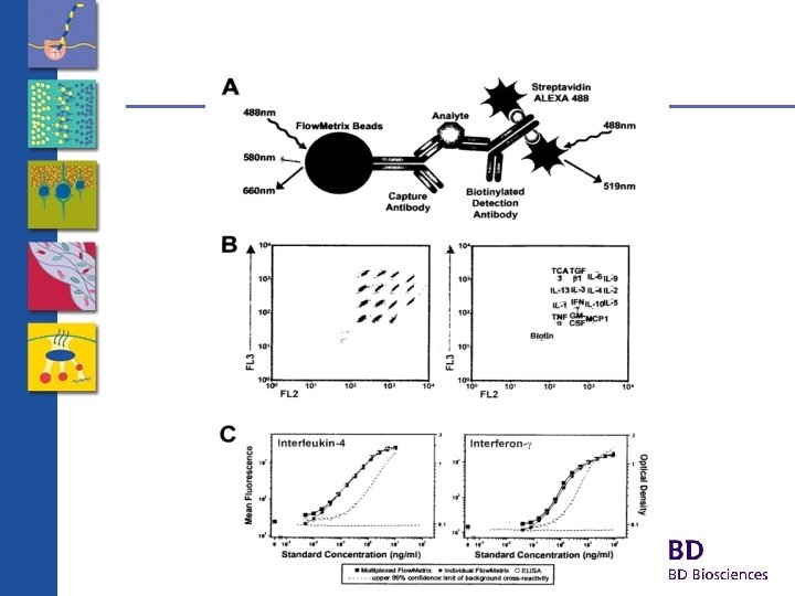
Multiplexed Beads Various analytes Antibody coupled beads, emitting at distinct FL 3 intensities Antibody coupled PE label, emitting at FL 2 intensity proportional to analyte conc. Shades of a color
FL 3 Beads Multiplexed Beads 0 pg/m. L FL 2 (PE) 80 pg/m. L 1250 pg/m. L 5000 pg/m. L
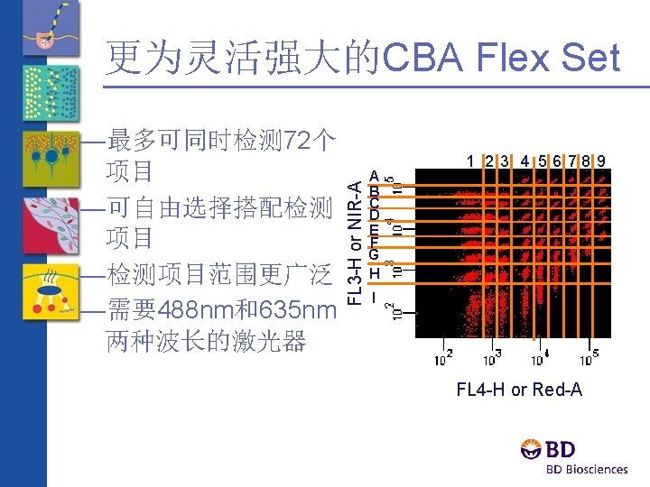
FL 3 -H or NIR-A BD CBA Flex Set A B C D E F G H 1 2 3 4 56 78 9 I FL 4 -H or Red-A BD CBA Flex Set Beads Old Format BD CBA Beads
9个指标同时检测活化T细胞 1 2 3 4 5 8 9 6 1. Itk (Y 511) 7 2. ERK (T 202/Y 204) 3. JNK (T 183/Y 185) 4. P 38 (T 180/Y 182) 5. PLCg (Y 783) 6. ZAP 70 (Y 319) 7. LAT (Y 171) 8. c-Jun (S 63) 9. RSK (S 380)
Kinetics of Jurkat Cell Activation With Anti-CD 3/CD 28 P-Specific Antibodies ERK (T 202/Y 204) JNK (T 183/Y 185) P 38 (T 180/Y 182) PLCg (Y 783) 10 ZAP-70 (Y 319) Itk (Y 511) LAT (Y 171) Jun (S 63) RSK (S 380) TIME (MINUTES) 20 15 10 5 1 0 FOLD INCREASE IN UNITS/ML 100
Monitoring the T Cell Activation Pathway by CBA (Control) Lck Itk P P PLC P V A V LAT P ZAP-70 P P LAT SLP-76 S O Grb 2 S DAG Ras PKC Rac P P MAPKKK Ras P P MEK IKK P P MEKK 1 -4 P MKK 4, 7 P MEKK 4 P MKK 3, 6 P P P ERK JNK p 38 P P P Elk Jun ATF 2 NF-k. B P RSK Cells were activated with anti-CD 3/CD 28 for 2 minutes. SDS was added to a final concentration of 1% and the material was placed in a boiling water bath for 5 minutes.
Monitoring the T Cell Activation Pathway by CBA (PD-98059) Jurkat cells were pre-incubated with 200 m. M PD 98059 (MEK inhibitor) for 20 minutes before being activated with anti-CD 3/CD 28 for 2 minutes. SDS was added to a final concentration of 1% and the material was placed in a boiling water bath for 5 minutes.
Monitoring the T Cell Activation Pathway by CBA (PP 2) Jurkat cells were pre-incubated with 10 m. M PP 2 (Src family kinase inhibitor) for 20 minutes before being activated with anti-CD 3/CD 28 for 2 minutes. SDS was added to a final concentration of 1% and the material was placed in a boiling water bath for 5 minutes.
Fluorescent Proteins Ds. Red Zs. Yellow Hc. Red Am. Cyan