FOOT The Arches of the Foot A segmented


 and arise from a single metatarsal as unipennate")


 ligaments strengthen the capsule. The interosseous (talocalcaneal)")


- Slides: 20
FOOT
The Arches of the Foot A segmented structure can hold up weight only if it is built in the form of an arch. The foot has three such arches, which are present at birth: the medial longitudinal, lateral longitudinal, and transverse arches. In the young child, the foot appears to be flat because of the presence of a large amount of subcutaneous fat on the sole of the foot. The Bones of the Arches An examination of an articulated foot shows the bones that form the arches. ■■ Medial longitudinal arch: This consists of the calcaneum, the talus, the navicular bone, the three cuneiform bones, and the first three metatarsal bones.
■■ Lateral longitudinal arch: This consists of the calcaneum, the cuboid, and the 4 th and 5 th metatarsal bones. ■■ Transverse arch: This consists of the bases of the metatarsal bones and the cuboid and the three cuneiform bones.
The foot, distal to the ankle, provides a platform for supporting the weight of the body when standing and has an important role in locomotion. The skeleton of the foot consists of 7 tarsals, 5 metatarsals, and 14 phalanges. The foot and its bones may be considered in terms of three anatomical and functional parts: • The hindfoot: talus and calcaneus. • The midfoot: navicular, cuboid, and cuneiforms. • The forefoot: metatarsals and phalanges
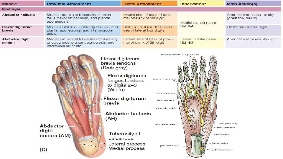
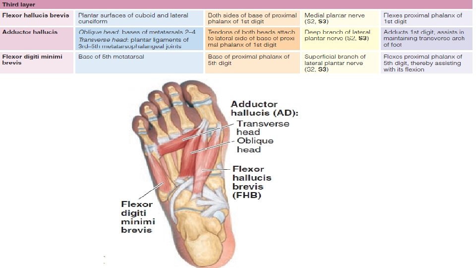
Muscles of Foot From the plantar aspect, muscles of the sole arranged in four layers within four compartments. Despite their compartmental and layered arrangement, the plantar muscles function primarily as a group during the support phase of stance to maintain the arches of the foot They basically resist forces that tend to reduce the longitudinal arch as weight is received at the heel (posterior end of the arch), and is then transferred to the ball of the foot and great toe (anterior end of the arch).
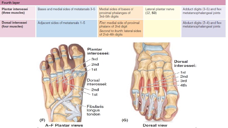
• Plantar interossei ADduct (PAD) and arise from a single metatarsal as unipennate muscles. • Dorsal interossei ABduct (DAB) and arise from two metatarsals as bipennate muscles. • Two closely connected muscles on the dorsum of the foot are the extensor digitorum brevis (EDB) and extensor hallucis brevis (EHB). The EHB is actually part of the EDB. These muscles form a fleshy mass on the lateral part of the dorsum of the foot, anterior to the lateral malleolus, and aid the extensor digitorum and extensor hallucis longus in extending digits one through four.
Tarsal Joints The joints of the foot involve the tarsals, metatarsals, and phalanges. The important intertarsal joints are the subtalar (talocalcaneal) joint and the calcaneocuboid and talonavicular joints). Inversion and eversion of the foot are the main movements involving these joints Subtalar Joint The subtalar joint is the posterior joint between the talus and the calcaneum. Articulation : Articulation is between the inferior surface of the body of the talus and the facet on the middle of the upper surface of the calcaneum. The articular surfaces are covered with cartilage. Type : These joints are synovial, of the plane variety. Capsule : The capsule encloses the joint and is attached to the margins of the articular areas of the two bones.
• Ligaments Medial and lateral (talocalcaneal) ligaments strengthen the capsule. The interosseous (talocalcaneal) ligament is strong and is the main bond of union between the two bones. It is attached above to the sulcus tali and below to the sulcus calcanei. • Synovial Membrane The synovial membrane lines the capsule. • Movements Gliding and rotatory movements are possible.
Talocalcaneonavicular Joint The talocalcaneonavicular joint is the anterior joint between the talus and the calcaneum and also involves the navicular bone. • Articulation : Articulation is between the rounded head of the talus, the upper surface of the sustentaculum tali, and the posterior concave surface of the navicular bone. The articular surfaces are covered with hyaline cartilage. • Type : The joint is a synovial joint. • Capsule : The capsule incompletely encloses the joint.
• Ligaments The plantar calcaneonavicular ligament is strong and runs from the anterior margin of the sustentaculum tali to the inferior surface and tuberosity of the navicular bone. The superior surface of the ligament is covered with fibrocartilage and supports the head of the talus. • Synovial Membrane The synovial membrane lines the capsule. • Movements Gliding and rotatory movements are possible
Calcaneocuboid Joint • Articulation is between the anterior end of the calcaneum and the posterior surface of the cuboid. The articular surfaces are covered with hyaline cartilage. • Type The calcaneocuboid joint is synovial, of the plane variety with capsule encloses the joint. • Ligaments The long plantar ligament is a strong ligament on the lower surface of the joint. It is attached to the undersurface of the calcaneum behind and to the undersurface of the cuboid and the bases of the third, fourth, and fifth metatarsal bones in front. It bridges over the groove for the peroneus longus tendon, converting it into a tunnel. The short plantar ligament is a wide, strong ligament that is attached to the anterior tubercle on the undersurface of the calcaneum and to the adjoining part of the cuboid bone.
Movements in the Subtalar, Talocalcaneonavicular, and Calcaneocuboid Joints • The important movements of inversion and eversion of the foot take place at the subtalar and transverse tarsal joints. Inversion is the movement of the foot so that the sole faces medially. Eversion is the opposite movement of the foot so that the sole faces in the lateral direction. The movement of inversion is more extensive than eversion. • Inversion is performed by the tibialis anterior, the extensor hallucis longus, and the medial tendons of extensor digitorum longus; the tibialis posterior also assists. • Eversion is performed by the peroneus longus, peroneus brevis, and peroneus tertius; the lateral tendons of the extensor digitorum longus also assist.
Cuneonavicular Joint The cuneonavicular joint is the articulation between the navicular bone and the three cuneiform bones. It is a synovial joint of the gliding variety. The capsule is strengthened by dorsal and plantar ligaments. The joint cavity is continuous with those of the intercuneiform and cuneocuboid joints and also with the cuneometatarsal and intermetatarsal joints, between the bases of the 2 nd and 3 rd and the 3 rd and 4 th metatarsal bones.
Cuboideonavicular Joint The cuboideonavicular joint is usually a fibrous joint, with the two bones connected by dorsal, plantar, and interosseous ligaments. Intercuneiform and Cuneocuboid Joints The intercuneiform and cuneocuboid joints are synovial joints of the plane variety. Their joint cavities are continuous with that of the cuneonavicular joint. The bones are connected by dorsal, plantar, and interosseous ligaments.
Tarsometatarsal and Intermetatarsal Joints The tarsometatarsal and intermetatarsal joints are synovial joints of the plane variety. The bones are connected by dorsal, plantar, and interosseous ligaments. The tarsometatarsal joint of the big toe has a separate joint cavity. Metatarsophalangeal and Interphalangeal Joints The metatarsophalangeal and interphalangeal joints closely resemble those of the hand. The deep transverse ligaments connect the joints of the five toes. The movements of abduction and adduction of the toes, performed by the interossei muscles, are minimal and take place from the midline of the second digit and not the third, as in the hand.
Thank You