FOOT TARSALS METATARSALS PHALANGES The human foot is


![Metatarsus � 5 metatarsal bones: numbered I – V [ 1 – 5] medial](https://slidetodoc.com/presentation_image_h/95249591d97bac6ad12d03e9b9641709/image-5.jpg "Metatarsus � 5 metatarsal bones: numbered I – V [ 1 – 5] medial")
![Phalanges [digits] �Numbered I – V medial to lateral �Each phalanx : proximal base,](https://slidetodoc.com/presentation_image_h/95249591d97bac6ad12d03e9b9641709/image-7.jpg "Phalanges [digits] �Numbered I – V medial to lateral �Each phalanx : proximal base,")
![Phalanges [digits] �Numbered I – V medial to lateral �Each phalanx : proximal base,](https://slidetodoc.com/presentation_image_h/95249591d97bac6ad12d03e9b9641709/image-8.jpg "Phalanges [digits] �Numbered I – V medial to lateral �Each phalanx : proximal base,")
 �MUSCLES �NERVES �VESSELS")


- Slides: 16
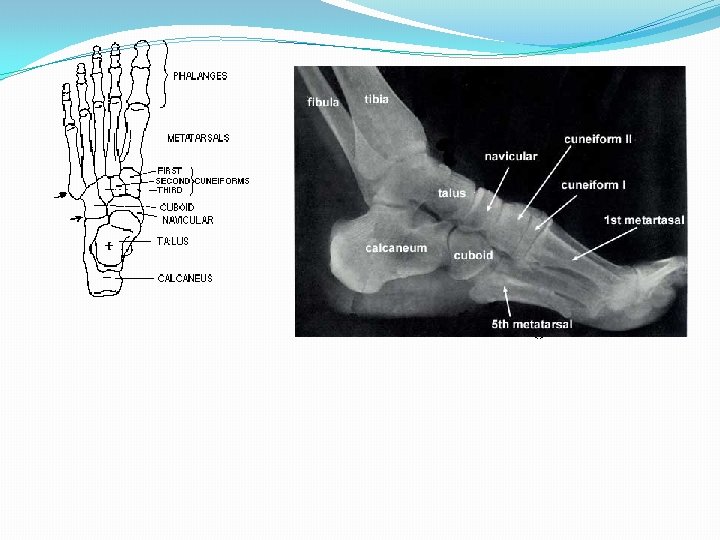
FOOT TARSALS, METATARSALS & PHALANGES The human foot is a complex structure containing 26 bones, 33 joints, many tendons, muscles, and ligaments.
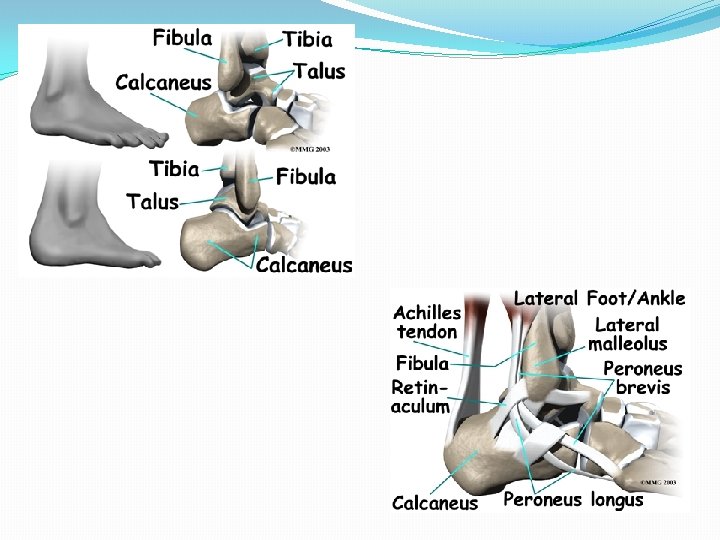
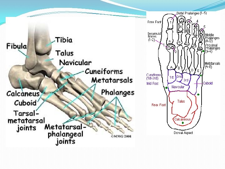
Tarsus = ankle �Proximal region of the foot � 7 tarsal bones �Talus: ankle bone �Calcaneus: heel bone �Navicular: ‘like a little boat’ � 3 Cuniform bones: wedge shaped - lateral, intermediate, medial �Cuboid: cube shaped
Metatarsus � 5 metatarsal bones: numbered I – V [ 1 – 5] medial to lateral �Each has a proximal base, an intermediate shaft and a distal head �articulate proximally with the first second and third cuneiform bones and the cuboid to form the tarsometatarsal joints �Articulate distally with the phalanges to form the metatarsophalangeal joint
Phalanges [digits] �Numbered I – V medial to lateral �Each phalanx : proximal base, intermediate shaft and distal head. �Hallux: has two phalanges [proximal & distal] �Other toes have three phalanges: proximal, middle and distal �Interphalangeal joints [between phalanges]
Phalanges [digits] �Numbered I – V medial to lateral �Each phalanx : proximal base, intermediate shaft and distal head. �Hallux: has two phalanges [proximal & distal] �Other toes have three phalanges: proximal, middle and distal �Interphalangeal joints [between phalanges]
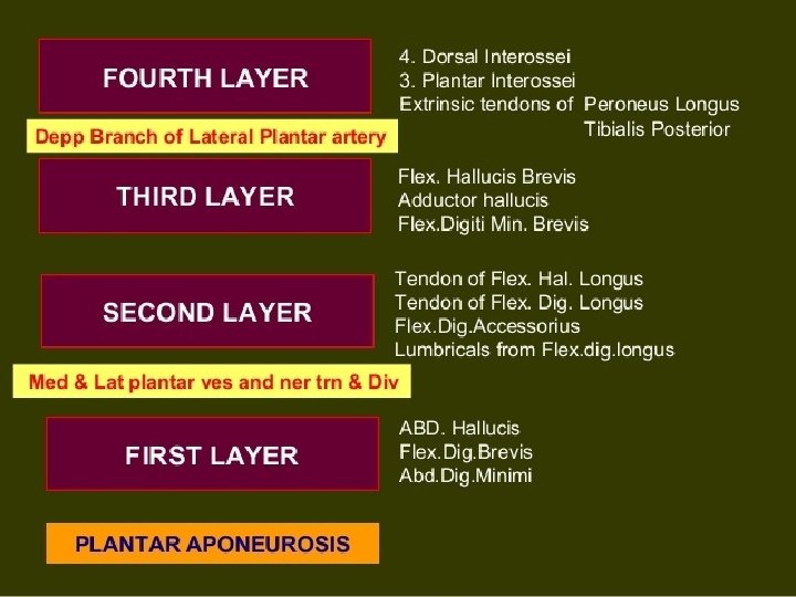
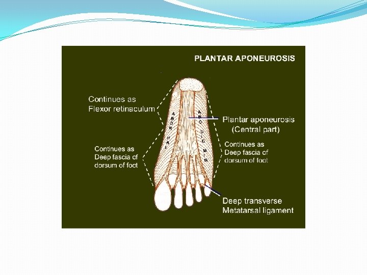
SOLE �SKIN �SUPERFICIAL FASCIA �DEEP FASCIA(plantar aponeurosis) �MUSCLES �NERVES �VESSELS
SOLE FIRST LAYER �ABDUCTOR HALLUCIS �FLEXOR DIGITORUM BREVIS �ABDUCTOR DIGITI MINIMI
SOLE 2 ND LAYER � TENDON OF FLEXOR HALLUCIS LONGUS � TENDON OF FLEXOR DIGITORUM LONGUS � FLEXOR DIGITORUM ACCESSORIUS � LUMBRICALS
SOLE THIRD LAYER � FLEXOR HALLUCIS BREVIS � ADDUCTOR HALLUCIS � FLEXOR DIGITI MINIMI BREVIS
SOLE 4 TH LAYER � 4 DORSAL INTEROSSEI � 3 PALMAR INTEROSSEI � TENDON OF TIBIALIS POSTERIOR � TENDON OF PERONEUS LONGUS
Applied anatomy � Plantar Fasciitis: "heel spurs“: an overuse injury affecting the sole or flexor surface (plantar) of the foot. A diagnosis of plantar fasciitis means you have inflamed the tough, fibrous band of tissue (fascia) connecting your heel bone to the base of your toes. � Higher risk: female, overweight, a job that requires a lot of walking or standing on hard surfaces; walk or run for exercise, especially if you have tight calf muscles that limit how far you can flex your ankles. People with very flat feet or very high arches are also more prone to plantar fasciitis. • starts gradually with mild pain at the heel bone often referred to as a stone bruise. • more likely to feel it after (not during) exercise. • The pain classically occurs again after arising from a midday lunch break. � If you don't treat plantar fasciitis, it may become a chronic condition. You may not be able to keep up your level of activity and you may also develop symptoms of foot, knee, hip and back problems because of the way plantar fasciitis changes the way you walk.