Foot ankle anatomy By Fatemeh Javadi Bones Calcaneus

Foot & ankle anatomy By: Fatemeh Javadi

Bones § Calcaneus: § forms the heel • calcaneal tuberosity • upper surface has articular facets for the talus • anterior end articulates with the cuboid bone. • sustentaculum tali • Tarsal sinus

Talus • rests on the superior surface of the calcaneus • upper part of the articular surface articulates with the end of the tibia • medial part, with the medial malleolus of the tibia • lateral part, with the lateral malleolus of the fibula • anterior end articulates with the navicular bone and inferiorly with the calcaneus

Navicular • The navicular lies anterior to the talus on the medial side of the foot. • It has a proximal articular surface for the head of the talus and a distal articular surface for the three cuneiform bones. • Its lateral surface is attached to the cuboid

cuneiforms • medial, intermediate, and lateral • Their proximal ends articulate with the navicular through a synovial joint • lateral cuneiform articulates with the cuboid

cuboid • The cuboid has a proximal articular surface for the calcaneus • distal one for the two lateral metatarsals • small medial one for the lateral cuneiform

Metatarsals and phalanges • bases of the metatarsals articulate with each other and with the cuneiform and cuboid bones • their heads articulate with the bases of the proximal phalanges • There are often two sesamoid bones at the metatarsophalangeal joint of the big toe.

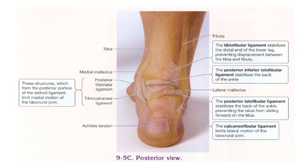
Ankle Joint • Hinge-type joint; distal ends of the tibia and fibula + Talus • Medial (deltoid) ligament: ant & post tibiotalar, tibiocalcaneal, and tibionavicular. Resists eversion of foot. • Lateral ligament: ant talofibular ligament, post talofibular ligament, and calcaneofibular ligament. Check inversion of the foot.
")
Joints of foot • Intertarsal joints; subtalar, talocalcaneonavicular, calcaneocuboid, cuneonavicular, transverse tarsal (chopart’s joint) • Tarsometatarsal joint Lisfranc joint • Intermetatarsal joints • Metatarsophalangeal joints • Interphalangeal joints

Foot ligaments • plantar calcaneonavicular ligament: resisting downward movement of the head of the talus, helps support the highest part of the arch • long plantar ligament: supports lateral part of the arch

• Talocalcaneal interosseous ligament

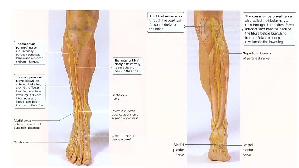
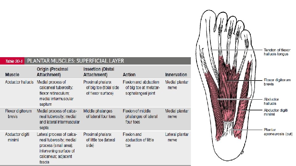
SUPERFICIAL NERVES • cutaneous innervation of the sole of the foot is through branches of the medial and lateral plantar nerves. • medial plantar nerve: branches to the medial side of the sole of the foot and three and a half toes. • The lateral plantar nerve lateral side of the plantar surface of the foot and to the lateral one and a half toes.

• skin on the medial side of the foot is innervated by the saphenous nerve • skin on the lateral border of the foot is supplied by the sural nerve. • skin of the dorsum of the foot is innervated mostly by superficial fibular nerve • adjacent sides of the first and second toes are innervated by the deep fibular nerve.

The dorsum of the foot. A, interphalangeal joint; B, distal interphalangeal joint (note transverse skin crease); C, proximal interphalangeal joint; D, toenail; E, lunula; F, first meratarsophalangeal joint; G, fifth metatarsal; H , first metatarsal-cuneiform joint; I, dorsalis pedis artery; J, navicular; K, medial cuneiform; L, typical site of second metatarsal stress fracture; M, typical site of Jones fracture

Prominence of extensor tendons of toes increased by active extension. A, extensor hallucis longus; landmark for dorsalis pedis artery B, extensor digitorum longus to fifth toe

Anterior aspect of the ankle joint. A, medial malleolus; B, lateral malleolus; C, tibialis anterior tendon: dorsiflex D, extensor hallucis longus; lat to ant. tibialis tendon E, extensor digitorum longus; F, anterior inferior tibiofibular ligament; G, peroneus tertius.

Lateral aspect of the foot and ankle. A, base of the fifth metatarsal; B, lateral malleolus; C, sinus tarsae; D, extensor digitorum brevis; E, peroneus brevis tendon; F, Achilles'tendon; G, sural nerve; H, typical site of fibular stress fracture I; anterior talofibular ligament J, calcaneofibular ligament K, peroneal tubercle L, tuberosity of the calcaneus; M, anterior process of the calcaneus N, cuboid O, subcutaneous bursa

Posterior aspect of the foot and ankle. A, plantar fat pad B, calcaneai tuberosity; C, Achilles'tendon; D, soleus; E, sural nerve; F, medial malleolus; G, lateral malleolus.

Medial aspect of the foot and ankle. A , medial longitudinal arch B , head of the first metatarsal; C, calcaneal tuberositv; D, medial malleolus; E, navicular tuberosity; F, saphenous vein G, post tibial tendon; inversion & plantar flex H, flexor digitorum longus tendon I, posterior tibial artery; J typical site of stress fracture of the medial malleolus; K, deltoid ligament.

Everting the foot increases the prominence of the peroneal tendons.

Plantar aspect of the foot' A, medial sesamoid; B, lateral sesamoid; C, typical site of plantar fasciitis; D‘ plantar fat pad of the heel.

Thanks for your attention
- Slides: 29