Follow up Of Bilateral Adrenalectomy In Cushing Disease

Follow up Of Bilateral Adrenalectomy In Cushing Disease Dr. Ghasemzade Ordibehesht 1394 – May 2015

Case § A 48 year old woman with history of bilateral adrenalectomy’s surgery in 1372/05/09 (because of Cushing disease) is now presented with poor control DM. § Her skin is hyperpigmented. § She has no headache, galactorhea, visual fields defect. § 24. h. U. Free cortisol : 77 ᶙg/24 hrs (50 – 190) § FSH : 79. 1 m. IU/ml § LH : 46. 1 m. IU/ml § Prolactin : 13. 64 ng/ml (4. 2 – 18. 4) § Cortisol. morning : 3. 3 ᶙg/dl § IGF-1 : 233. 4 ng/ml (93 – 245)

§ In 1958, Don Nelson et al. described the first case of the eponymous syndrome in a 33 -year-old woman who had undergone total bilateral adrenalectomy (TBA) 3 years previously for the treatment of refractory Cushing’s disease. § Nelson’s syndrome does not occur infrequently and is a potentially lifethreatening complication of TBA for the treatment of refractory Cushing’s disease with an incidence of 8– 43%in adults and 25– 66%in children. § Nelson’s syndrome can develop up to 24 years post TBA, with mean development at 15 years post TBA. § Hyperpigmentation occurs in up to 42%.
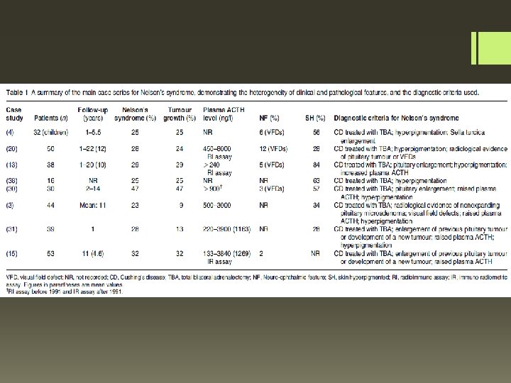

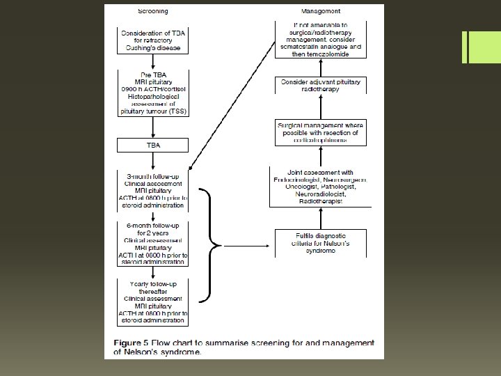
Diagnostic criteria § For a diagnosis of Nelson’s syndrome to be made, a patient must have had prior treatment with TBA for Cushing’s disease (regardless of prior TSS) in addition to at least one of the following two criteria : 1)An expanding pituitary mass lesion post TBA surgery (shown on MRI or CT scan) compared with MRI of the pituitary prior to TBA surgery 2)An elevated level of ACTH to>500 ng/l from a single plasma sample taken at 0800 h prior to steroid administration and post TBA surgery, in addition to progressive elevations of ACTH levels from plasma samples taken on at least three consecutive occasions at different time-points post TBA surgery (a rise of ACTH by O 30% of the initial post-TBA sample)

What is the aetiology of Nelson’s syndrome § However, there is less evidence in support of the ‘released negative feedback’ hypothesis in humans. Although cortisol has been shown to lower the proliferation rate of human pituitary adenoma cells in vitro , secretion of POMC-derived peptides from patients with Cushing’s disease is not attenuated by glucocorticoids. § corticotrophs from Nelson’s syndrome tumours in humans exhibit an attenuated negative feedback response to glucocorticoids in vivo. § there are inconsistencies in that not all post-TBA patients develop Nelson’s syndrome after long-term follow-up, and most patients who develop Nelson’s syndrome also have adequate exogenous steroid replacement therapy. § An important clue for the aetiology of Nelson’s syndrome is the observation that this condition occurs in those patients with Cushing’s disease and aggressive forms of corticotrophinomas. This subgroup of patients also tend to be those who relapse following TSS and who subsequently require TBA.

§ residual cortisol secretion due to accessory adrenal tissue or adrenal remnants was found in 3 – 34%(5 studies, 236 patients), less than 2 % had a relapse of CS. § Nelson’s syndrome occurred in 21 % (0 - 47) of the patients (24 studies, 768 patients)

Endpoint 2: Biochemical outcome § A total of 7 studies investigated the reappearance of cortisol secretion after BADx. Two investigators stated that all patients achieved biochemical resolution of hypercortisolism. § The other 5 studies (n=236 patients) reported evidence for persistent or recurrent endogenous cortisol secretion at follow up. § Only 3 patients ( 2%) experienced a relapse of CS with clinical signs of hypercortisolism

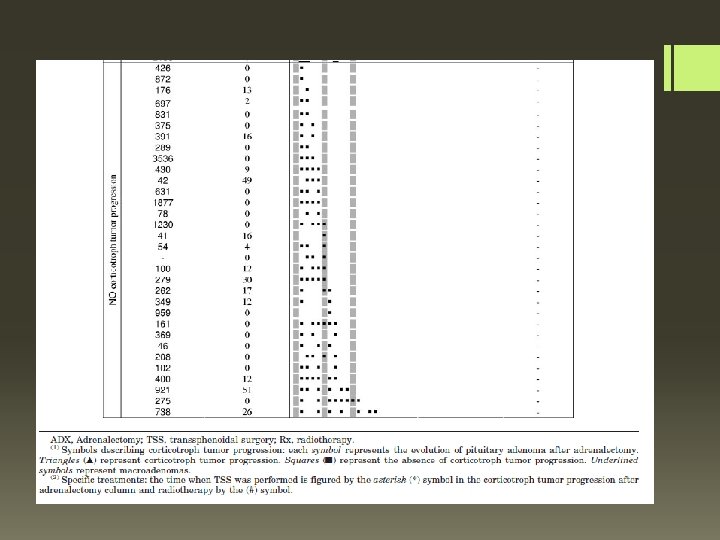
§ Patients included 53 Cushing’s disease patients treated by adrenalectomy between 1991 and 2002, without previous pituitary irradiation. § Results: Corticotroph tumor progression ultimately occurred in half the patients, generally within 3 yr after adrenalectomy. § A shorter duration of Cushing’s disease (adjusted HR: 0. 884/yr), and § a high plasma ACTH concentration in the year after adrenalectomy [adjusted HR per 100 pg/ml (22 pmol/liter): 1. 069] were predictive of corticotroph tumor progression. § During follow-up, corticotroph tumor progression was associated with the increase of corresponding ACTH concentrations (odds ratio per 100 pg/ml of ACTH variation: 1. 055).

Corticotroph tumor progression was defined as the occurrence of an adenoma in cases in which no adenoma was visible on previous MRI scans or by the progression of an existing adenoma

Thanks!
- Slides: 13