Focus Identify the cells using the correct Epithelium


")
")


 Bone (osseous tissue) · Composed of: · Bone cells in")
 Hyaline cartilage · Most common cartilage · Composed of: ·")
 Dense connective tissue · Main matrix element is collagen fibers")
 Adipose tissue · Matrix is an areolar tissue in")
 Blood · Blood cells surrounded by fluid matrix · Fibers")
 Elastic cartilage · Provides elasticity · Example: supports the external")
 Fibrocartilage · Highly compressible · Example: forms cushion-like discs between")
 Areolar connective tissue · Most widely distributed connective tissue ·")
 Reticular connective tissue · Delicate network of interwoven fibers ·")


- Slides: 43
Focus: • Identify the cells using the correct Epithelium classification:
Specialized Structures
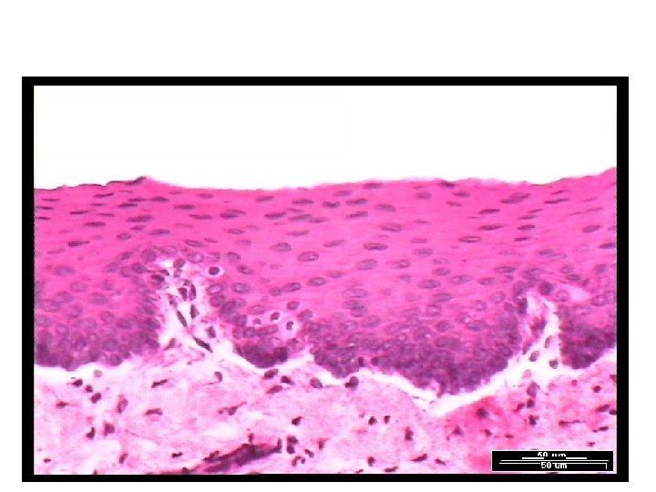
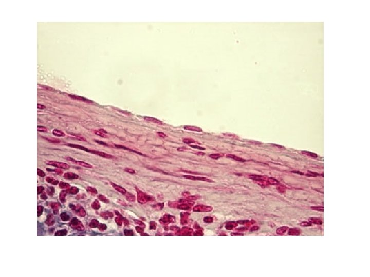
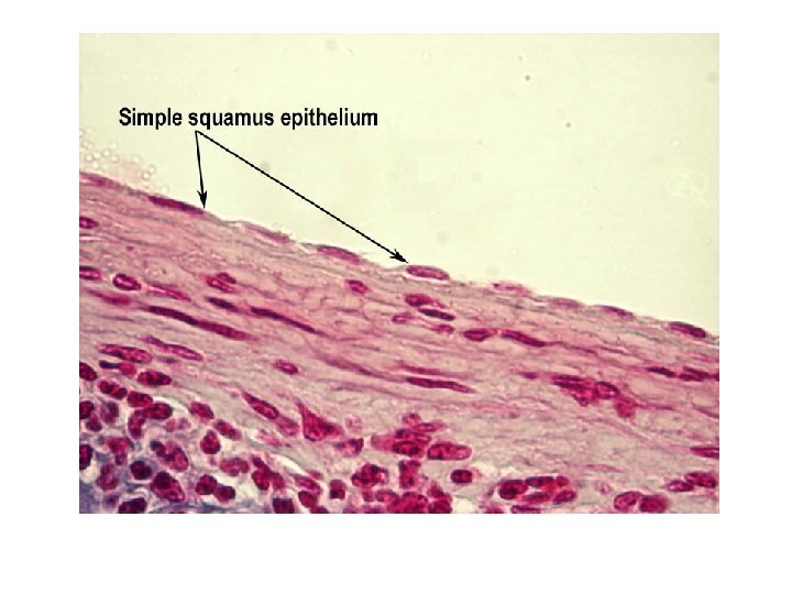
Simple Squamous Epithelium
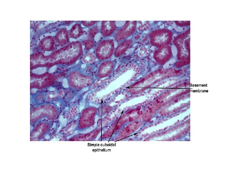
Simple Cuboidal
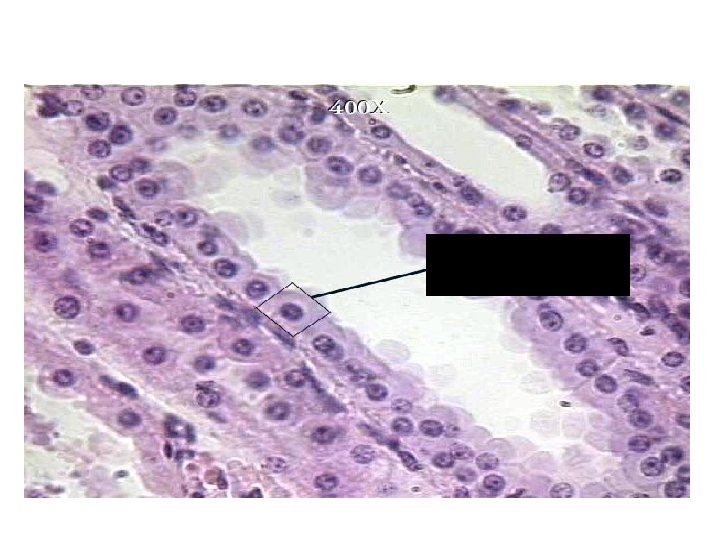
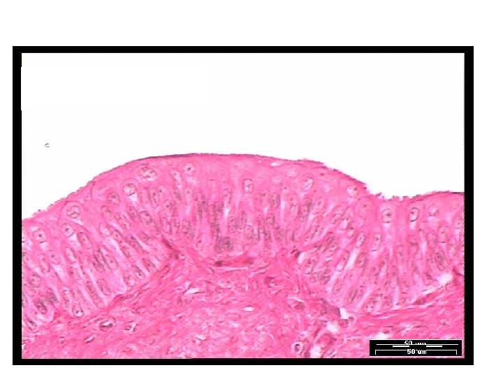
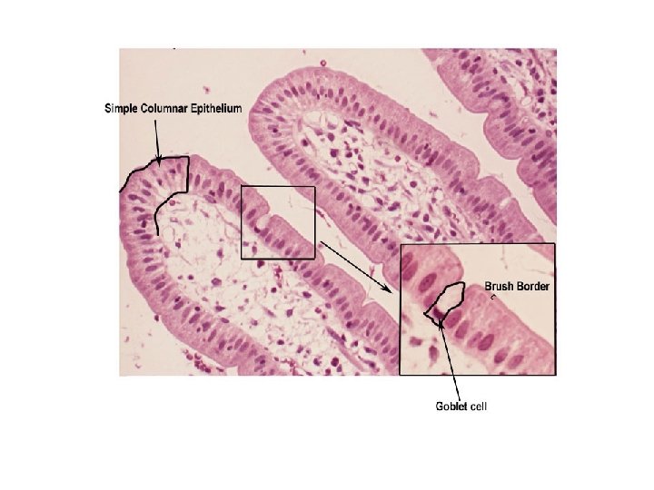
Simple Columnar
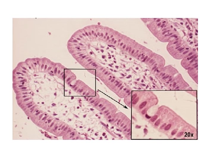
Goblet Cells in simple columnar
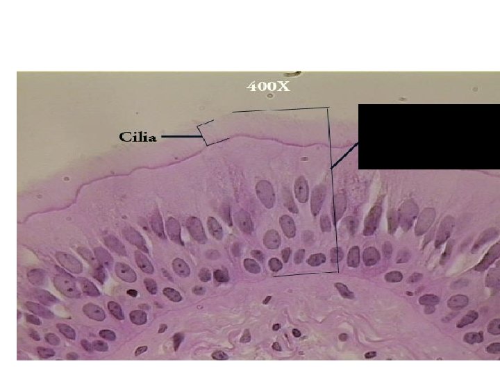
Pseudostratified Columnar
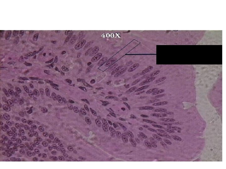
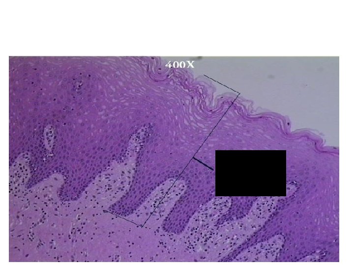
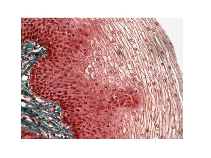
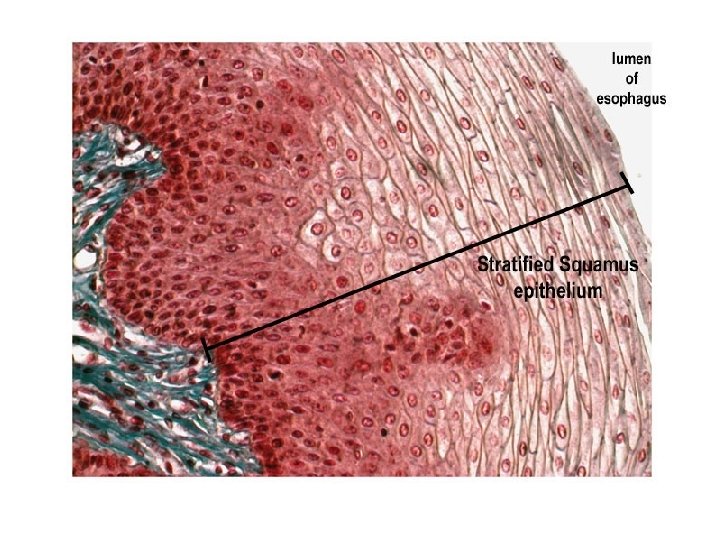
Stratified Squamous
Stratified squamous (Tongue)
Transitional Epithelium (Bladder)
Today’s Objectives • Classify types of Connective Tissue • Identify chief locations of each type of connective tissue • Compare/Contrast connective tissue to epithelial tissue
Connective Tissue: Characteristics · Found everywhere in the body · Includes the most abundant and widely distributed tissues · Functions · Binds body tissues together · Supports the body · Provides protection Copyright © 2003 Pearson Education, Inc. publishing as Benjamin Cummings
Where located • Commonly found under epithelium
Special Characteristics · Variations in blood supply · vascularized · avascular · Extracellular matrix · Non-living material that surrounds living cells Copyright © 2003 Pearson Education, Inc. publishing as Benjamin Cummings
Extracellular Matrix · Two main elements · Ground substance – mostly water along with adhesion proteins and polysaccharide molecules · Fiber · Three types of fibers · Collagen fibers- thick, appear parallel, and are stron. Not stretchy · Elastic fibers- cells spaced apart, stretchy · Reticular fibers. Copyright © 2003 Pearson Education, Inc. publishing as Benjamin Cummings
Connective Tissue Types-Lab 1) Bone (osseous tissue) · Composed of: · Bone cells in lacunae (cavities) · Hard matrix of calcium salts (make rigid) · Large numbers of collagen fibers · Used to protect and support the body Copyright © 2003 Pearson Education, Inc. publishing as Benjamin Cummings
Connective Tissue Types-Lab 2) Hyaline cartilage · Most common cartilage · Composed of: · Abundant collagen fibers · Covers bone and joints · Entire fetal skeleton is hyaline cartilage Copyright © 2003 Pearson Education, Inc. publishing as Benjamin Cummings Figure 3. 18 b
Connective Tissue Types-Lab 3) Dense connective tissue · Main matrix element is collagen fibers · Cells are fibroblasts · Examples · Tendon – attach muscle to bone · Ligaments – attach bone to bone Copyright © 2003 Pearson Education, Inc. publishing as Benjamin Cummings
Connective Tissue Types-Lab 4. ) Adipose tissue · Matrix is an areolar tissue in which fat globules predominate · Many cells contain large lipid deposits · Functions · Insulates the body · Protects some organs · Serves as a site of fuel storage Copyright © 2003 Pearson Education, Inc. publishing as Benjamin Cummings
Connective Tissue Types-Lab 5) Blood · Blood cells surrounded by fluid matrix · Fibers are visible during clotting · Functions as the transport vehicle for materials Copyright © 2003 Pearson Education, Inc. publishing as Benjamin Cummings
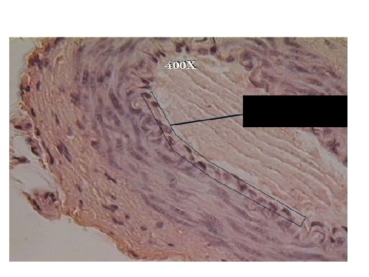
Connective Tissue Types 6) Elastic cartilage · Provides elasticity · Example: supports the external ear Copyright © 2003 Pearson Education, Inc. publishing as Benjamin Cummings Slide 3. 58 a
Connective Tissue Types 7) Fibrocartilage · Highly compressible · Example: forms cushion-like discs between vertebrae Figure 3. 18 c Copyright © 2003 Pearson Education, Inc. publishing as Benjamin Cummings Slide 3. 58 b
Connective Tissue Types 8) Areolar connective tissue · Most widely distributed connective tissue · Soft, pliable tissue · Contains all fiber types · Can soak up excess fluid Copyright © 2003 Pearson Education, Inc. publishing as Benjamin Cummings Figure 3. 18 e Slide 3. 60
Connective Tissue Types 9) Reticular connective tissue · Delicate network of interwoven fibers · Forms stroma (internal supporting network) of lymphoid organs · Lymph nodes · Spleen · Bone marrow Copyright © 2003 Pearson Education, Inc. publishing as Benjamin Cummings Figure 3. 18 g Slide 3. 62
Epithelial membranes - combinations of epithelial and connective tissues which have specific functions • Serous membranes - combined simple squamous epithelium and areolar connective tissue. – Secretes serous fluid as a lubricant for sliding of the tissues. – Found as the pericardial sack which prevents friction when the heart beats, pleural membranes around the lungs, mesenteries attaching the intestines, peritoneum lining the abdominal cavity and covering its organs. • Mucous membranes - combined of columnar (may be ciliated or p. c. c. e. ) epithelium and areolar connective and smooth muscle. – Forms the structure of the GI and respiratory passageways. – Specialized glands, or cells called goblet cells, secrete mucus to protect the lining, lubricate the propulsion of food, and remove particulates form the respiratory tract.
Microscope Classification Lab #3 part two • Each person should have a Hand out • Match-the pictures to the correct slide (label the correct type of connective tissue) • Sketch-portion of tissue to show the structure • Label- tissue type, power and Field Diameter • Write-function and location of tissue in human body
Place the following in the correct place on the Ven Diagram 1. 2. 3. 4. 5. 6. 7. 8. 9. 10. 11. 12. 13. Cells packed together Individual cells Extracellular matrix Throughout body Protection Binds tissue together Epithelial Tissue Support Avascular Vascular Cover Filler Apical surface Basement membrane Connective Tissue
• • • • Cells packed together= epi Individual cells= conn Extracellular matrix= conn Throughout body= both Protection =both Binds tissue together= conn Support= both Avascular= both Vascular= Conn Cover= epi Filler= con Apical surface= epi Basement membrane= epi