FMRI DIFFICULTIES IN MIND READING f MRI functional

 magnetic resonance imaging � Neuroimaging: get the structure of")


 usable image")


- Slides: 24
FMRI – DIFFICULTIES IN MIND READING
� f. MRI: functional (nuclear) magnetic resonance imaging � Neuroimaging: get the structure of the brain �Want to know how it works: connection brain parts and brain functions �Aim: measure the local „thinking activity” � Usage (criticism): �Lie detector �Neural- and psychological-modell checking (think/know experiment)
How we use it � Human attempts frequent � Well-planned tasks or questions � Measures: order of minutes �One measure: order of 5 seconds �Measure with and without tasks or question, further investigation based on the difference � Overlap image the intensity map and brain
The basics of f. MRI � MRI: interaction between spins and magnetic field � QM based phenomena �Classical view is almost satisfactory � Find a „think-activity”-sensitive MRI measureable quantity, measure it, and then reflect to think activity
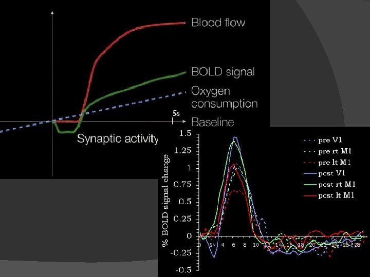
Get the signal - BOLD � Blood-oxygen-level dependence � Hemoglobin: Fe 2+ can absorb O 2 �Hemoglobin + O 2 �Hemoglobin – O 2 � Measure : diamagnetic molecule : paramagnetic (S=2) the oxygen-flow differencies in vein � Determine the connection between BOLD singal and toughts
Get the signal – O 2 flow � The oxygen flow depends on the communicating intenstiy � Communication needs energy �Neuron cells don’t have repository �Increased activity needs more energy �BOLD signal decreasing and then increasing
Time, accuracy and resolution � Time: depends on the BOLD signal, about less than half a minute �Do experiments with the same patient, same time � Accuracy: easily detect maximum of BOLD � Space: resolution: in order of mm×mm×mm �Problem: the motion of the patient
Increasing space resolution No new information with increasing resolution Signal comes from multiple capillars Solution: BOLD signal minimum: more localised Longer time or Bigger magnetic field No (? ) news: cerebral tasks are not well localised
Temporal resolution � � Increasing time resolution does count � (more details) usable image � Two tricks to improve: �Spin echo – gradient echo �EPI: echo planar imaging
Measurement: EOM � First: apply � Short RF pulse: � � �
Measurement: relaxation and decoherence � Solutions after „excitation”: � We can measure � Important: � New rotating cylindrical coordinates with frequency:
Measurement: spin-echo
Measurement: spin-echo
Measurement: spin-echo
Measurement: sliceexcitation
Measurement: slice-excitation and spin-echo � Find for the desired excitationdistribution:
Measurement: another way, the gradient echo
Measurement: EPI � Echo Planar Imaging: increase time resolution even more!
What we get: images
An experiment: caffeine � caffeine as a contrast booster
Limits � We cannot answer any why question – only answer the question where � The brain is 3 D – the image is 2 D, so more experiments needed � We can measure only the neurons firing, but not the real actyvity (block or stimulate? ) � High degree of cerebral plasticity: places may vary
Mind reading The map of optic nerves to vissual cortex is so „localised”, „continous” � Training set: known videos and those f. MRI signal � Unknown video: f. MRI signal → video images � Ability to guess what patient think � Numerical problem � Further aim: movement of implants � Success: camera-eye �
� http: //sites. google. com/site/gallantlabucb /publications