Fluorescence Microscopy fluorescence imaging has become the mainstay

Fluorescence Microscopy “fluorescence imaging has become the mainstay of microscopy” -- Nat. Methods, 2005 Objectives: 1. Understand the basics of fluorescence 2. Understand how fluorophores are used in microscopy 3. Understand how a microscope is set up to do fluorescence 4. Understand basics of immunofluorescence

Have you used a fluorescence microscope before? A. Yes B. No

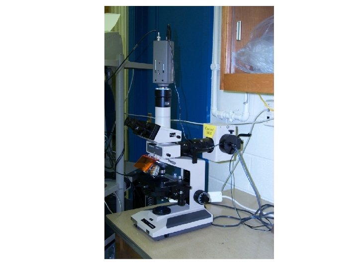
Light is white

Light is white But is composed of a mixture of colors.

R O Y G B I V 700 650 600 550 500 450 400 wavelengths in nm E = h h/f

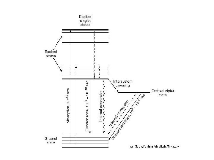

FITC Fluorescein isothiocyanate

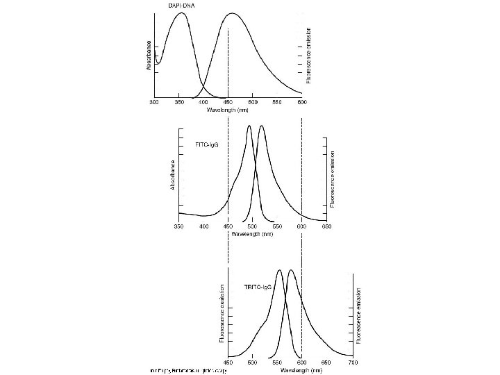
FITC Amax = 490 nm Emax = 520 nm

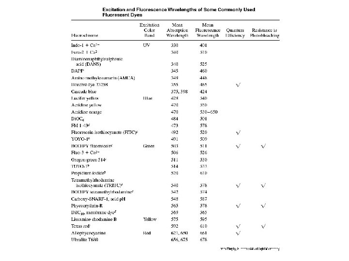
How fluorophores are used 1. Some bind to biological specimens 2. Others can be coupled (conjugated) to other non-fluorescent molecules that bind to specimens

http: //probes. invitrogen. com/

http: //fluorescence. nexus-solutions. net/frames 6. htm

What color of light do we want a fluorescein excitation filter to pass? A. Blue only B. Green only C. Red only D. Any color light

What color of light do we want a fluorescein emission filter to pass? A. Blue only B. Green only C. Red only D. Any color light

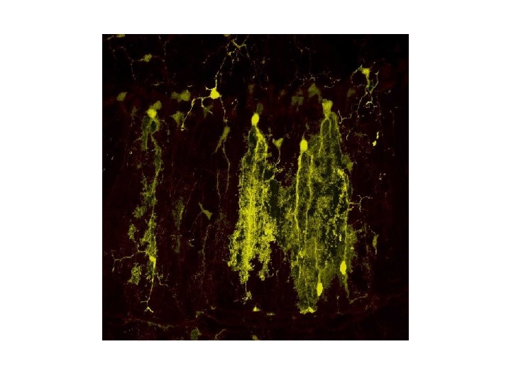
What do we see when we look down a fluorescence microscope at a specimen stained with fluorescein? A. Blue background with green stain B. Green background with blue stain C. Red background with green stain D. Black background with green stain

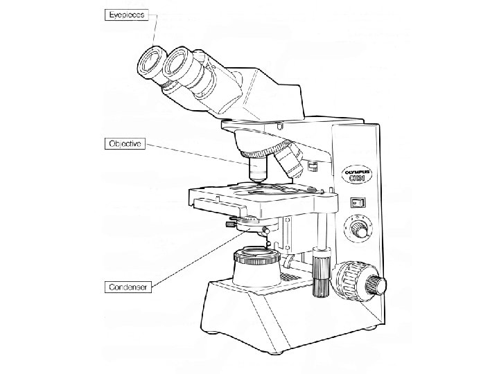
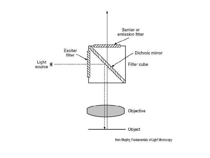
Epifluorescence

Immunofluorescence 1. Direct 2. Indirect

Imagine you made an antibody to protein Y by injecting a goat. Which of the following would be okay as a secondary antibody to localize protein Y? A. A goat anti-rabbit-immunoglobulin B. A horse anti-chicken-immunoglobulin C. A sheep anti-mouse-immunoglobulin D. A cow anti-goat-immunoglobulin

GFP Green fluorescent protein


J. W. Lichtman and J. A. Conchello, 2005. Fluorescence microscopy Nature Methods 2: 910 -919. http: //www. nature. com/nmeth/journal/v 2/n 12/full/nmeth 817. html
- Slides: 27