FLOW CYTOMETRY introduction Flow cytometry is a technique


 was developed in 1968 by")
 of a single wavelength is directed")


: FITC, Alexa Fluor 488, GFP. Orange (usually")


- Slides: 26
FLOW CYTOMETRY
introduction Flow cytometry is a technique for counting and examining microscopic particles, such as cells and chromosomes, by suspending them in a stream of fluid and passing them by an electronic detection apparatus. It allows simultaneous multiparametric analysis of the physical and/or chemical characteristics of up to thousands of particles per second. Flow cytometry is routinely used in the diagnosis of health disorders, especially blood cancers, but has many other applications in both research and clinical practice.
History The first fluorescence-based flow cytometry device (ICP 11) was developed in 1968 by Wolfgang Göhde. Some of the flow cytometry instruments developed were cytofluorograph, PAS 8000, and EPICS. The original name of the flow cytometry technology was "pulse cytophotometry"
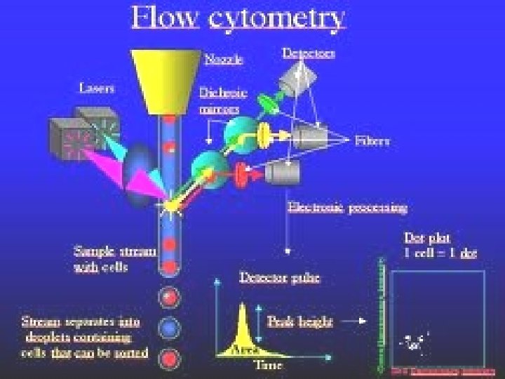
PRINCIPLE A beam of light (usually laser light) of a single wavelength is directed onto a hydrodynamically-focused stream of fluid. No. of detectors aims at a point where the stream passes through the beam of light parallel to it is Forward Scatter (FSC )and perpendicular to is Side Scatter(SSC). Particle passing through the beam scatters the ray, and fluorescent chemicals found in the particle may be excited into emitting light at a longer wavelength than the light source.
Contd… This combination of scattered and fluorescent light is picked up the detectors and based upon the peak formed. The physical and chemical structure of each individual particle can be determined. FSC depends on cell volume and SSC depends on inner complexity of the particle.
Instrumentation A flow cytometer has 5 main components: • a flow cell - liquid stream (sheath fluid) • Light source- mercury, Xenon • a detector • an amplification system - linear or logarithmic • a computer for analysis of the signals.
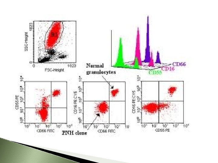
The process of collecting data from samples using the flow cytometer is termed 'Acquisition'. Acquisition is mediated by a computer physically connected to the flow cytometer, and the software which handles the digital interface with the cytometer. Data analysis The data generated by flow-cytometers can be plotted in a single dimension, to produce a histogram, or in twodimensional dot plots or even in three dimensions.
Contd… The regions on these plots can be sequentially separated, based on fluorescence intensity, by creating a series of subset extractions, termed "gates. " The plots are often made on logarithmic scales. Because different fluorescent dyes' emission spectra overlap, signals at the detectors have to be compensated electronically as well as computationally.
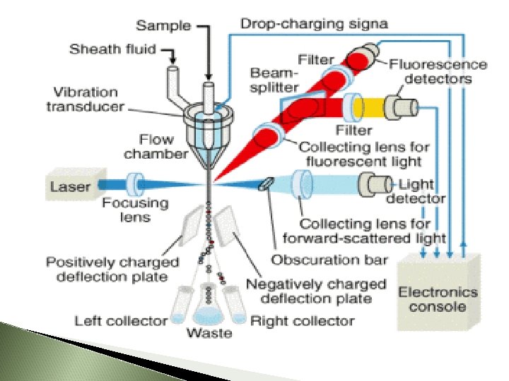
Fluorescence-activated cell sorting It provides a method for sorting a heterogeneous mixture of biological cells, based upon the specific light scattering and fluorescent characteristics of each cell. The cell suspension is entrained in the center of a narrow, rapidly flowing stream of liquid. The flow is arranged so that there is a large separation between cells relative to their diameter. A vibrating mechanism causes the stream of cells to break into individual droplets.
cont. D… The system is adjusted so that there is a low probability of more than one cell per droplet. Just before the stream breaks into droplets, the flow passes through a fluorescence measuring station where the fluorescent character of interest of each cell is measured. An electrical charging ring is placed just at the point where the stream breaks into droplets. A charge is placed on the ring based on the immediatelyprior fluorescence intensity measurement.
The opposite charge is trapped on the droplet as it breaks from the stream. The charged droplets then fall through an electrostatic deflection system that diverts droplets into containers based upon their charge. In some systems, the charge is applied directly to the stream, and the droplet breaking off retains charge of the same sign as the stream. The stream is then returned to neutral after the droplet breaks off.
Fluorescent labels Green (usually labelled FL 1): FITC, Alexa Fluor 488, GFP. Orange (usually FL 2): PE Red channel (usually FL 3): Per. CP, PE-Cy 5. Infra –red (usually FL 4) : PE-Cy 7. Red diode laser : APC, APC-Cy 7
Measurable parameters volume and morphological complexity of cells cell pigments such as chlorophyll or phycoerythrin total DNA content total RNA content DNA copy number variation chromosome analysis and sorting protein expression and localization Protein modifications, phospho-proteins nuclear antigens enzymatic activity
applications In molecular biology it is useful when used with fluorescent tagged antibodies. In the field of medicine in transplantation, hematology, tumor immunology and IVF In marine biology, photosynthetic plankton can be studied. In protein engineering , it is used to identify cell surface – displayed protein variants.
Use of flow cytometry to measure copy number variation of a specific DNA sequence (Flow-FISH)
Flow Cytometry Antibodies & Secondary Detection Extensive selection primary and secondary antibodies, streptavidin conjugates and antibody labeling kits using Molecular Probes® dyes and nanocrystals, all validated for flow cytometry
Cell Health and Viability Assays for Flow Cytometry Find a diverse array of flow cytometry-validated reagents and kits for analyzing apoptosis, cell viability, cell cycle and cell proliferation, including the new Cell. Trace™ Reagents for Flow Cytometry, LIVE/DEAD® Fixable and SYTOX® Dead Cell Stains.
Apoptosis Assays for Flow Cytometry Invitrogen offers a wide array of apoptosis assays for flow cytometry.
Cell Proliferation Assays for Flow Cytometry Cell Proliferation assays are an important set of fluorescence based tests that can monitor cell health and cell division by either evaluating DNA synthesis through thymidine incorporation or population doubling using dye dilution cell tracing stain. These assays provide a rich data set that can be analyzed to answer complex questions
Cell Cycle Assays for Flow Cytometry nvitrogen offers a series of fluorescent dyes to allow accurate cell cycle analysis in either live or fixed cell populations.
Cell Viability Assays for Flow Cytometry Removing dead cells from your flow cytometry data is a critical step to ensure accurate results and analysis.
THANK YOU