Flow Cytometry FACS FluorescenceActivated Cell Sorter Flow Cytometry
")

, kromě NK buněk CD-4 helperské")


 n Historically 1 -2")

 and lenses")

 n n n n Clone Isotype Mouse, rat, rabbit, goat Reactivity Specificity")


")
")


- Slides: 28
Flow Cytometry FACS (Fluorescence-Activated Cell Sorter)
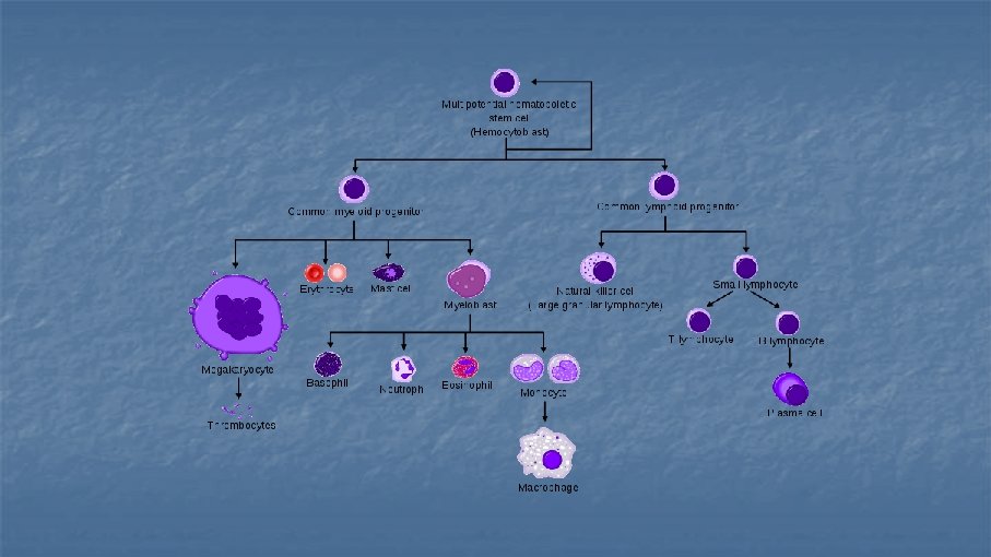
Flow Cytometry Leonard Herzenberg n n technique for analyzing populations of cells are measured individually but in large numbers cells are incubated with fluorescently labeled monoclonal antibodies directed against different antigens (cell surface, intracellular, nuclear) CD numbers (clusters of differentiation) n CD 1 -CD 364 (Nov 2014) n Lineage specific markers n Used in phenotyping (leukaemias)
CD-2 nezralé T-lymfocyty CD-3 všechny T-lymfocyty (součást Tc. R), kromě NK buněk CD-4 helperské T-ly CD-7 T-ly nacházející se v thymu CD-8 cytotoxické T-ly CD-14 monocyty a makrofágy CD-15 neutrofily, eosinofilní granulocyty CD-16 NK buňky, neutrofily CD-19 B-lymfocyty CD-34 lymfoidní a myeloidní progenitorové buňky CD-38 plazmatické buňky CD-40 B-ly (izotypový přesmyk za přítomnosti CD-40 ligandu) CD-45 panleukocytární antigen CD-56 NK buňky CD-58 endothelie, buňky prezentující antigen T-lymfocytům (adhese s CD-2) CD-64 Fc γ receptor (makrofágy, neutrofily…) CD-68 dendritické buňky CD-80 a 86 buňky prezentující antigen T-lymfocytům CD-95 FAS receptor CD-203 bazofilní granulocyty
Common phenotypes and AML FAB Subtyp Common Phenotype e M 0 DR, CD 13, CD 34, CD 7−/+, Td. T−/+ M 1 Similar to MO except CD 15−/+ M 2 DR, CD 13, CD 33, more CD 15 and less CD 34 than M 1 M 3 DR(−), CD 13, CD 15, CD 33, CD 34−/+, CD 2 occasionally M 4, M 5 DR, CD 15, CD 14+/−, CD 33 > CD 13, CD 34−/+, CD 4 weak M 6 DR, CD 13−/+, CD 33+/−, CD 34, CD 45 weak M 7 DR−/+, CD 33+/−, CD 34, CD 41, Blood CD 61 October 15, 1997 vol. 90 no. 8 2863 -2892
Flow Cytometer Instrumentation n n four general components n Fluidics n Optics n Detectors n Electronics Understanding how a flow cytometer operates is critical to the design of your experiments
Flow Cytometry Fluidics n n n The cell sample is injected into a stream of sheath fluid Labeled cells are accelerated and individually pass through a laser beam its resulting fluorescence and angle deflection detected by a photocell
Flow Cytometer Optics n n n Light emitted from the interaction between the cell and the laser beam is collected by a lens The light moves through a system of optical mirrors and filters Specified wavelengths are then routed to optical detectors
What can you measure with a Flow Cytometer? n 1. Size and complexity of cells n Forward-scattered light (FSC) n n Side-scattered light (SSC) n n n is proportional to the surface area or size of a cell is proportional to the granularity or internal complexity of a cell 2. Cell surface molecules 3. Nuclear antigens 4. Protein expression/localization 5. Copy number variation (Flow-FISH) 6. Cell pigments
Fluorochrome Emission n n The laser beam excites the fluorochrome at a specific wavelength and the fluorochrome emits light at a separate wavelength (emission) Property of any fluorescent dye; 2 things: n Excitation spectra n Emission spectra n If your laser functions at 488 nm, find dyes that have excitation spectra at that λ
Flourochromes Have Overlapping Emission Wavelengths
Technical Components n n Detection Systems n Photomultiplier Tubes (PMTs) n Historically 1 -2 n Current Instruments 3 -9 Illumination Systems n Lasers (Blue 488 nm, Green 532 nm, Red 640 nm, violet 405 nm) n BD FACS Calibur n n n BD FACS Canto II n n Argon ion (488) He. Ne (633) Solid state (488) He. Ne (633) Violet (405) FACS Aria sorter
Electrostatic Flow Sorting
How do we detect the signal? n n n Photomultiplier tubes (PMT) and lenses FL-1 lets light through at 500 -560 nm FL-2 lets light in at 560 -611 nm FL-3 610 -660 nm FL-4 >660 nm Picking dyes too close in λ results in no detection
Flow Cytometer Electronics n n The voltage pulse height, width, and area are determined by the particle’s size, speed, and fluorescence intensity parameters are acquired analyzed in real-time by a computer
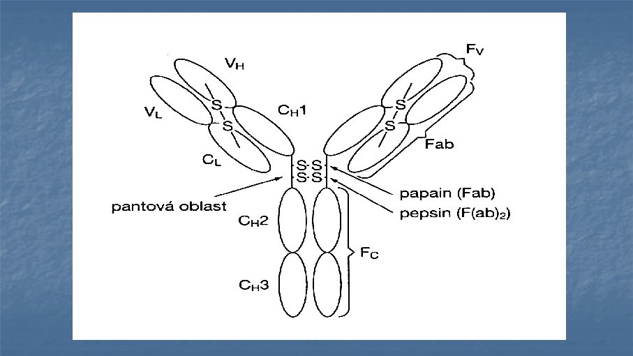
Antibodies (immunoglobulins) n n n n Clone Isotype Mouse, rat, rabbit, goat Reactivity Specificity Fluorochrome Catalogue number and cost: $$$ £££ €€
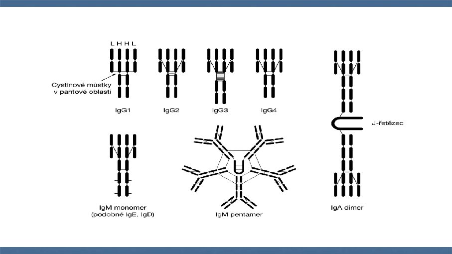
Polyclonal vs Monoclonal antibodies
-Cells incubated at 4°C, 37°C or room temperature -avoid non-specific binding -always have a negative control
Flow Cytometry (Data)
Flow Plots (1 D histogram, 2 D scatter plots, 2 D contour plots)
Gating
Application of FC n n n Phenotyping Cell function; proliferation, apoptosis, cell cycle Intracellular staining; IFN Nuclear staining Clinical n detection of malignant cells