FIRST PASS RADIONUCLIDE ANGIOCARDIOGRAPHY Assoc prof A Punda


 right ventricul which can fill out retrosternal space Left atrium isn’t")


, after the intravenous")


- Slides: 34
FIRST PASS RADIONUCLIDE ANGIOCARDIOGRAPHY Assoc. prof. A. Punda, MD. , Ph. D. Assoc. prof. V. Marković, MD. , Ph. D. S. Gračan, MD. , nucl. med. spec.
• diagnostic imaging method which uses intravenously injecticed radiopharmaceutical for dynamic scintigraphy of the first pass of radiopharmaceutical through the heart, lungs and blood vessels by gamma camera over the heart • analasys of sequantial scintigraphs and generating “time activity curve” allows for diagnosis of heart abnormalities with pathologic communication between heart chambers or between pulmonary artery and aorta, and for quantification of left-to-right and right-to-left shunts
Radiopharmaceuticals and biodistribution - Tc-99 m-pertechnetate or Tc-99 -m labeled albumin or red blood cells - 8 -10 m. Ci/m², adults 20 m. Ci; minimal dose 2 m. Ci, volume < 0, 5 ml, bolus injection flushed with 10 ml saline - v. jugularis interna or v. jugularis externa
- Supine position, gamma camera precardial - collimator: multihole parallel, high sensitivity or convergating - 2 -20 frame/sec (1 frame: 0, 05 -0, 5 sec) = better time resolution, during 30 sec - Images are stored to a computer - Premedication with Na-perchlorate (Irrenat)
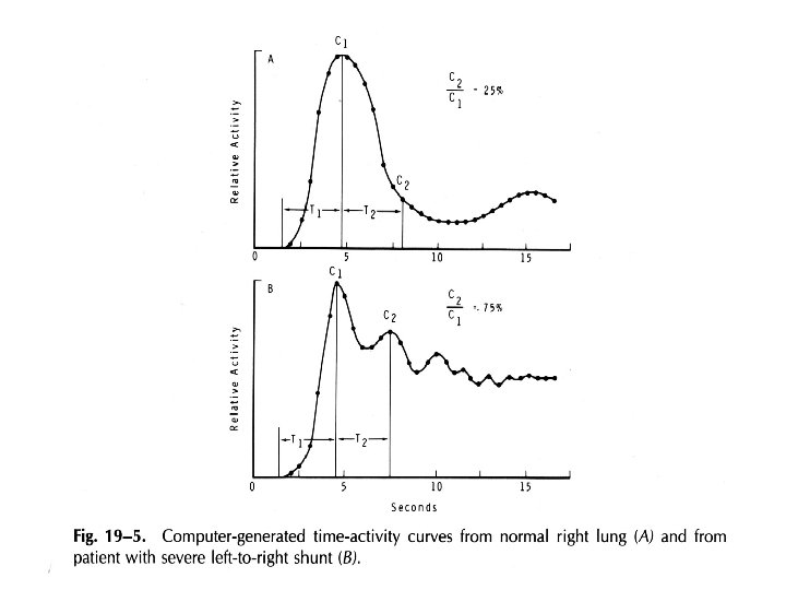
Normal lung curve of radioactive indicator; left peak, which is higher and thiner, is showing first passthrough the heart, and right peak is showing recirculation of the systemic blood pool
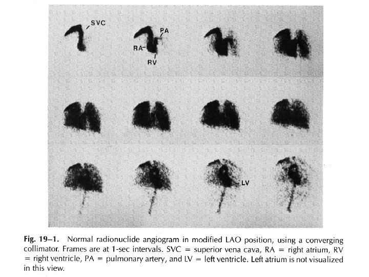
Normal findings
L→R shunt - Early recirculation of radiopharmaceutical in the left to right shunt - Delayed elimination of radiopharmaceutical from the lung (higher blood-flow through lungs) - Poor image of the left heart - Identification and quantification of the shunt on the curve
Radionuclide angiocardiography in a patient with a ventricule septal defect Midium big left-right shunt. Individual scintigrams represent 0, 8 sec sequences. There is a delay of activity in the lungs due to early recirculation of radiopharmaceutical.
Normal lung curve Lung curve in patient with left-to-right shunt: On descending slope of the curve, which represents first passage of the radionuclide, there is peak of early recirculation
L-R shunt
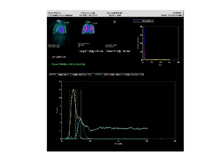
Quantification
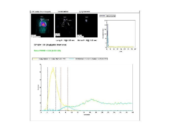
Cardiac shunt lung curves normal lung TAC See physiological recirculation at about 18 s Pathological lung TAC See bump at the falling half Of the lung curve
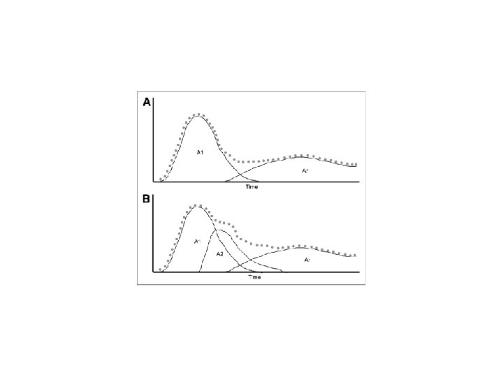
Modelling of ideal bolus injection Gamma-Fit of Normal curve pathological curve: same input function
Analysis of difference Difference between pathological Curve and Gamma. Fit equals shunt + Phyisiological recirculation Gamma. Fit of difference curve equals shunt
Indications for the FP-RA: 1. Detection, localization and quantification of the intracardiac shunt and shunt between large blood vessels 2. Follow-up of patients for the evaluation of the shunt’s size, as well as for the detection of postsurgical residual shunt
Congenital heart defects with left-to-right shunt and higher blood flow through the lungs • ASD: 10 % of congenital heart defect in the childhood; ostium secundum (most often), ostium primum and sinus venosus • VSD: most common congenital heart defect in the childhood (30%) • Ductus Botalli (ductus arteriosus persistens), 8 -10%; conjuntion between aorta and left branch of the pulmonary artery
ASD Prominent (enlarged) right ventricul which can fill out retrosternal space Left atrium isn’t enlarged because left atrium decompression is being formed in the right atrium through the defect
VSD Enlarged heart shadow Enlarged truncus pulmonale Enlarged pulmonary heart vessels Congestive heart disease Pan sistolic murmor
DUCTUS ARTERIOSUS PERSISTENT DA – normal conjunction between aorta and pulmonary artery which is normaly closed within 24 hours after birth or remains persistent untill the second month of life, or less common untill the end of the first year Small conjunction – normal size of the heart Wide ductus bigger suply and blood flow load of the LA and LV expanded main branch of the pulmonary artery, depending of the flow wight of the perifer arteries is determined, sizing from the normal wight to the plethora INCREASE OF THE BLOOD PRESSURE – BIDIRECETION SHUNT EXERCISE CIANOSIS FURTHER INCREASE OF THE BLOOD PRESSURE – RIGHT TO LEFT SHUNT CIANOSIS, EISENMENGER SY
Congenital heart defects with right-to-left shunt, cianosis and diminished blood flow through the lungs • Tetralogy Fallot (10%): pulmonary aretry stenosis, VSD, hypertrophy of the right ventricle, ante – and dextraposition of the aorta origin • Tricuspidal atresia, TA (2%): shunt in the atria (and ventriculs), blood arrives to the lungs through the other way; or through the ventricular septum in the right atrium and lungs, or throuhg the persistent ductus Botally • Ebstein anomaly: anomaly formed tricuspidal valve, lies deep in the right ventricule, most often with defect in the atria with right-to-left shunt
Congenital defects with right-to-left shunt and cianosis • Transposition of the large arteries, d-TVA (5, 4%) • Adjusted transposition of the large arteries, l-TVA • Exit of both arteries from the right ventricul, “double outlet” • joint ventricul • Truncus arteriosus persistens
Schematic representation of curve activities of the heart, lung and aorta: A. Normal, B. Left-to-right shunt, C. Right-to-left shunt
RIGHT-LEFT shunt - Early pass of radiopharmaceutical from the right to the left heart and it’s arrival to the aorta (and sistemic circulation) at the same time as the arrival of the radiopharmaceutical to the lung
• Tc-99 m-MAA (size of the particels 10 -20 µm), after the intravenous injection they are allmost entirely “captured” in the first capillary system (lungs), dosage 100 – 400 µCi • >3% pass – caracteristic for the right-to-left shunt % shunt = (whole body counts – lung counts/ whole body counts) x 100
MAA- normal finding, no right to leftt shunt R-L shunt
R→L shunt
R→L shunt • Tc-99 m-MAA • Acumulation of the radiopharmaceuticals in the brain and kidneys.
Dextrocardia
Dextrocardia, Tetralogia Fallot, dilatated right atrium and ventricul The end