First lecture Immune system structure and function Immune


")


 which serves as a recognition")


 adaptive immune responses.")

 or Neutrophils: Predominant type of white blood cell, rapidly migrate")


 and maturation")


 B cells CLP Myeloid DC (CD 11 c+, CD 11 b+,")


 * tingible body")
 The paracortex contains lymphocytes and accessory cells along with supporting")


 and blood is a region of")


 This is comprised of: * tonsils, adenoids (Waldeyer's ring) *")
 cells, they collect Ag. Peyer's patches facilitate the generation")

 In addition to the lymphoid tissue concentrated within the lymph")

 DC Epidermis Skin Dermis Denstritic")


 Inflammation (TLR-PAMP, IL-1, TNF-")
 Skin draining lymph node DC Skin draining LN T")


- Slides: 95
First lecture: Immune system structure and function
Immune system like any other system in the body includes: Organs, tissues, cells, molecules and some times fluids. The tissue of the immune system are called lymphoid tissue. The cells of the immune system are all blood cells except the RBCs. The molecules of the immune system are: antibodies. complement, cytokine, chemokines etc. The fluid of immune system is called lymph. The lymph: is plasma components.
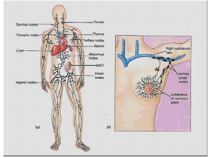
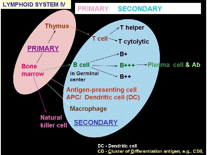
OVERVIEW OF THE IMMUNE SYSTEM Organized similarly to nervous system: Cells of the immune system (IS) found throughout the body, but also found in specialized organs. Cells: lymphocytes, macrophages & monocytes, dendritic cells, granulocytes. All arise from pluripotent hematopoietic progenitor cells in bone marrow. Organs: lymph nodes (found in various locations), thymus, spleen - these constitute the lymphoid organs.
Lymphatic system
Immune system organs and tissues (Lymphoid tissue)
LOCATION OF MAJOR LYMPHOID ORGANS THROUGHOUT THE BODY (not truly a lymphoid organ, but the source of IS progenitor cells)
Lymphoid tissues: Are the sites where the ; – Generation, – Maturation, – Habitation and – Activation of the immune cells take place.
Lymphocytes: Major subtypes are T and B cells, responsible for immunological memory. T cells mature in thymus; B cells in avian Bursa of Fabricius but mammalian fetal liver & bone marrow. Cells participate in cell-mediated immunity & regulation responses; B cells synthesize Abs. NK cells are morphologically similar to T & B cells; are cytotoxic in absence of prior stimulation.
lymphocyte from blood smear, Wright-Giemsa stain, 1000 x
LOCATION OF THE BURSA OF FABRICIUS T and B cells have specific antigen receptors, which play roles in developing immunological memory and in specificity of the immune response to antigens. Both T and B cells secrete proteins called cytokines, which form the communication system among and between cells and cell types.
T versus B Cells Ag receptor Ig T cells B cells TCR related to Ig BCR is membrane-bound but not Ig Ag recognition in context of MHC on APC or accessory cells can recognize Ag alone Functional subsets Th (helper) and Tc (cytolytic) subsets of B cells not different in function Secrete Cytokines Ig (as Ab) and cytokines Surface markers CD 4 and CD 8 (among many others) Ig (among many others) When Agthen activated Become (proliferating) Become lymphoblasts, lymphoblasts become plasma cells Costimulation required? Yes No
TWO MAJOR TYPES OF T CELLS Th 1 & Th 2 Effectors cells help B cells make Ab Class I MHC expression – ubiquitous (every where). Class II MHC expression - constitutive: restricted to B cells, a proportion of monos / macs & DCs after activation: induced on most cell types.
MONOCYTES AND MACROPHAGES Monocytes are immature macrophages, circulate in blood & accumulate at sites of inflammation. MQs may differentiate in tissue in absence of antigen (e. g. Kupffer cells in liver) or differentiate in response to Ag. They are Ag-presenting cells (APC) and cooperate with B and T cells in mounting immune responses. Also phagocytose microbes; contain bactericidal mechanisms. macrophage in tissue, H&E stain, 400 x mono in blood smear Wright Giemsa, 1000 x
MONOS AND MQS-CONTINUED Express a myeloid receptor (CD 14) which serves as a recognition molecule for a wide variety of bacterial envelope molecules, such as LPS from Gram -ve organisms and components of Mycobacterial and Gram +ve cell walls. Ligation of this receptor leads to MQs activation.
Also they’re activated by T cell derived cytokines leading to increased phagocytosis and microbicidal activity (increased activity of degradative enzymes, nitrogen and oxygen free radical production and prostaglandins etc. ).
NOTE: T cell derived cytokines increase the antigen presenting activity of macrophages which, in turn, are able to present antigen to T cells. This cycle will continue as a positive feedback loop until the antigen is eliminated.
DENDRITIC CELLS DCs are the APCs for primary (1 st time) adaptive immune responses. Also constitute major components of the innate immune system and the bridge to adaptive immunity. Two sites of origin, plasmacytoid DCs (periphery & spleen) and myeloid DCs (bone marrow). So far no major functional differences noted. LPS-activated DCs
THE DENDRITIC CELL THAT’S NOT A DENDRITIC CELL Follicular DCs are found in primary & secondary lymphoid follicles but are not of the same origin as plasmacytoid or myeloid DCs. In fact, their exact origin is unknown, though they may be myeloid. FDCs play a role in controlling B cells responses. FDCs in the spleen
GRANULOCYTES Polymorphonuclear leukocytes (PMNs) or Neutrophils: Predominant type of white blood cell, rapidly migrate to sites of infection or inflammation. Phagocytic, they have special enzymatic pathways for enhanced bactericidal action. Also called azurophils, due to bluestained (azurophilic) granules.
azurophilic staining of PMNs PMN, note tri-lobed nucleus Wright-Giemsa, 1000 x Mono Comparison of mono to PMN
BASOPHILS AND MAST CELL Have basophilic granules, which contain mediators, especially of allergic responses. Basophils circulate, mast cells found in tissue. Basophil Degranulating mast cell intact mast cells
EOSINOPHILS Have granules that stain red with eosin. Mediate late phase of allergic response, active in immune response to parasites & tumors (antibodydependent cell-mediate cytotoxicity). Granules contain toxic proteins of high p. H. Eosinophil eosino Eosinophil PMNs Comparison of PMNs to eosinophil
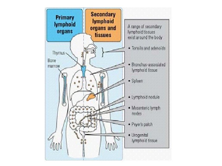
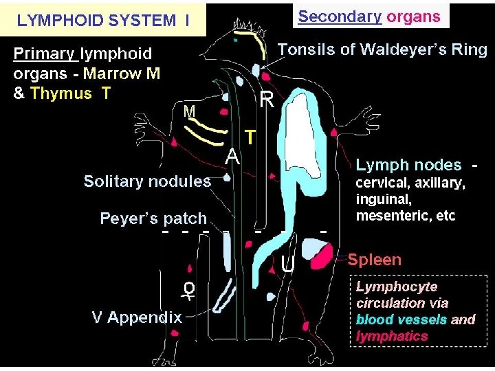
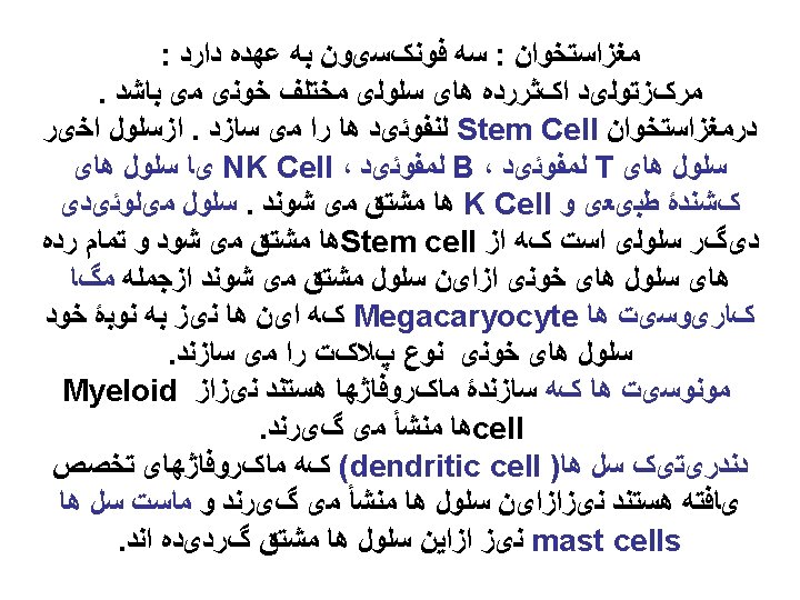
ORGANS OF THE IMMUNE SYSTEM PRIMARY LYMPHOID ORGANS Primary lymphoid organs are where lymphocytes arise and mature in the absence of antigenic stimuli. They are the bone marrow and thymus. Bone marrow: Source of all hematopoietic progenitor (stem) cells, site of B cell maturation post-birth in mammals. Hematopoietic stem cell Stromal stem cell
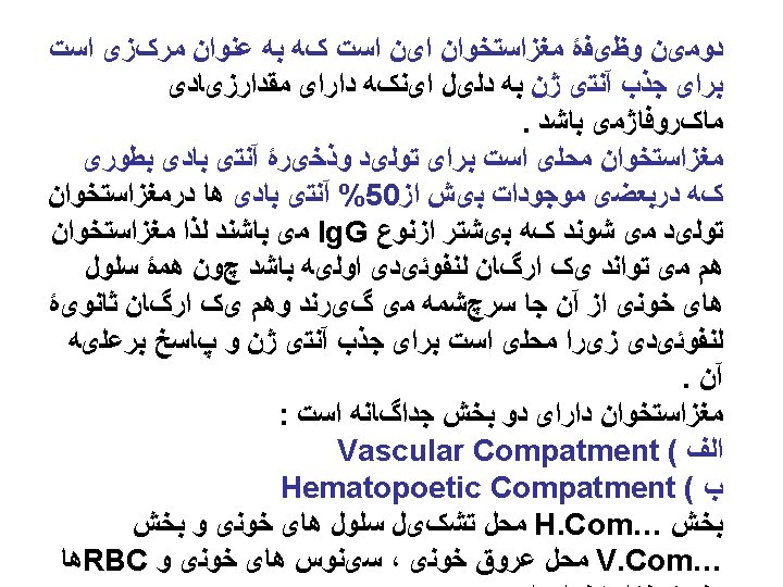
Primary lymphoid tissues The sites where the blood cells, generation ( haematopoiesis) and maturation take place Haematopoiesis takes place: • In yolk sac (first 5 weeks of fetus’s age) • In fetal liver (5 -8 weeks of age) • In the whole bone (after 4 months of fetus’s age) In adult the haematopoiesis takes place in the flat bones only (sternum, scapula, skull, pelvis) – Bone marrow: the site where all blood cells generation and maturation (except T cells maturation) take place.
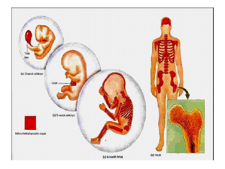
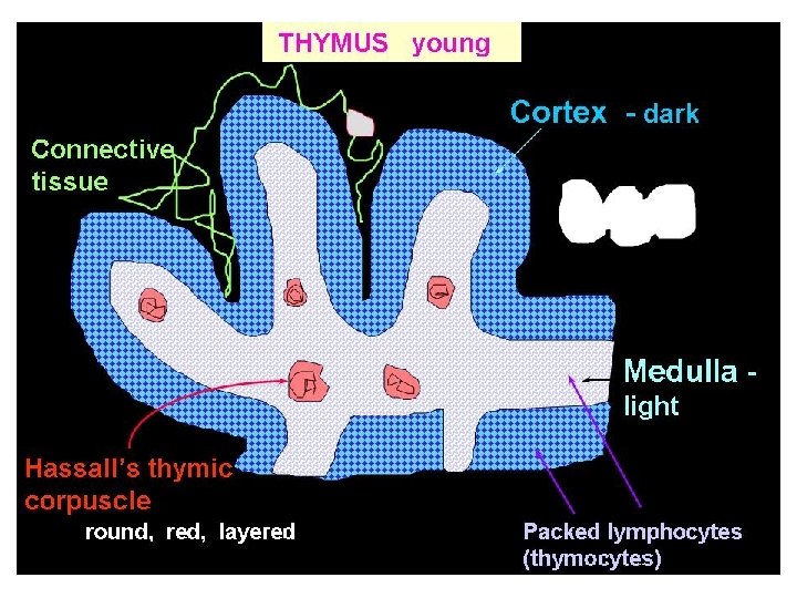
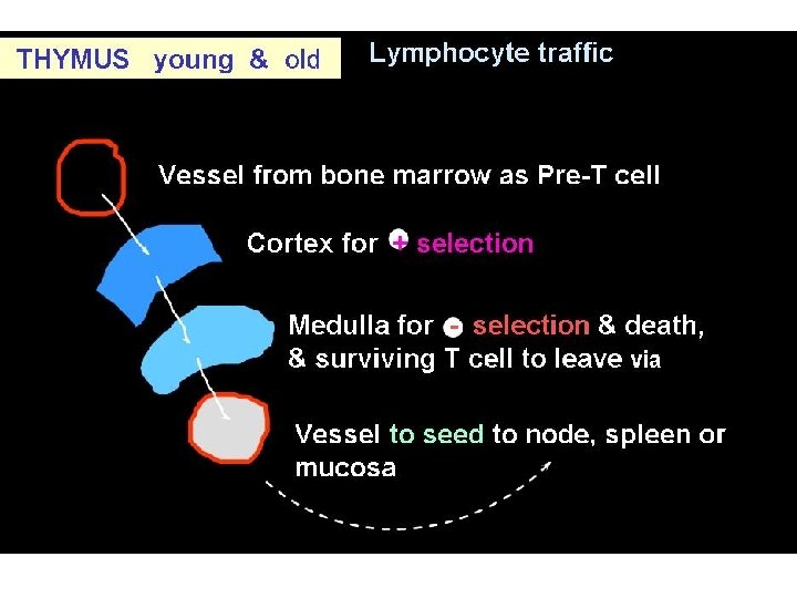
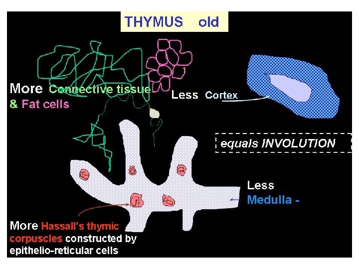
PRIMARY LYMPHOID ORGANS: THYMUS The thymus is the site where lymphoid cells undergo maturation and education into T cells prior to release into the circulation. This process allows T cells to develop the important attribute known as self-tolerance. The thymus is found in the thorax in the anterior mediastinum. It gradually enlarges during childhood but after puberty it undergoes a process of involution resulting in a reduction in the functioning mass of the gland. It continues to function throughout life, however. The thymus is arranged into an outer cellular cortex and an inner medulla. Immature lymphoid cells enter the cortex, where they proliferate, mature, and move to the medulla, from where mature T lymphocytes enter the circulation.
Lymphoid tissues-continued
THYMUS young Cortex - dark Connective tissue Lobules Medulla light Hassall’s thymic corpuscle round, red, layered Packed lymphocytes (thymocytes)
PRINCIPAL THYMIC CELLS EPITHELIORETICULAR CELL NA ﺩ VE LYMPHOCYTES (THYMOCYTES( desmosome MF disposing of unapproved thymocyte
Haematopoesis
Dendritic cells (DC) B cells CLP Myeloid DC (CD 11 c+, CD 11 b+, CD 8 -) T cells Langerhans cells (skin) (CD 11 c+, CD 11 b+, CD 8 +/-, Langerin) Dendritic cells HSC Lymphoid DC (CD 11 c+, CD 11 b-, CD 8 +) CMP Monocytes Plasmacytoid DC (CD 11 c+, B 220+) granulocytes Monocyte-derived DC (CD 11 c+/-, CD 11 b+, CD 8 -) inflammation erythrocytes Megakaryoctes
Lymphoid progenitor
Myeloid progenitor
Monocytes progenitor
Stem cell differentiation
Ag PRESENTATION IN LYMPH NODES from Itano & Jenkins. Nature Immunology 4, 733 - 739 (2003). Antigen presentation to naive CD 4 T cells in the lymph node.
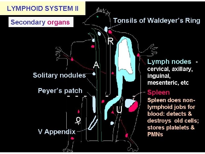
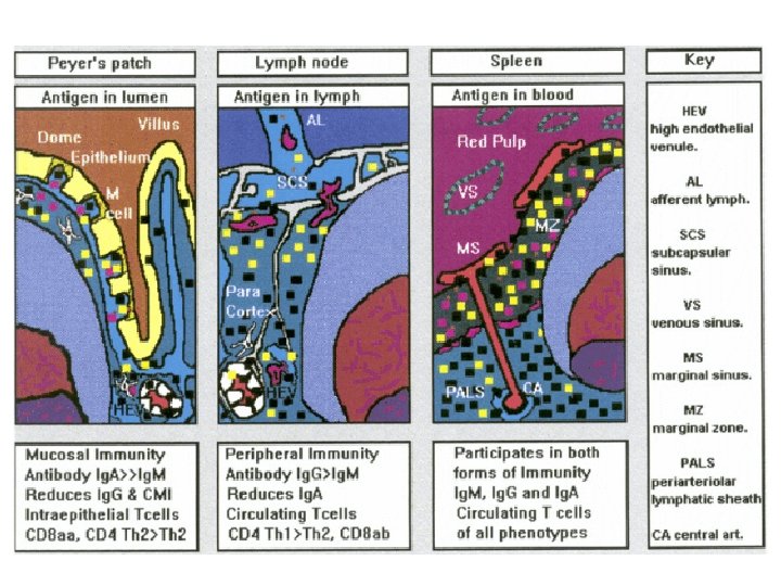
SECONDARY LYMPHOID ORGANS These are the peripheral lymphoid organs: lymph nodes, spleen, tonsils, adenoids, and lymphoid tissue associated with other organ systems (MALT for mucosa, GALT for gut, BALT for bronchus and SALT in skin. LYMPH NODES: filter lymphatic fluid; sites of Ag presentation & cell traffic Lymph nodes have a fibrous capsule from which trabeculae extend towards the center, forming a framework for the lymphatic sinuses, blood vessels, and parenchyma (cortex, paracortex, and medulla). Cortical nodules (follicles) Cortex Medullary sinuses
LYMPH NODES, CONTINUED Functions of structural elements of lymph nodes The lymphatic system is a series of vessels which drain and filter the tissue fluids. Lymph enters the node via afferent lymphatics, passes through the sinuses lined with macrophages and leaves via efferent lymphatic (ultimately all drain into the portal vein). Lymphocytes enter the node primarily from the blood via HEV and leave via efferent lymphatics. DCs migrating from tissue enter the node into the T cell areas. B cells entering nodes from blood must cross the T rich area in transit to the B cell rich areas thus optimizing T-B cooperation. The B cell rich areas contain mature, resting B cells organized into structures around follicular dendritic cells (primary follicles). Sub-capsular Sinus
Lymph node parenchyma is made up of three components: * cortex * paracortex * medulla Cortex (B cell area) B cells enter the lymph node via HEVs and pass to the follicles. If activated by antigenic stimulation, they proliferate and remain in the node. Unstimulated B cells, however, pass out rapidly from the node to return to the general circulation.
Activated B cells within the lymphoid follicles are known as follicle centre cells. The pale staining central area of a secondary follicle is known as a germinal centre and this is surrounded by a mantle zone consisting of small, naive B cells and a few T cells. The follicle centre cells within the germinal centres consist of cells with cleaved nuclei (centrocytes) and cells with larger more open nuclei and several nucleoli (centroblasts).
Stimulated mature B cells responding to antigen change into centrocytes and then centroblasts. The centroblasts leave the follicle and pass to the paracortex and medullary sinuses, where they become immunoblasts. The immunoblasts divide to give rise to plasma cells or memory B cells which are ready for their next encounter with specific antigen. B cells alone are not able to mount immune responses.
They are assisted by accessory cells: * sinus macrophages (highly phagocytic) * tingible body macrophages (ingest cellular debris in germinal centres) * marginal zone macrophages (found beneath the subcapsular sinus) * follicular dendritic cells
Paracortex (T cell area) The paracortex contains lymphocytes and accessory cells along with supporting cells. It is the predominant site for T cells within the lymph node. The various types of T cell enter the node from the blood via the HEVs. When activated they form lymphoblasts, which divide to produce a clone of T cells responding to a specific antigen.
Activated T cells then pass into the circulation to reach peripheral sites. Accessory cells: Interdigitating cells are numerous in the paracortex and act as Agpresenting cells. Medulla The medulla is comprised of: * large blood vessels * medullary cords * medullary sinuses
The medullary cords are rich in plasma cells, which produce Ab that pass out of the node via the efferent lymphatic. Macrophages are also numerous within the medulla. Lymph passes into the node through the afferent lymphatic into the marginal sinus, though the cortical sinuses to reach the medullary sinuses before leaving via the efferent lymphatic. Particulate matter in the lymph is removed by macrophages.
Antigens are taken up by antigen presenting cells and these facilitate the specific immune response. Less than 10% of lymphocytes enter the node in the lymph, the large majority entering from the blood via the HEVs.
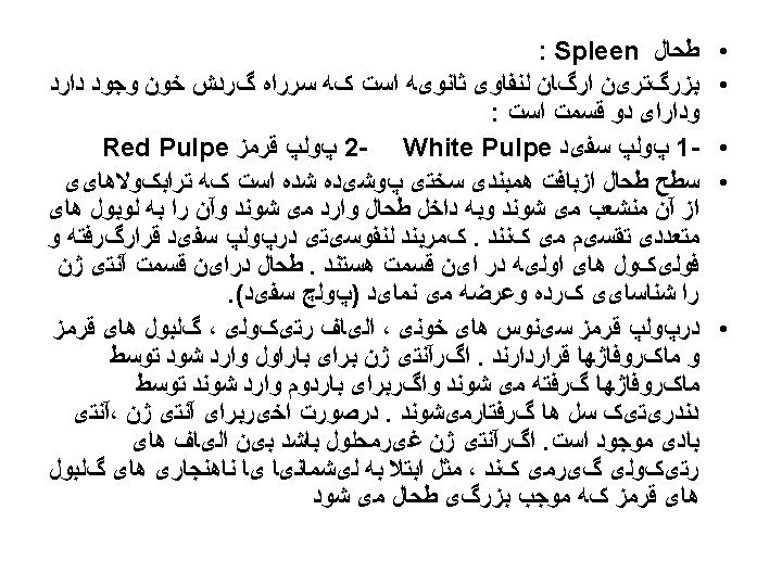
PALS There are two distinct components of the spleen, the red pulp and the white pulp. The red pulp consists of large numbers of sinuses and sinusoids filled with blood and is responsible for the filtration function of the spleen. The white pulp consists of aggregates of lymphoid tissue and is responsible for the immunological function of the spleen.
The spleen serves two major functions: *It is responsible for the destruction of old red blood cells (RBCs); *It is a major site for mounting the immune response. The spleen behaves similar to a lymph node but instead of filtering lymph, it filters blood. Blood entering the spleen travels through progressively smaller arterioles until it is deposited in the red pulp, where the RBCs are processed.
The interface between PALS (peri artherial lymphoid sheath) and blood is a region of intense phagocytic activity and sets the stage for immune responses. The immune reactivity of the spleen is especially effective for dealing with blood-borne antigens such as bacteria.
INSIDE THE SPLEEN Red pulp The red pulp has a complex system of blood vessels within it, arranged to facilitate removal of old or damaged RBCs from the circulation. A small proportion of splenic blood flow passes through more rapidly without undergoing filtration.
INSIDE THE SPLEEN CONTINUED Capsule Trabecula Primary follicle Vascular sinusoids Marginal zone White pulp Periarterial lymphatic sheath (PALS) Germinal center Red pulp Vein Artery White pulp The white pulp contains T cells, B cells and accessory cells. There are many similarities with lymph node structure. The purpose of the white pulp is to mount an immunological response to antigens within the blood. The white pulp is present in the form of a periarteriolar lymphoid sheath. This sheath contains B cell follicles and T cells. At the edge of the T zone is a region known as the marginal zone where larger lymphocytes and antigen presenting dendritic cells are located.
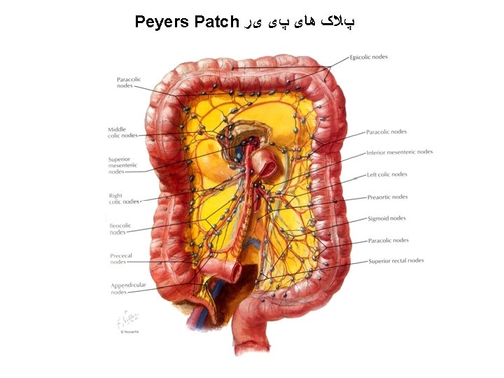
GUT-ASSOCIATED LYMPHOID TISSUE (GALT) This is comprised of: * tonsils, adenoids (Waldeyer's ring) * Peyer's patches * lymphoid aggregates in the appendix and large intestine * lymphoid tissue accumulating with age in the stomach * small lymphoid aggregates in the esophagus * diffusely distributed lymphoid cells and plasma cells in the lamina propria of the gut Large aggregates of GALT have distinct B cell follicles and T cell areas. Antigen presenting accessory cells are also present. Peyer's patches are large aggregates of lymphoid tissue found in the small intestine. Lymphocytes form domed follicles of B cells surrounded by T cells. Some epithelial cells have complex microfolds in their surfaces.
Known as M (multi-fenestrated, arrangement) cells, they collect Ag. Peyer's patches facilitate the generation of an immune response within the mucosa. B cell precursors and memory cells are stimulated by Ag in Peyer's patches. Cells pass to the mesenteric lymph nodes where the immune response is amplified. Activated lymphocytes pass into the blood stream via the thorasic duct. These cells then home in the gut and carry out their final effector functions. HEVs are not present in Peyer's patches and the mechanism by which cells home in on mucosal sites is unknown. Cell surface molecules known as addressins may have a role.
ORGANIZATION OF GALT M cell villi follicle-associated epithelium lymphatic network lamina propria high endothelial venule T cells gut lumen Peyer’s patch follicular DC B cells in domed follicle centroblast B cells
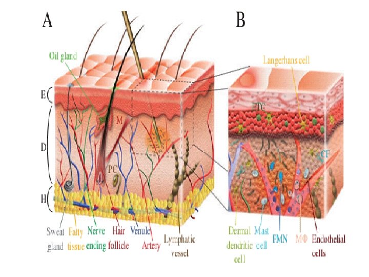
MUCOSA-ASSOCIATED LYMPHOID TISSUE (MALT) In addition to the lymphoid tissue concentrated within the lymph nodes and spleen, lymphoid tissue is also found at other sites, most notably the gastrointestinal tract, respiratory tract and urogenital tract. MALT consists of aggregates of lymphocytes, macrophages, DCs, and other accessory cells. In the gut, these aggregates are scattered throughout the lamina propia, although Peyer’s patches (which resemble lymph nodes in that they have germinal centers and B cell-rich follicles) are also present in the gut. SKIN-ASSOCIATED LYMPHOID TISSUE (SALT) Skin is an active participant in host defense. It has the capability to generate and support local immune and inflammatory responses to foreign Ags that enter the body via the skin. Cells of SALT include keratinocytes, Langerhans cells (immature DCs found in skin), intraepiethelial T cells, and melanocytes. Langerhans cells form a continuous epidermal meshwork: they capture Ag, then migrate to draining lymph nodes, where they act as Ag-presenting cells. The majority of T cells are found in the dermal layer of skin.
SALT
Antigen presentation in skin infection Langerhans cells Interstitial (dermal) DC Epidermis Skin Dermis Denstritic cells undergo maturation upon antigen capture. TLR-PAMP TNF- , IL-1 Immature DC Low surface MHC-II retained in lysosomes (lamp is a lysosomal protein) Mature DC High surface MHC-II High co-stimulation Low co-stimulation (CD 80, CD 86) Active internalization of antigens Inefficient internalization of antigens
Animal skin separates the inner world of the body from the largely hostile outside world and is actively involved in the defense against microbes. However, the skin is no perfect defense barrier and many micro organisms have managed to live on or within the skin as harmless passengers or as disease-causing pathogens. Microbes have evolved numerous strategies that allow them to gain access to the layers underneath the epidermis where they either multiply within the dermis or move to distant destinations within the body for replication.
A number of viruses, bacteria and parasites use arthropod vectors, like ticks or mosquitoes, deliver them in to the dermis While taking their blood meal. Within the dermis, successful pathogens subvert the function of a variety of Skin resident cells or cells of the innate immune System that rush to the site of infection.
Immature DCs efficiently capture antigens. DCs can internalize diverse antigens. B cells only internalize antigens that bind to BCR. Fc R, CR, Mannose receptors phagocytosis macropinocytosis endocytosis BCR Endosomes/lysosomes DC maturation decreases antigen uptake. DCs internalized carbon particles.
Inflammation induces DC maturation. LPS + HEL Immature DC internalizes HEL. 4 hr 9 hr 22 hr HEL peptide +LPS Mature DC presents HEL peptides / MHC complex on cell surface. -LPS HEL peptide/MHC II moves to cell surface after LPS treatment. Mature DCs have higher levels of surface MHC-II than B cells and macrophages.
Immature dendritic cells can retain antigens in endosomes/lysosomes. Immature DC in peripheral tissues Cystatin C inhibits cathepsins to prevent degradation of antigens and invariant chain. Invariant chain retains MHC II in lysosomes. Mature DC in lymphoid tissues Cystatin C level decreases during maturation. Degradation of antigen and invariant chain allows peptide loading and exit of MHC-II from lysosomes. Strong proteolytic activity in macrophage lysosomes cause excessive degradation of antigens.
DC maturation increases surface expression of co-stimulatory molecules and MHC-I. Co-stimulation B 7 and class I MHC move from ER to cell surface. (B 7) Cell number Immature DC TNF- LPS Mature DC Co-localization of peptide and B 7 with MHCII on cell surface facilitates T cell activation.
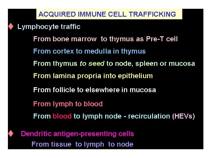
LYMPHOCYTE RECIRCULATION Lymphocytes and some monos can recirculate between lymphoid and non-lymphoid tissues. This helps lymphocytes to be exposed to the antigens which they recognize and is valuable for the distribution of effector cells to sites where they are needed. The recirculation is a complex process depending on interactions between the cells of the immune response and other cell types such as endothelial cells. Naive lymphocytes move from the primary to secondary lymphoid tissue via the blood. .
Activated lymphocytes move from the spleen, lymph nodes, and other lymphoid tissue (e. g. , MALT) into the blood and then to other lymphoid and non-lymphoid tissues. APCs may carry Ag back to lymphoid tissues from the periphery. The complex patterns of recirculation depend on the state of activation of the lymphocytes, the adhesion molecules expressed by endothelial cells, and the presence of chemotactic molecules, which selectively attract particular populations of lymphocytes or macrophages
Naïve T cells circulate between blood and secondary lymphoid organs. Afferent lymph Efferent lymph Lymph node Efferent lymph Lymphocytes (B and T cells) 25 -33% leukocytes thymus Peyer’s patch Mucosal tissues spleen
T cells are recruited to secondary lymphoid tissues by chemokines. Spleen white pulp B cell follicle CXCL 13 CCL 21 T cell region CCL 21 CCR 7 B cell CXCR 5 T cell: CCR 7 CXCL 13 CCL 21 Lymph node HEV B cell follicle CCL 21 CXCL 13 CCL 21
Inflammation induces DC migration into lymph node. Peripheral tissue (skin) Inflammation (TLR-PAMP, IL-1, TNF- ) induces CCR 7 expression. Langerhans cells Interstitial DCs Monocyte-derived DCs Afferent lymphatic vessel CCL 19 CCL 21 Lymph node Macrophages do not migrate to lymphoid tissues.
inflammation DC skin DC (blue) Skin draining lymph node DC Skin draining LN T cells (brown, CD 3 staining) B cell follicle (CD 19 staining) DCs migrating through lymphatic vessels. Langerhans cells (blue, langrin staining) from skin localize in T cell area.
Antigen presentation by migrant and resident DCs in lymph node Migrant DCs capture antigens locally at low concentrations. Antigen transported to lymph nodes through afferent lymph Particular antigen may need to be processed and presented by migrant DCs. Later and persistent T cell activation Resident DCs capture lymph-borne antigens for initial T cell activation Spleen white pulp B cell follicles T cell B cell follicle CXCL 13 T cell region (CD 11 c+, Langerhans cells, myeloid DCs potent activators of CD 4 T cells. CD 11 b+) are the most Blood-borne antigens are captured by DCs in spleen
No antigenic challenge T cells 12 -18 hours Antigenic challenge TCR recognizes antigen. B cells 24 hours Activation and proliferation Secondary lymphoid organs facilitate the encounter of rare antigen-specific lymphocytes with antigen.
The antigen receptor molecules There are three groups of molecules • that specifically recognize foreign antigen for the adaptive immune system. • B cell receptor(BCR or the antibody) • T cell receptor (TCR) • Major histocompatibility complex(MHC), this cluster of genes is known as human leukocyte antigen (HLA).
MHC This group of antigen receptor is represented • by the proteins encoded by the MHC genes (located on chromosome 6) and it is known as Human leukocyte antigen (HLA). • There are two main classes of molecules, which were initially named because of their role in tissue (histo-) graft rejection (compatibility). • Class I MHC molecules are found essentially on all cells except RBCs, and Class II MHC molecules are found chiefly on • APC (B cells and Monocytes). • The main function of MHC molecules is to present antigens to T cells.
Antigen recognition molecules