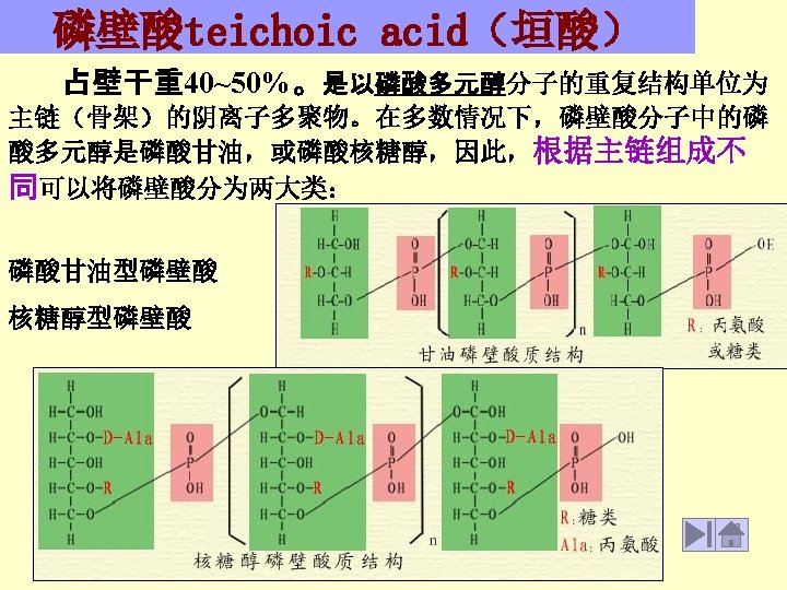
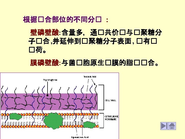
Figure 8 The structure of teichoic acid Figure


- Slides: 15
Figure 8 - The structure of teichoic acid
Figure 7. Muramic acid subunit of the peptidoglycan of Escherichia. coli. This is the type of peptidoglycan found in most Gram-negative bacteria. The glycan backbone is a repeat polymer of two amino sugars, N-acetylglucosamine (Gl. NAc) and N-acetylmuramic acid (Mur. NAc). Attached to the Mur. NAc is a tetrapeptide consisting of L-ala-D-glu. DAP-D-ala. In the polymeric form of the molecule, nearby tetrapeptide side chains may be linked to one another by an interpeptide bond between DAP on one chain and D-ala on the other.
Figure 9 a. Schematic illustration of the outer membrane, cell wall and plasma membrane of a Gramnegative bacterium. Note the structure and arrangement of molecules that constitute the outer membrane.
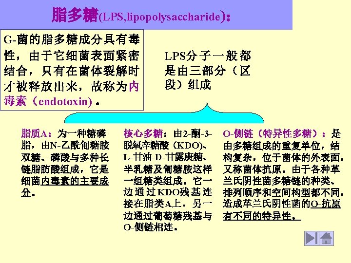
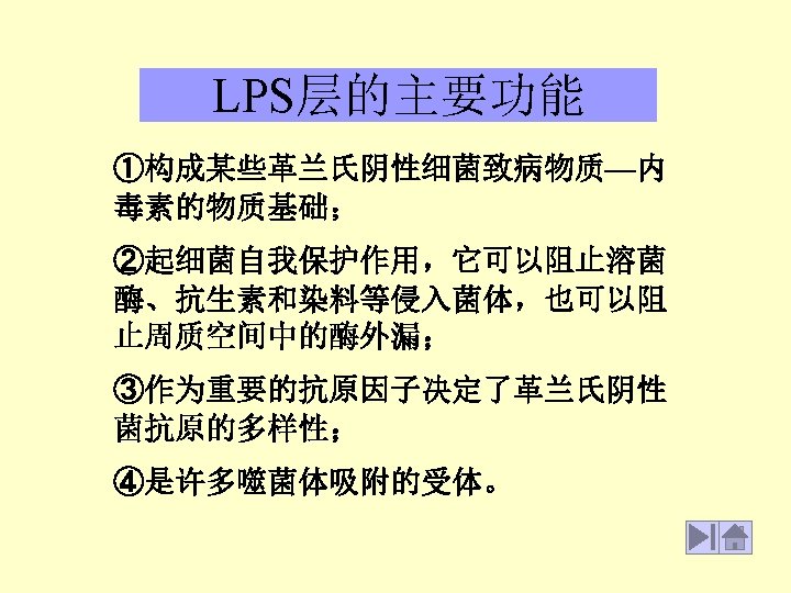
Figure 9 b. Structure of LPS
LPS, lipopolysaccharide Figure. Structure of LPS
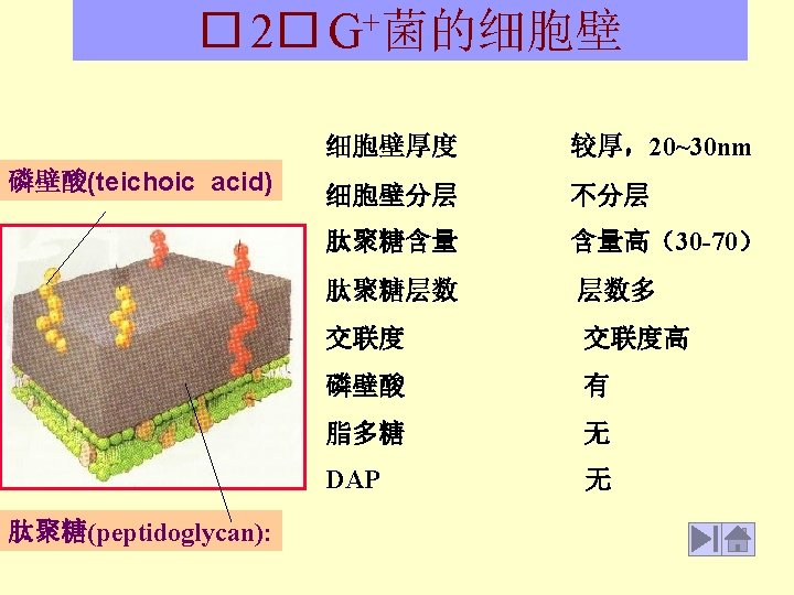
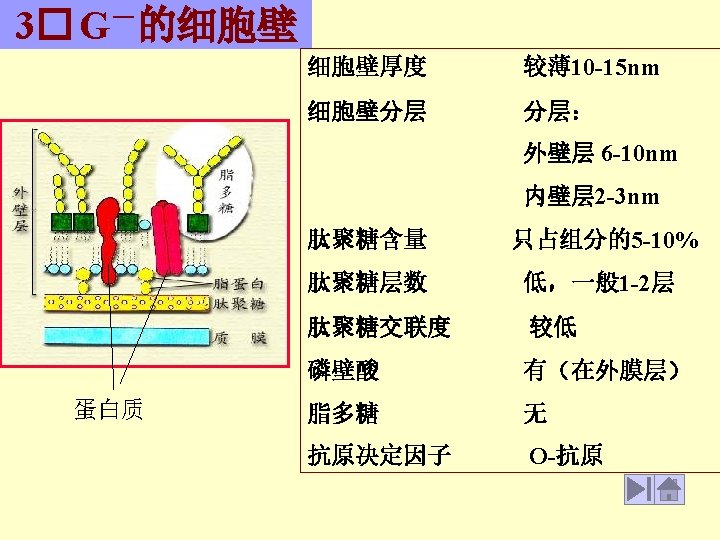
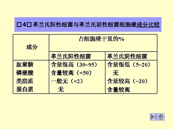
Table 5. Correlation of Grams stain with other properties of Bacteria. Property Gram-positive Gram-negative Thickness of wall thick (20 -80 nm) thin (10 nm) Number of layers Peptidoglycan (murein) content Teichoic acids in wall 1 2 >50% 10 -20% present absent Lipid and lipoprotein content 0 -3% 58% Protein content 0 9% Lipopolysaccharide content 0 13% Sensitivity to Penicillin G yes no (1) Sensitivity to lysozyme yes no (2)