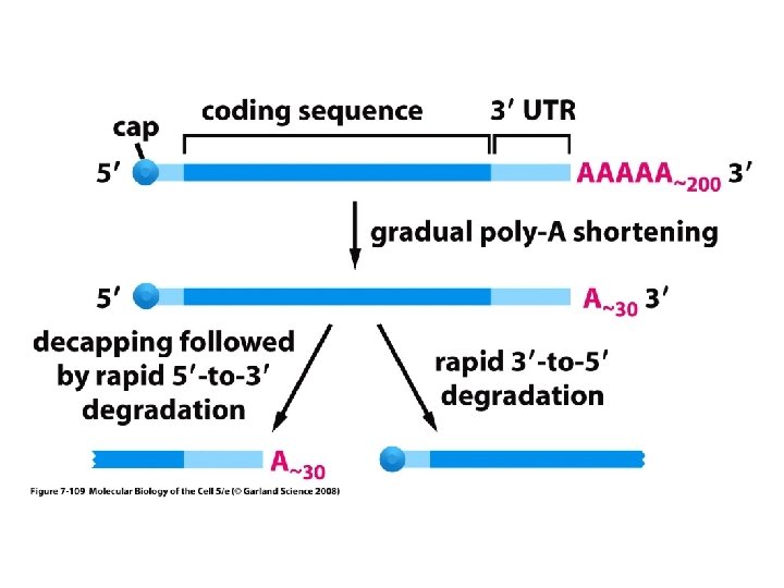
Figure 7 108 Molecular Biology of the Cell
")
Figure 7 -108 Molecular Biology of the Cell (© Garland Science 2008)

Resolution of biological objects

Capturing and interpreting light images

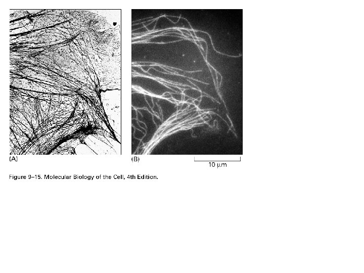
Different methods to visualize cellular morphology and objects

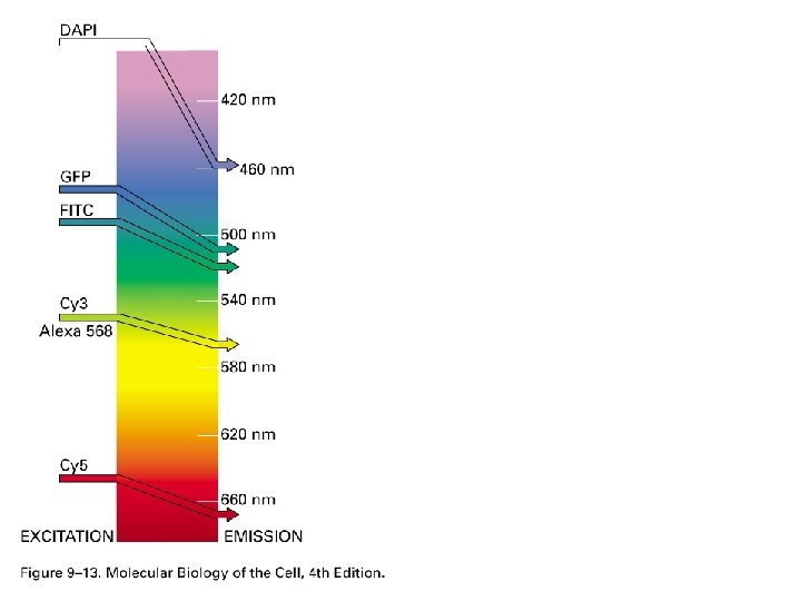
Light and fluorescence microscopy

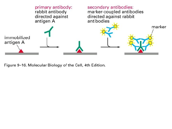
Detection methods for subcellular structures

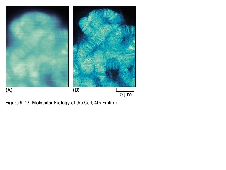
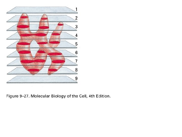
Methods to better resolve objects in 3 D

Relative sizes On the microscopic to Macroscopic scale

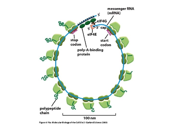
Cellular proteins can be visualized in Real time in living cells GOLGI GREEN AND RED FLUORESCENT PROTEINS S. CEREVISCIAE - BAKERS YEAST

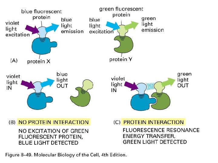
How Flourescence works
 Isolate protein - green fluorescent")
Making Fluorescent Cells A. VICTORIA (AKA - GLOWING JELLYFISH) Isolate protein - green fluorescent protein (GFP) Abs. Max = 488 nm Now can fuse protein of interest with GFP 5’ UTRAUG Getmeoutaherenow GFP Getmeoutaherenow DNA plasmid UAG UAA UGA UTR 3’

Making Fluorescent Cells GFP is a Beta-can chromophore -helices -sheets Cerulean GFP Banana Orange……. Ta da…. . The Brainbow

Resolving power d = 0. 61 n sin D = minimum distance of 2 points = wavelength n = numerical aperture = angle of cone of light (1/2)

Specific stains Methods to increase contrast Bright field Phase contrast CFP Nomarski Light microscope comparisons YFP axons

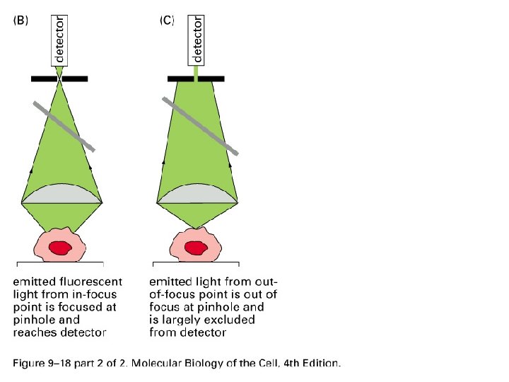
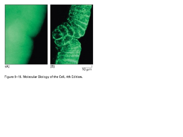
Out of focus light can be removed by computers Raw Deconvolved

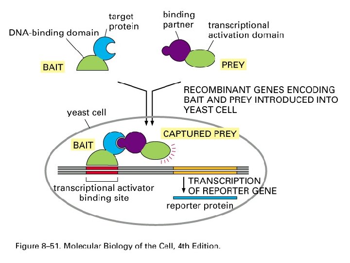
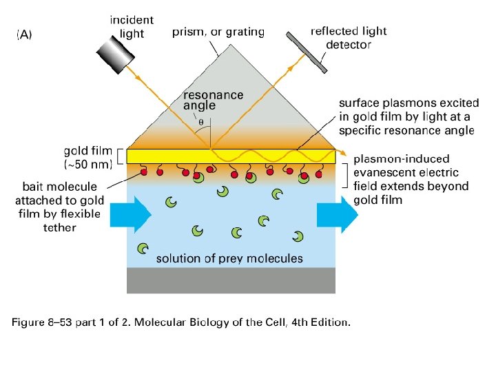
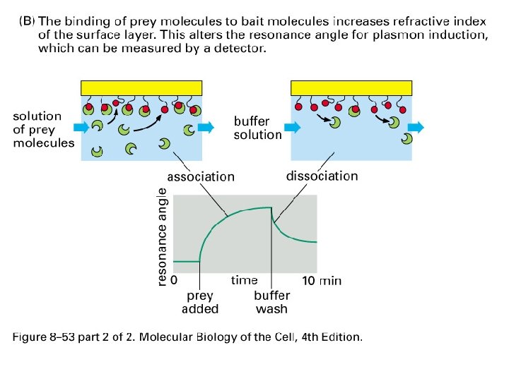
Other uses of Fluorescent proteins FRET - fluorescence resonance energy transfer -can detect changes in interactions -donor FL energy reduced while acceptor increased Bi-molecular complementation Caged proteins

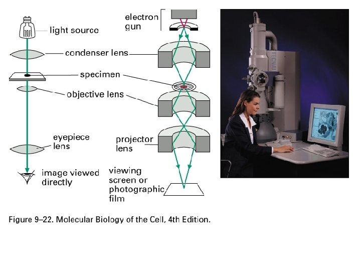
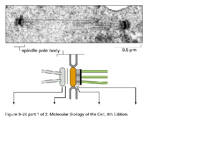
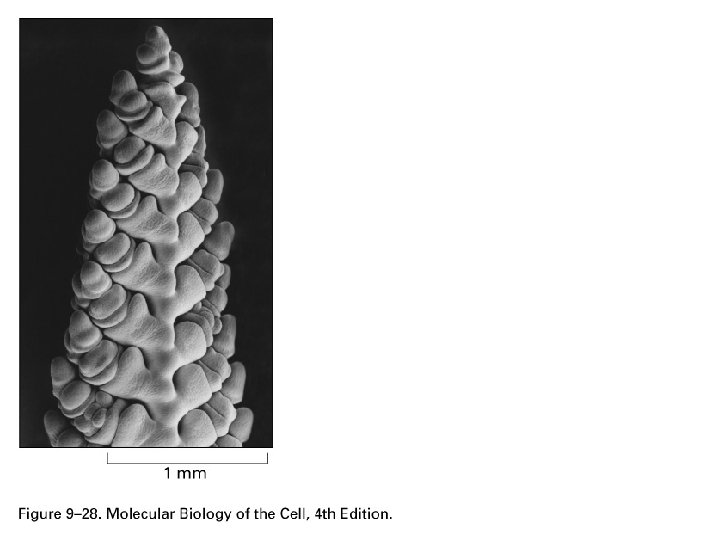
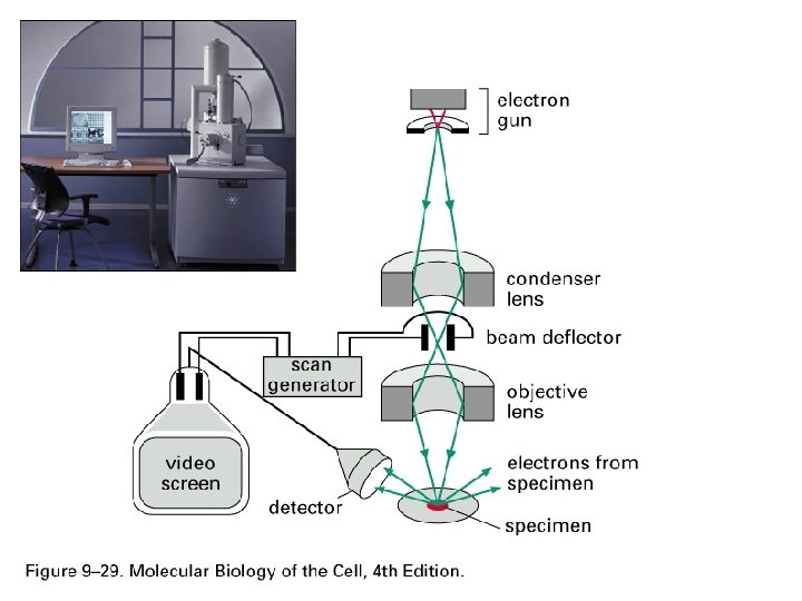
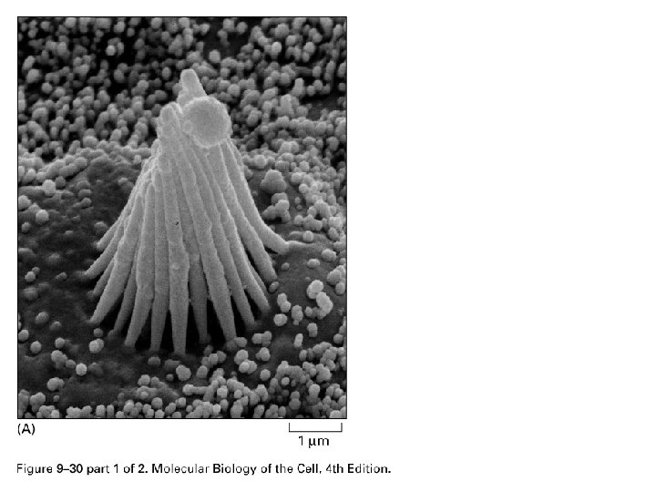
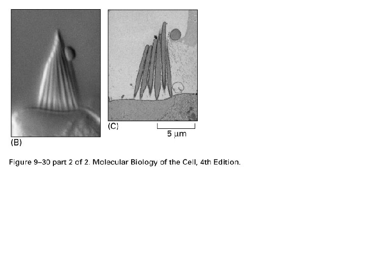
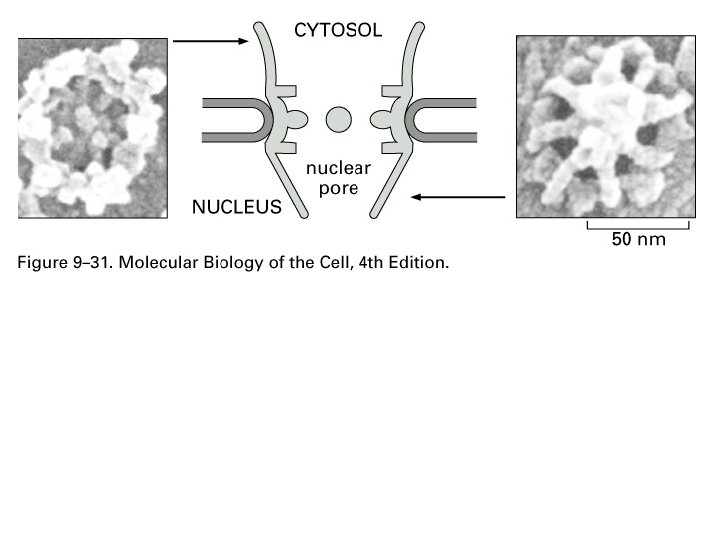
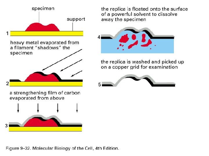
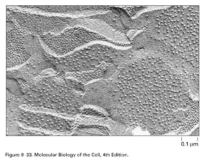
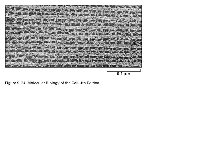
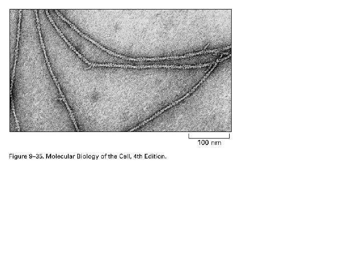
Electron microscopy Light microscope 1 nm resolution Electron microscope Negative stain Shadow casting

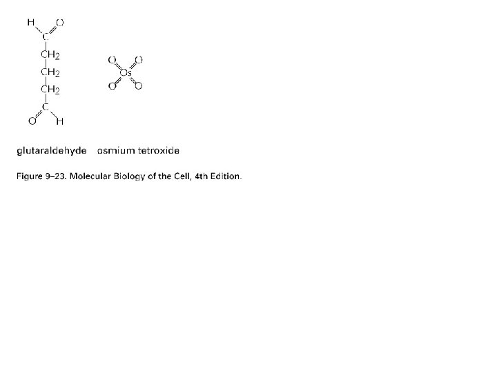
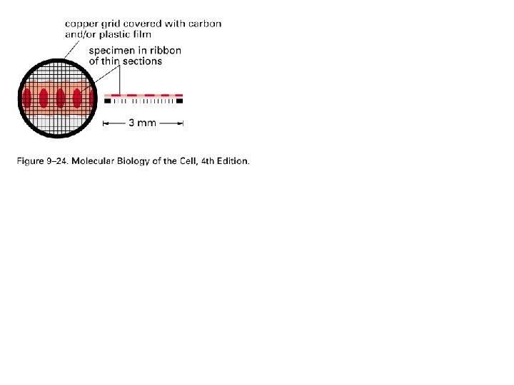
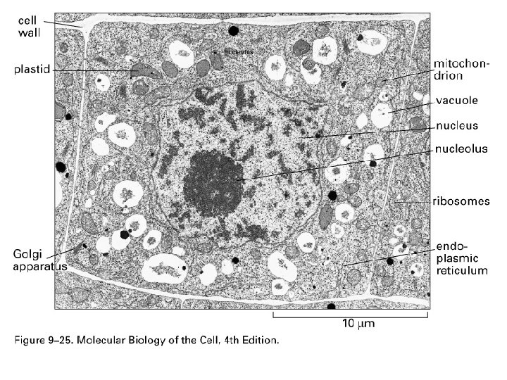
Sample preparation is time consuming
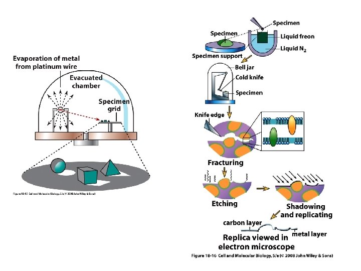

Ciliary axoneme - deep etch Onion root cell Freeze-fractured Insect head - SEM - bacteriophage

- Slides: 54