Figure 35 11 Primary growth in stems Epidermis

 A plant can")
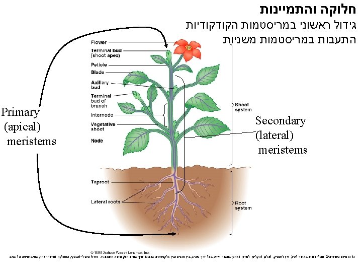
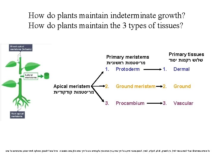
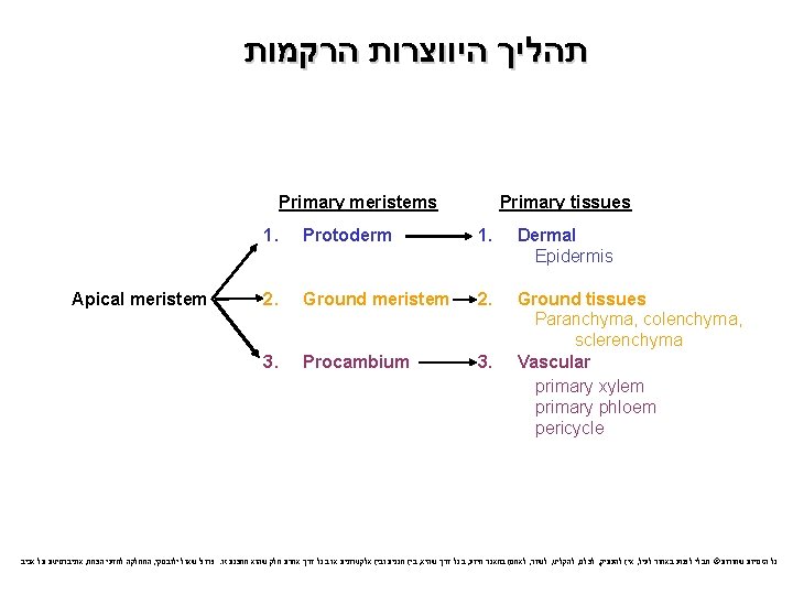
 There are two main types of meristems: apical meristems and lateral meristems b)Apical")
 Lateral meristems add thickness to woody plants, a process called secondary growth b)")
Initials, also called")


 Angiosperms comprise more than 250, 000 living species b) Previously, angiosperms")


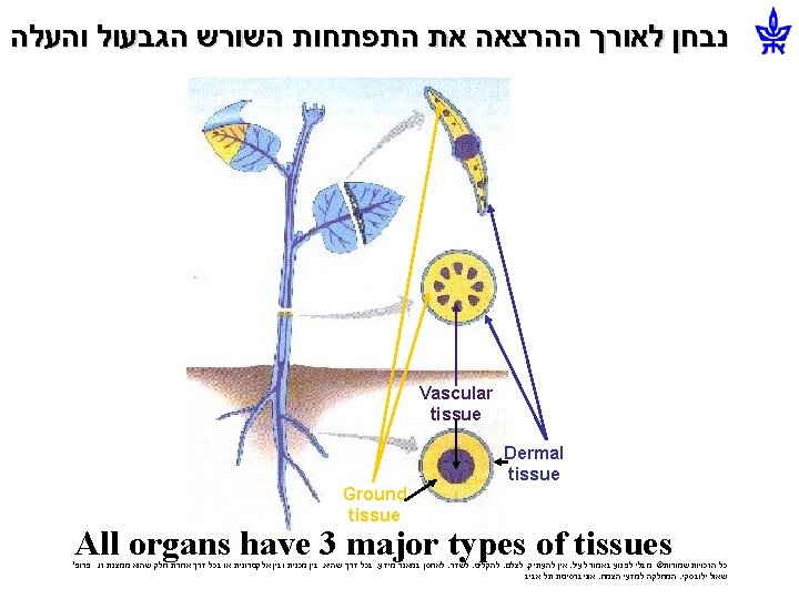
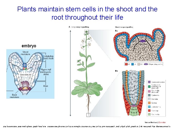
 The primary growth of roots produces the epidermis, ground tissue, and vascular tissue")
 The ground tissue, mostly parenchyma cells, fills the cortex, the region between the")


 A shoot apical meristem is")
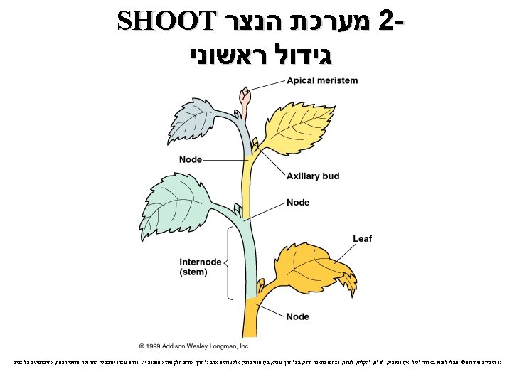
 The closer an axillary bud is to the active")
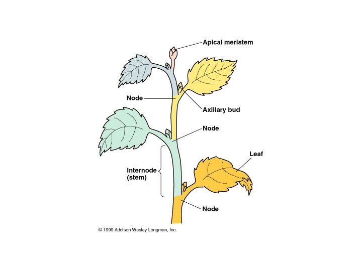
 Lateral shoots develop from axillary buds on the stem’s surface b) In most")
 Phloem Xylem Ground")
 In most monocot stems,")


 The epidermis in leaves is interrupted by stomata, pores that allow CO 2")
 The ground tissue in a leaf, called mesophyll, is sandwiched between the upper")
 The vascular tissue of each leaf is continuous with the vascular tissue of")
 By")
 New cell walls form in a")
 Leaf growth results from a combination of transverse and longitudinal cell divisions b)")

 The symmetry")


 Polarity is the condition of having structural or chemical differences at")


 Plant cells grow rapidly and “cheaply”")

 Primary and secondary growth in a two-year-old woody stem Pith")

 Secondary growth occurs in stems and roots of woody plants")
")
 In a typical woody stem, the vascular")
 In cross section, the vascular cambium appears as")


 Elongated initials produce tracheids, vessel elements, fibers of")
 Secondary xylem accumulates as wood and consists of tracheids,")
 Tree rings are visible where late and early wood meet, and can be")
 As a tree or woody shrub ages, the")
 Cork cambium gives rise to")
 Lenticels in the periderm allow for gas exchange between living stem or root")

- Slides: 87
Figure 35. 11 גידול ראשוני ומשני Primary growth in stems Epidermis Cortex Shoot tip (shoot apical meristem and young leaves) Axillary bud meristem Primary phloem Primary xylem Pith Vascular cambium Cork cambium Lateral Secondary growth in stems meristems Cork cambium Periderm Pith Root apical meristems © 2015 Pearson Education Ltd Primary xylem Secondary xylem Cortex Primary phloem Secondary phloem Vascular cambium
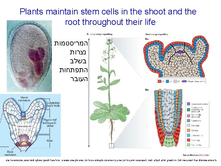
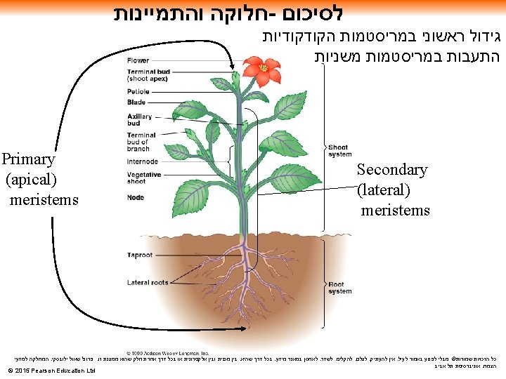
Different meristems generate new cells for primary and secondary growth a) A plant can grow throughout its life; this is called indeterminate growth b) Meristems are perpetually embryonic tissue and allow for indeterminate growth c) Some plant organs cease to grow at a certain size; this is called determinate growth © 2015 Pearson Education Ltd
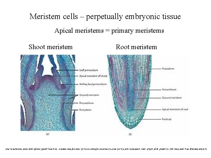
a) There are two main types of meristems: apical meristems and lateral meristems b)Apical meristems are located at the tips of roots and shoots c) Apical meristems elongate shoots and roots, a process called primary growth © 2015 Pearson Education Ltd
a) Lateral meristems add thickness to woody plants, a process called secondary growth b) There are two lateral meristems: the vascular cambium and the cork cambium c) The vascular cambium adds layers of vascular tissue called secondary xylem (wood) and secondary phloem d) The cork cambium replaces the epidermis with periderm, which is thicker and tougher © 2015 Pearson Education Ltd
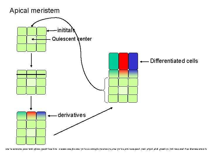
תאי גזע - תאים מריסטמתים A. Meristems give rise to a)Initials, also called stem cells, which remain in the meristem b)Derivatives, which become specialized in mature tissues B. In woody plants, primary growth and secondary growth occur simultaneously but in different locations © 2015 Pearson Education Ltd
Angiosperms-Flowering plants אבולוציה של מכוסי הזרע Chlorophytes Bryophytes Vascular Plants Gymnosperms Angiosperms - מחלקות פסיגים - דו פסיגים - חד 150 MYA צמחים בעלי זרעים צמחים ווסקולרים Embryophytes First- oxygen; chloroplast; stem; leaves: Ohad N. , Dep. of Plant Sciences, TAU כול הזכויות שמורות צמחים בעלי עוברים seeds; flowers
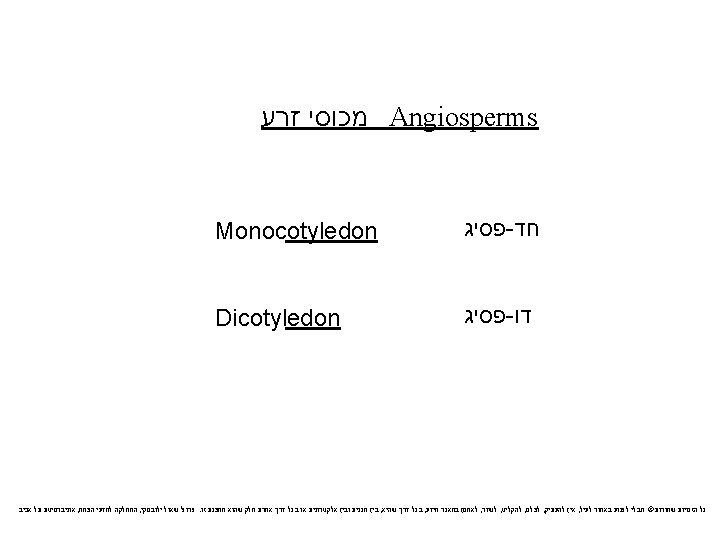
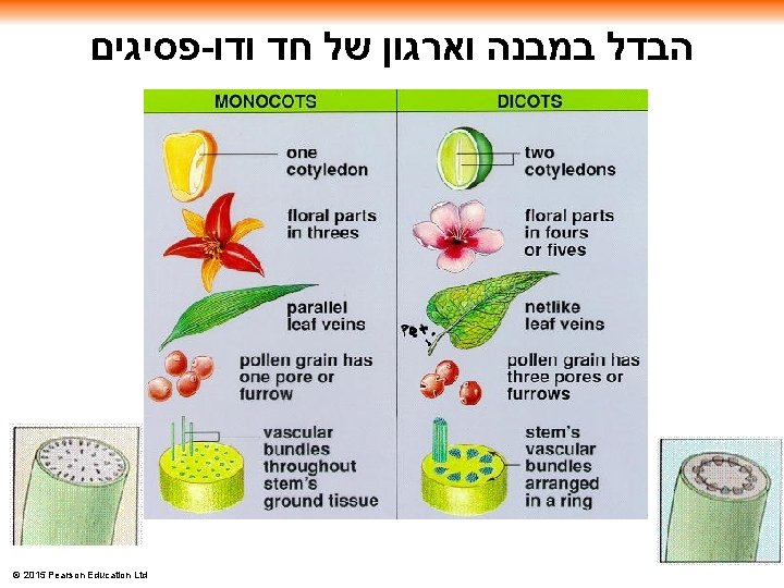
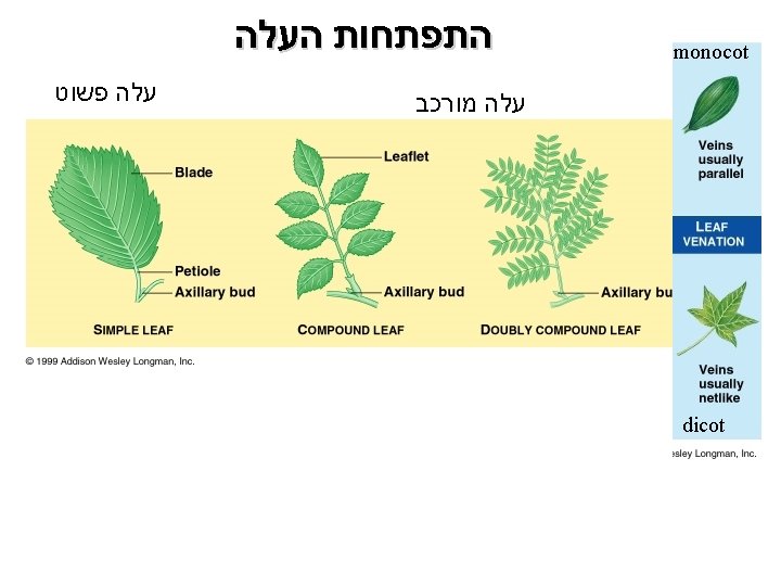
Angiosperm Diversity a) Angiosperms comprise more than 250, 000 living species b) Previously, angiosperms were divided into two main groups a) Monocots (one cotyledon) חד פסיגים b) Dicots (two dicots) פסיגים - דו c) DNA studies suggest that dicots are paraphyletic פסיג עלה עוברי ראשון © 2015 Pearson Education Ltd
פסיגיים - מחלקת הדו © 2015 Pearson Education Ltd
פסיגיים - מחלקת החד © 2015 Pearson Education Ltd
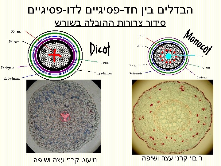
Figure 35. 14 Epidermis Cortex Endodermis Vascular cylinder Pericycle Core of parenchyma cells Xylem 100 μm Phloem (a) Root with xylem and phloem in the center (typical of eudicots פסיג - ) דו Endodermis 100 μm (b) Root with parenchyma in the center (typical of monocots ) חד פסיג Pericycle Xylem מבנה וארגון השורש פסיגים - נבדל בין חג פסיגים לדו Phloem 70 μm © 2015 Pearson Education Ltd Dermal Ground Vascular
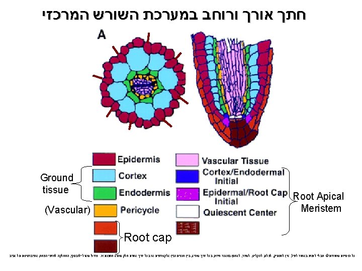
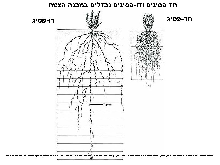
a) The primary growth of roots produces the epidermis, ground tissue, and vascular tissue b) In angiosperm roots, the stele is a vascular cylinder c) In most eudicots, the xylem is starlike in appearance with phloem between the “arms” d) In many monocots, a core of parenchyma cells is surrounded by rings of xylem then phloem © 2015 Pearson Education Ltd
a) The ground tissue, mostly parenchyma cells, fills the cortex, the region between the vascular cylinder and epidermis b) The innermost layer of the cortex is called the endodermis c) The endodermis regulates passage of substances from the soil into the vascular cylinder © 2015 Pearson Education Ltd
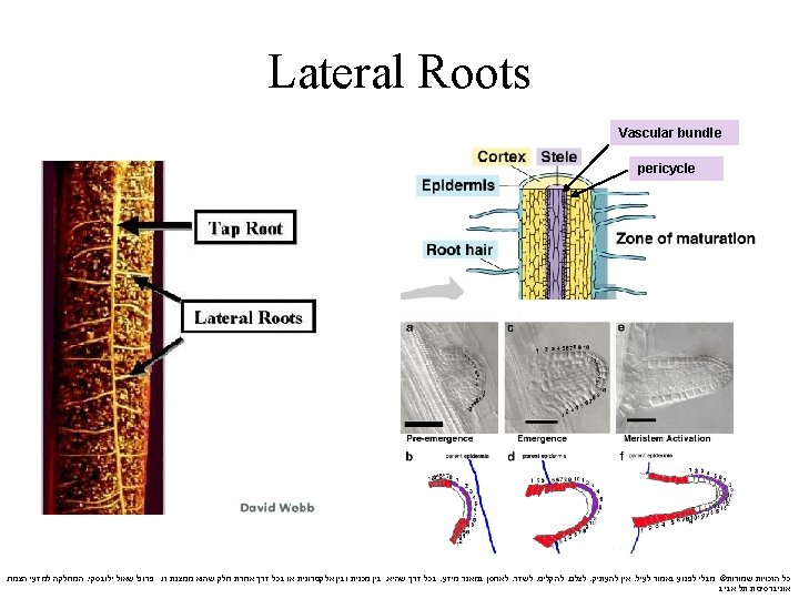
כיצד מתפתחים שורשים צדדים ? האם יש בכך בעיה חתך רוחב בשורש Emerging lateral root 100 μm Epidermis Lateral root Cortex Vascular cylinder 1 Pericycle 2 3 Lateral roots arise from within the pericycle, the outermost cell layer in the vascular cylinder © 2015 Pearson Education Ltd
מריסטמת הנצר Leaf primordia Young leaf Shoot apical meristem Developing vascular strand Axillary bud meristems Figure 35. 16 0. 25 mm © 2015 Pearson Education Ltd
Primary Growth of Shoots גדילה ראשונית של הנצר a) A shoot apical meristem is a dome-shaped mass of dividing cells at the shoot tip b) Leaves develop from leaf primordia along the sides of the apical meristem c) Axillary buds develop from meristematic cells left at the bases of leaf primordia © 2015 Pearson Education Ltd
Apical dominance שלטון קודקודי a) The closer an axillary bud is to the active apical bud, the more inhibited it is b) Axillary buds are released from this apical dominance if the shoot tip is removed or shaded c) In some monocots, meristematic activity occurs at the bases of stems and leaves © 2015 Pearson Education Ltd
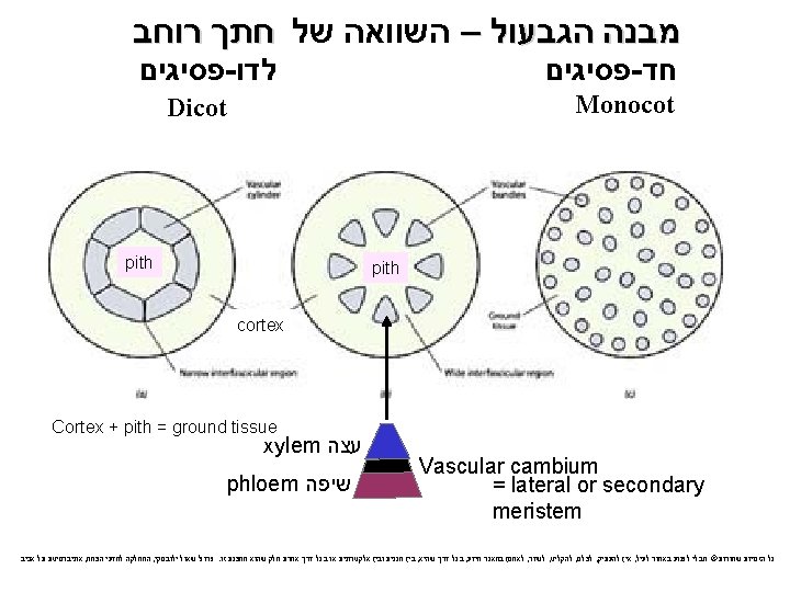
a) Lateral shoots develop from axillary buds on the stem’s surface b) In most eudicots, the vascular tissue consists of vascular bundles arranged in a ring © 2015 Pearson Education Ltd
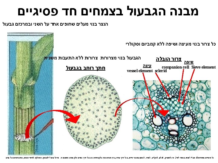
Figure 35. 17 חתך רוחב בגבעול דו פסיגים Sclerenchyma (fiber cells) Phloem Xylem Ground tissue connecting pith to cortex חד פסיגים Ground tissue Pith Epidermis Cortex Epidermis Vascular bundle (a) Cross section of stem with vascular bundles forming a ring (typical of eudicots) (LM) © 2015 Pearson Education Ltd Vascular bundles 1 mm Dermal Ground Vascular (b) Cross section of stem with scattered vascular bundles (typical of monocots) (LM)
בחד פסיגים מערכות ההובלה פזורות לרוחב חתך הגבעול a) In most monocot stems, the vascular bundles are scattered throughout the ground tissue, rather than forming a ring גבעול שורש © 2015 Pearson Education Ltd
The Leaf העלה. 3 Cuticle 50 μm Guard cells Stomatal pore Epidermal cell Sclerenchyma fibers Stoma (b) Surface view of a spiderwort (Tradescantia) leaf (LM) Upper epidermis Palisade mesophyll 100 μm Spongy mesophyll Lower epidermis Bundlesheath cell Vein Xylem Phloem (a) Cutaway drawing of leaf tissues Figure 35. 18 © 2015 Pearson Education Ltd Guard cells Cuticle Dermal Ground Vascular שלוש רקמות היסוד Vein Air spaces Guard cells (c) Cross section of a lilac (Syringa) leaf (LM)
Trichomes Guard cells Stomata
After germination, the shoot apical meristem makes leaves Leaves Shoot apex at germination Post-embryonic leaf formation Leaves Cotyledons Reprinted by permission from Macmillan Publishers, Ltd: NATURE. Long, J. A. , Moan, E. I. , Medford, J. I. , and Barton, M. K. (1996) A member of the KNOTTED class of homeodomain proteins encoded by the STM gene of Arabidopsis. Nature 379: 66 -69. 44 כל הזכויות אוניברסיטת תל אביב , המחלקה למדעי הצמח , פרופ' שאול ילובסקי. בין מכנית ובין אלקטרונית או בכל דרך אחרת חלק שהוא ממצגת זו , בכל דרך שהיא , לאחסן במאגר מידע , לשדר , להקליט , לצלם , אין להעתיק , שמורות© מבלי לפגוע באמור לעיל
Phyllotaxy פילוטקסיס • The pattern of organ initiation at the shoot apical meristem Alternate Opposite Whorled Spiral 46
BUNDLE SHEATH M E S O P H Y L L
a) The epidermis in leaves is interrupted by stomata, pores that allow CO 2 and O 2 exchange between the air and the photosynthetic cells in a leaf b) Stomata are also major avenues for evaporative loss of water c) Each stomatal pore is flanked by two guard cells, which regulate its opening and closing © 2015 Pearson Education Ltd
a) The ground tissue in a leaf, called mesophyll, is sandwiched between the upper and lower epidermis b) The mesophyll of eudicots has two layers a)The palisade mesophyll in the upper part of the leaf b)The spongy mesophyll in the lower part of the leaf; the loose arrangement allows for gas exchange © 2015 Pearson Education Ltd
a) The vascular tissue of each leaf is continuous with the vascular tissue of the stem b) Veins are the leaf’s vascular bundles and function as the leaf’s skeleton c) Each vein in a leaf is enclosed by a protective bundle sheath © 2015 Pearson Education Ltd
Growth: Cell Division and Cell Expansion התרחבות תא וחלוקת תא : גדילה a) By increasing cell number, cell division in meristems increases the potential for growth b) Cell enlargement accounts for the actual increase in plant size 7 μm © 2015 Pearson Education Ltd
The Plane and Symmetry of Cell Division a) New cell walls form in a plane (direction) perpendicular to the main axis of cell expansion b) The plane in which a cell divides is determined during late interphase c) Microtubules become concentrated into a ring called the preprophase band that predicts the future plane of cell division © 2015 Pearson Education Ltd
a) Leaf growth results from a combination of transverse and longitudinal cell divisions b) It was previously thought that the plane of cell division determines leaf form c) A mutation in the tangled-1 gene that affects longitudinal divisions does not affect leaf shape © 2015 Pearson Education Ltd
30 μm Figure 35. 27 Leaf epidermal cells of wild-type maize © 2015 Pearson Education Ltd Leaf epidermal cells of tangled-1 maize mutant
דוגמה לחלוקה לא סימטרית התמיינות של תאי שמירה בהתפתחות פיונית a) The symmetry of cell division, the distribution of cytoplasm between daughter cells, determines cell fate b) Asymmetrical cell division signals a key event in development a)For example, the formation of guard cells involves asymmetrical cell division and a change in the plane of cell division © 2015 Pearson Education Ltd
The plane and symmetry of cell division influence development of form Plane of cell division (a) Planes of cell division Developing guard cells Unspecialized epidermal cell (b) Asymmetrical cell division © 2015 Pearson Education Ltd Guard cell “mother cell”
Figure 35. 28 Asymmetrical cell division Unspecialized epidermal cell Guard cell “mother cell” Developing guard cells © 2015 Pearson Education Ltd
What determines stomata spacing in the developing leaf? B B A C Day 2 A A Day 3 C Day 6 C Mature
Guard cells differentiation in Arabidopsis Meristemoid Mother Cell Asymmetric division Differentiation Guard Mother Cell Guard Cell Pair Symmetric division Protodermal cell Guard cells are formed through a tightly controlled series of cell divisions Barton, M. K. (2007) Making holes in leaves: Promoting cell state transitions in stomatal development. Plant Cell 19: 1140 - 1143.
Wild type “TOO MANY MOUTHS”
Guard cells differentiate through a tightly controlled series of cell divisions SPEECHLESS Asymmetric division Meristemoid Mother Cell Differentiation Guard Mother Cell Guard Cell Pair Symmetric division Protodermal cell SPEECHLESS is necessary for the initiation of the guard cell developmental pathway. Wild-type speechless SPEECHLESS-OX Barton, M. K. (2007) Making holes in leaves: Promoting cell state transitions in stomatal development. Plant Cell 19: 1140 - 1143.
Guard cells differentiate through a tightly controlled series of cell divisions SPEECHLESS Asymmetric division Meristemoid Mother Cell Differentiation Guard Mother Cell Guard Cell Pair Symmetric division MUTE Protodermal cell Loss of Function Gain of Function MUTE promotes differentiation of guard mother cells. In plants that over- express MUTE, all epidermal cells differentiate as guard cells. Barton, M. K. (2007) Making holes in leaves: Promoting cell state transitions in stomatal development. Plant Cell 19: 1140 - 1143.
Guard cells differentiate through a tightly controlled series of cell divisions SPEECHLESS Asymmetric division Meristemoid Mother Cell Differentiation MUTE Protodermal cell In fama loss-of-function mutants, continued GMC division generates rows of undifferentiated guard cell precursors Guard Mother Cell Guard Cell Pair Symmetric division FAMA Loss of Function Gain of Function Barton, M. K. (2007) Making holes in leaves: Promoting cell state transitions in stomatal development. Plant Cell 19: 1140 - 1143.
פולריות a) Polarity is the condition of having structural or chemical differences at opposite ends of an organism a)For example, plants have a root end a shoot end b) Asymmetrical cell divisions play a role in establishing polarity © 2015 Pearson Education Ltd
Figure 35. 30 Expansion Cellulose microfibrils Nucleus Vacuoles 5 μm © 2015 Pearson Education Ltd
Figure 35. 30 a © 2015 Pearson Education Ltd
Orientation of Cell Expansion התרחבות התא הצמחי a) Plant cells grow rapidly and “cheaply” by intake and storage of water in vacuoles b) Plant cells expand primarily along the plant’s main axis c) Cellulose microfibrils in the cell wall restrict the direction of cell elongation © 2015 Pearson Education Ltd
גדילה והתעבות משנית Secondary growth increases the diameter of stems and roots in woody plants a) Many land plants display secondary growth, the growth in thickness produced by lateral meristems b) Secondary growth is characteristic of gymnosperms and many eudicots, but not monocots © 2015 Pearson Education Ltd
Figure 35. 19 (a) Primary and secondary growth in a two-year-old woody stem Pith Primary xylem Vascular cambium Primary phloem Epidermis Cortex Epidermis Primary phloem Vascular cambium Primary xylem wth Gro Vascular ray Secondary xylem Pith Secondary phloem First cork cambium Cork th w Gro Periderm (mainly cork cambia and cork) Secondary phloem Vascular cambium Bark Cork Secondary xylem Bark Cork cambium Late wood Early wood Layers of periderm Cork 1 mm Secondary phloem Most recent cork cambium Vascular ray Secondary xylem © 2015 Pearson Education Ltd 1. 4 mm Growth ring (b) Cross section of a three-yearold Tilia (linden) stem (LM) Periderm
Figure 35. 19 b חתך רוחב בגזע של עץ בן שלוש שנים Secondary phloem Vascular cambium Secondary xylem Bark Cork cambium Late wood Early wood Periderm 1 mm Cork Vascular ray 1. 4 mm © 2015 Pearson Education Ltd Growth ring (b) Cross section of a three-yearold Tilia (linden) stem (LM) עץ טיליה – תרזה ממשפחת החלמתיים
גדילה משנית a) Secondary growth occurs in stems and roots of woody plants but rarely in leaves b) Secondary growth consists of the tissues produced by the vascular cambium and cork cambium c) Primary growth and secondary growth occur simultaneously © 2015 Pearson Education Ltd
The Vascular Cambium and Secondary Vascular Tissue הקמביום הווסקולרי והתפתחות מערכות ההובלה המשניות a) The vascular cambium, a cylinder of meristematic cells one cell layer thick, is wholly responsible for the production of secondary vascular tissue b) It develops from undifferentiated parenchyma cells © 2015 Pearson Education Ltd
פסיגים - התעבות משנית בדו a) In a typical woody stem, the vascular cambium is located outside the pith and primary xylem and to the inside of the primary phloem and the cortex b) In a typical woody root, the vascular cambium forms exterior to the primary xylem and interior to the primary phloem and pericycle © 2015 Pearson Education Ltd
מבנה הגזע בחתך רוחב a) In cross section, the vascular cambium appears as a ring of meristematic cells b) Division of these cells increases the vascular cambium’s circumference and adds secondary xylem to the inside and secondary phloem to the outside © 2015 Pearson Education Ltd
Figure 35. 20 כיצד נעשה הגידול המשני Vascular cambium Growth X X C P P X X C P Secondary xylem Vascular cambium Secondary phloem X C P פנים X C C After one year of growth C =תא cambium © 2015 Pearson Education Ltd After two years of growth חוץ
גזע / התעבות משנית של הגבעול Primary meristems Apical meristem Primary tissues 1. 2. Protoderm Ground meristem 1. 2. Epidermis Cortex Periderm Cork cambium Cork 3. Procambium 3. Vascular cylinder Primary Xylem Primary Phloem Vascular cambium Pericycle Secondary Xylem Secondary Phloem paranchyma Cork cambium Cork אוניברסיטת תל אביב , המחלקה למדעי הצמח , פרופ' שאול ילובסקי. בין מכנית ובין אלקטרונית או בכל דרך אחרת חלק שהוא ממצגת זו , בכל דרך שהיא , לאחסן במאגר מידע , לשדר , להקליט , לצלם , אין להעתיק , כל הזכויות שמורות© מבלי לפגוע באמור לעיל
Figure 35. 22 התעבות עצים רב שנתיים Growth ring Vascular ray Heartwood Secondary xylem Sapwood Vascular cambium Secondary phloem Bark Layers of periderm © 2015 Pearson Education Ltd
Figure 35. 23 © 2015 Pearson Education Ltd
התמיינות של תאי גזע a) Elongated initials produce tracheids, vessel elements, fibers of xylem, sieve-tube elements, companion cells, axially oriented parenchyma, and fibers of the phloem b) Shorter initials produce vascular rays, radial files of parenchyma cells that connect secondary xylem and phloem © 2015 Pearson Education Ltd
יצירת קליפת הגזע a) Secondary xylem accumulates as wood and consists of tracheids, vessel elements (only in angiosperms), and fibers b) Early wood, formed in the spring, has thin cell walls to maximize water delivery c) Late wood, formed in late summer, has thick-walled cells and contributes more to stem support © 2015 Pearson Education Ltd
a) Tree rings are visible where late and early wood meet, and can be used to estimate a tree’s age b) Dendrochronology is the analysis of tree ring growth patterns and can be used to study past climate change © 2015 Pearson Education Ltd
התפתחות ותפקיד חלקי הגזע a) As a tree or woody shrub ages, the older layers of secondary xylem, the heartwood, no longer transport water and minerals b) The outer layers, known as sapwood, still transport materials through the xylem c) Older secondary phloem sloughs off and does not accumulate © 2015 Pearson Education Ltd
The Cork Cambium and the Production of Periderm a) Cork cambium gives rise to cork cells that accumulate to the exterior of the cork cambium b) Cork cells deposit waxy suberin in their walls, then die c) The cork cambium and the tissues it produces compose a layer of periderm © 2015 Pearson Education Ltd
a) Lenticels in the periderm allow for gas exchange between living stem or root cells and the outside air b) Bark consists of all the tissues external to the vascular cambium, including secondary phloem and periderm © 2015 Pearson Education Ltd
השוואה בין חד פסיגים לדו פסיגים Embryos Leaf venation One cotyledon Veins usually parallel Stems Roots Pollen Flowers Pollen grain with one opening Floral organs usually in multiples of three Taproot Pollen grain (main root) with three usually present openings Floral organs usually in multiples of four or five Monocot Characteristics Root system Vascular tissue usually fibrous scattered (no main root) Eudicot Characteristics Vascular tissue Two Veins usually arranged cotyledons netlike in ring Figure 30. 16