
Figure 25 1 Anatomy of the ear External
- Slides: 6
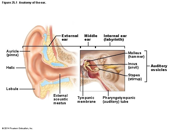
Figure 25. 1 Anatomy of the ear. External ear Middle ear Internal ear (labyrinth) Auricle (pinna) Malleus (hammer) Helix Incus (anvil) Stapes (stirrup) Lobule External acoustic meatus © 2014 Pearson Education, Inc. Tympanic membrane Pharyngotympanic (auditory) tube Auditory ossicles
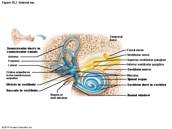
Figure 25. 2 Internal ear. Temporal bone Semicircular ducts in semicircular canals Facial nerve Vestibular nerve Anterior Posterior Superior vestibular ganglion Lateral Inferior vestibular ganglion Cochlear nerve Cristae ampullares in the membranous ampullae Maculae Spinal organ Utricle in vestibule Cochlear duct in cochlea Saccule in vestibule Stapes in oval window © 2014 Pearson Education, Inc. Round window
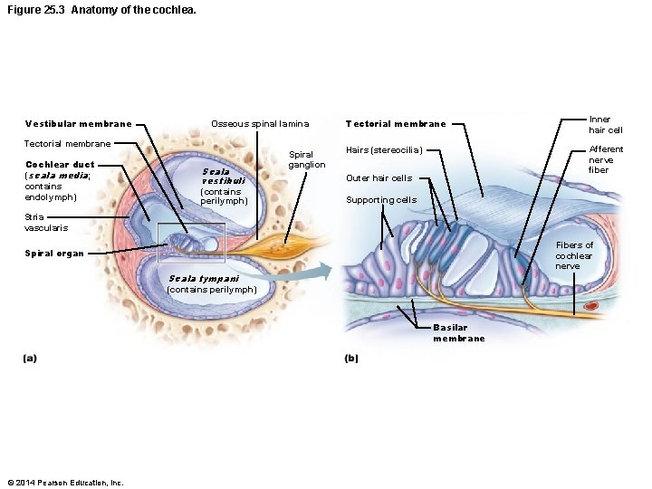
Figure 25. 3 Anatomy of the cochlea. Vestibular membrane Osseous spinal lamina Tectorial membrane Cochlear duct (scala media; contains endolymph) Scala vestibuli (contains perilymph) Spiral ganglion Tectorial membrane Inner hair cell Hairs (stereocilia) Afferent nerve fiber Outer hair cells Supporting cells Stria vascularis Fibers of cochlear nerve Spiral organ Scala tympani (contains perilymph) Basilar membrane © 2014 Pearson Education, Inc.
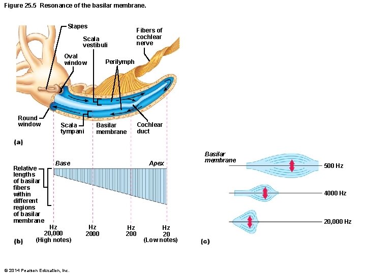
Figure 25. 5 Resonance of the basilar membrane. Stapes Fibers of cochlear nerve Scala vestibuli Oval window Round window Relative lengths of basilar fibers within different regions of basilar membrane Scala tympani Perilymph Basilar membrane Base Hz 20, 000 (High notes) © 2014 Pearson Education, Inc. Cochlear duct Apex Basilar membrane 500 Hz 4000 Hz Hz 2000 Hz 20 (Low notes) 20, 000 Hz
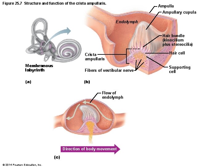
Figure 25. 7 Structure and function of the crista ampullaris. Ampullary cupula Endolymph Hair bundle (kinocilium plus stereocilia) Crista ampullaris Membranous labyrinth Fibers of vestibular nerve Flow of endolymph Direction of body movement © 2014 Pearson Education, Inc. Hair cell Supporting cell
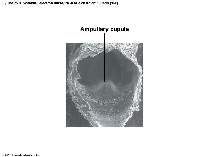
Figure 25. 8 Scanning electron micrograph of a crista ampullaris (14×). Ampullary cupula © 2014 Pearson Education, Inc.