Figure 13 3 Experiment Living S cells Living
 Heat-killed S cells")

 in phage protein Labeled phages infect cells.")
 in phage DNA Labeled phages infect cells.")

 Rosalind Franklin © 2016 Pearson Education, Inc. (b) Franklin’s X-ray")


 Conservative model (b) Semiconservative model (c) Dispersive model")
 Results Bacteria transferred to")


 Origin of replication in an E. coli cell Origin of replication Doublestranded DNA")
 Origins of replication in a eukaryotic cell Origin of replication Parental (template) strand")


- Slides: 33
Figure 13. 3 Experiment Living S cells Living R cells (control) Heat-killed S cells Mixture of heat(control) killed S cells and living R cells Results Mouse dies Mouse healthy Mouse dies Living S cells © 2016 Pearson Education, Inc.
Figure 13. 4 Phage head DNA Tail fiber Genetic material Bacterial cell © 2016 Pearson Education, Inc. 100 nm Tail sheath
Experiment Batch 1: Radioactive sulfur (35 S) in phage protein Labeled phages infect cells. Agitation frees outside phage parts from cells. Centrifuged cells form a pellet. Free phages and phage parts remain in liquid. Radioactivity (phage protein) found in liquid Radioactive protein DNA Centrifuge Pellet © 2016 Pearson Education, Inc.
Experiment Batch 2: Radioactive phosphorus (32 P) in phage DNA Labeled phages infect cells. Agitation frees outside phage parts from cells. Centrifuged cells form a pellet. Free phages and phage parts remain in liquid. Radioactive DNA Centrifuge Pellet © 2016 Pearson Education, Inc. Radioactivity (phage DNA) found in pellet
"It has not escaped our notice that the specific pairing we have postulated immediately suggests a possible copying mechanism for the genetic material. "
Figure 13. 7 (a) Rosalind Franklin © 2016 Pearson Education, Inc. (b) Franklin’s X-ray diffraction photograph of DNA
© 2016 Pearson Education, Inc.
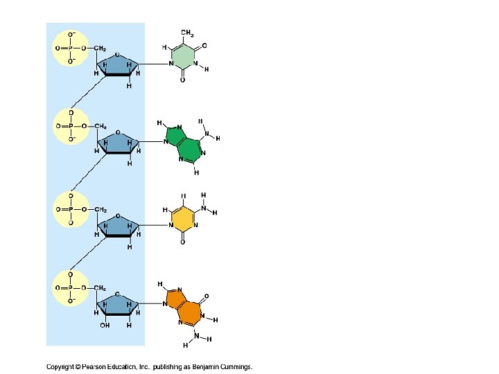
Which are purines? ? ? Which are pyrimidines?
Shows why Chargaff’s rules apply Hydrogen bonds 2 between A and T 3 between G and C
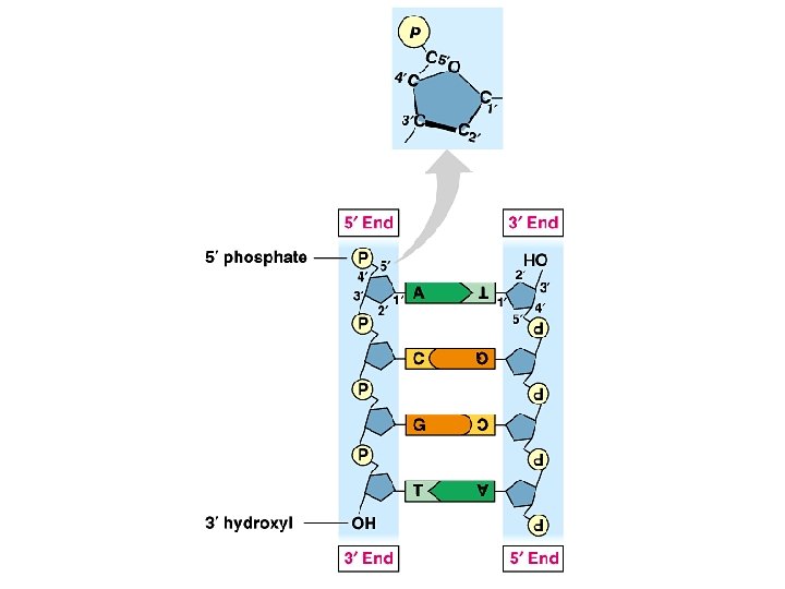
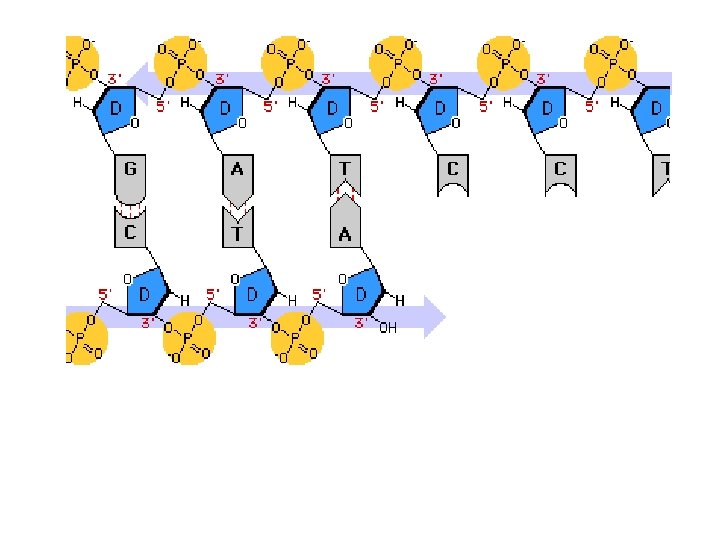
5¢ 3¢ 3¢ A T C G T A A T G C 5¢ (a) Parental molecule © 2016 Pearson Education, Inc.
5¢ 3¢ 3¢ 5¢ 3¢ A T C G T A A T G C 5¢ (a) Parental molecule 3¢ 5¢ (b) Separation of parental strands into templates © 2016 Pearson Education, Inc.
5¢ 3¢ 3¢ 5¢ 3¢ A T A T C G C G T A T A T G C G C 5¢ (a) Parental molecule 3¢ 5¢ (b) Separation of parental strands into templates © 2016 Pearson Education, Inc. 3¢ 5¢ (c) Formation of new strands complementary to template strands
Parent cell First Second replication (a) Conservative model (b) Semiconservative model (c) Dispersive model © 2016 Pearson Education, Inc.
Experiment Bacteria cultured in medium with 15 N (heavy isotope) Results Bacteria transferred to medium with 14 N (lighter isotope) DNA sample centrifuged after first replication © 2016 Pearson Education, Inc. DNA sample centrifuged after second replication Less dense More dense
Conclusion Predictions: Conservative model Semiconservative model Dispersive model © 2016 Pearson Education, Inc. First replication Second replication
Animation: DNA Replication Overview © 2016 Pearson Education, Inc.
(a) Origin of replication in an E. coli cell Origin of replication Doublestranded DNA molecule Parental (template) strand Daughter (new) strand Replication fork Replication bubble 0. 5 mm Two daughter DNA molecules © 2016 Pearson Education, Inc.
(b) Origins of replication in a eukaryotic cell Origin of replication Parental (template) strand Bubble Double-stranded DNA molecule Daughter (new) strand Replication fork 0. 25 mm Two daughter DNA molecules © 2016 Pearson Education, Inc.
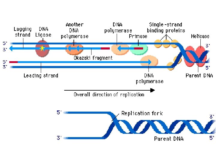
Primase Topoisomerase 3¢ 5¢ 5¢ 3¢ RNA primer Replication fork 3¢ 5¢ Helicase Single-strand binding proteins © 2016 Pearson Education, Inc.
DNA Polymerase • Enzyme that adds DNA nucleotides for replication • Has some “quirks” – Can only add to a pre-existing chain – Can only add to the 3’ end of a chain How are these problems overcome? -Use an RNA primer as the first segment of the new chain (later replaced by DNA) - PRIMASE is the enzyme that does this
Animation: Leading Strand © 2016 Pearson Education, Inc.
Overview Origin of replication Leading strand Lagging strand Primer Lagging strand Overall directions of replication Leading strand Origin of replication 3¢ 5¢ 5¢ 3¢ RNA primer 3¢ 5¢ Parental DNA pol III 3¢ 5¢ 5¢ 3¢ © 2016 Pearson Education, Inc. Continuous elongation in the 5¢ to 3¢ direction 3¢ 5¢
What about that second problem? Not a problem for one The other strand has of the templates to be synthesized (original DNA chain) backwards
Animation: Lagging Strand © 2016 Pearson Education, Inc.
Animation: DNA Replication Review © 2016 Pearson Education, Inc.
Leading strand template DNA pol III Parental DNA 3¢ 5¢ 5¢ 3¢ 3¢ 5¢ 5¢ Connecting protein 3¢ Helicase DNA pol III Lagging strand template Leading strand 3¢ 5¢ Lagging strand Overall direction of replication © 2016 Pearson Education, Inc.
Animation: DNA Replication © 2016 Pearson Education, Inc.