Figure 12 1 Embryonic development of the human
")
Figure 12. 1 Embryonic development of the human brain. Neural tube (contains neural canal) Anterior (rostral) Primary brain vesicles Secondary brain vesicles Adult brain structures Adult neural canal regions Telencephalon Cerebrum: cerebral hemispheres (cortex, white matter, basal nuclei) Lateral ventricles Prosencephalon (forebrain) Diencephalon (thalamus, hypothalamus, epithalamus), retina Third ventricle Mesencephalon (midbrain) Mesencephalon Brain stem: midbrain Cerebral aqueduct Metencephalon Brain stem: pons Rhombencephalon (hindbrain) Cerebellum Myelencephalon Posterior (caudal) © 2014 Pearson Education, Inc. Fourth ventricle Brain stem: medulla oblongata Spinal cord Central canal

Chapter Opener 12 © 2014 Pearson Education, Inc.

Figure 12. 2 c Brain development. Cerebral hemisphere Diencephalon Cerebellum Brain stem • Midbrain • Pons • Medulla oblongata Birth: Shows adult pattern of structures and convolutions. © 2014 Pearson Education, Inc.

Figure 12. 4 a Lobes, sulci, and fissures of the cerebral hemispheres. Anterior Longitudinal fissure Frontal lobe Cerebral veins and arteries covered by arachnoid mater Parietal lobe Left cerebral hemisphere Right cerebral hemisphere Occipital lobe Posterior Superior view © 2014 Pearson Education, Inc.

Figure 12. 4 c Lobes, sulci, and fissures of the cerebral hemispheres. Precentral gyrus Frontal lobe Central sulcus Postcentral gyrus Parietal lobe Parieto-occipital sulcus (on medial surface of hemisphere) Lateral sulcus Fissure (a deep sulcus) Occipital lobe Temporal lobe Transverse cerebral fissure Cerebellum Pons Medulla oblongata Spinal cord Gyrus Cortex (gray matter) Sulcus White matter Lobes and sulci of the cerebrum © 2014 Pearson Education, Inc.
 Cerebral cortex Cerebral white matter")
Figure 12. 9 b Basal nuclei. (2 of 2) Cerebral cortex Cerebral white matter Corpus callosum Anterior horn of lateral ventricle Head of caudate nucleus Putamen Globus pallidus Thalamus Third ventricle Inferior horn of lateral ventricle © 2014 Pearson Education, Inc.

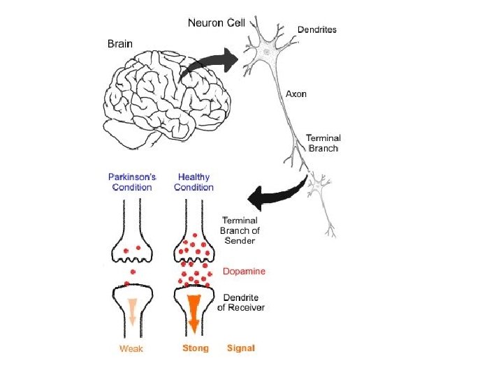
Parkinson’s Disease -Degeneration of dopamine-releasing neurons -What is dopamine? -Without dopamine basal nuclei become overactive-tremor

Huntingtin’s Disease -The Huntingtin gene provides the genetic information for a protein that is also called "huntingtin" -Fatal hereditary disorder -Huntington protein accumulates in basal nuclei -Autosomal dominant mutation -Any child of an affected person typically has a 50% chance of inheriting the disease

Figure 12. 10 a Midsagittal section of the brain. Cerebral hemisphere Corpus callosum Fornix Choroid plexus Septum pellucidum Interthalamic adhesion (intermediate mass of thalamus) Thalamus (encloses third ventricle) Interventricular foramen Anterior commissure Hypothalamus Optic chiasma Pituitary gland Mammillary body Pons Medulla oblongata Spinal cord © 2014 Pearson Education, Inc. Posterior commissure Pineal gland Epithalamus Corpora quadrigemina Cerebral aqueduct Midbrain Arbor vitae (of cerebellum) Fourth ventricle Choroid plexus Cerebellum
 and the diencephalon")
Figure 12. 13 a–b Three views of the brain stem (green) and the diencephalon (purple). Thalamus Diencephalon Hypothalamus Midbrain Pons Medulla oblongata Brain stem View (a) View (c) View (b) Optic chiasma Diencephalon • Thalamus • Hypothalamus Optic nerve (II) Thalamus Optic tract Mammillary body Oculomotor nerve (III) Trochlear nerve (IV) Infundibulum Pituitary gland Crus cerebri of cerebral peduncles (midbrain) Trigeminal nerve (V) Middle cerebellar peduncle Pons Facial nerve (VII) Abducens nerve (VI) Vestibulocochlear nerve (VIII) Pyramid Abducens nerve (VI) Glossopharyngeal nerve (IX) Hypoglossal nerve (XII) Superior colliculus Inferior colliculus Trochlear nerve (IV) Superior cerebellar peduncle Middle cerebellar peduncle Inferior cerebellar peduncle Vestibulocochlear nerve (VIII) Olive Vagus nerve (X) Ventral root of first cervical nerve Accessory nerve (XI) Decussation of pyramids Spinal cord © 2014 Pearson Education, Inc. Ventral view Left lateral view
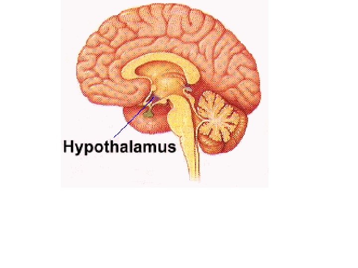

Figure 12. 15 b Cerebellum. Anterior lobe Cerebellar cortex Arbor vitae Cerebellar peduncles • Superior • Middle • Inferior Medulla oblongata © 2014 Pearson Education, Inc. Posterior lobe Flocculonodular lobe Choroid plexus of fourth ventricle

Figure 12. 22 Meninges: dura mater, arachnoid mater, and pia mater. Skin of scalp Periosteum Superior sagittal sinus Subdural space Subarachnoid space © 2014 Pearson Education, Inc. Bone of skull Dura mater • Periosteal layer • Meningeal layer Arachnoid mater Pia mater Arachnoid villus Blood vessel Falx cerebri (in longitudinal fissure only)
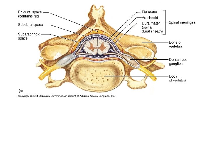

Figure 12. 24 a Formation, location, and circulation of CSF. 4 Superior sagittal sinus Arachnoid villus Choroid plexus Subarachnoid space Arachnoid mater Meningeal dura mater Periosteal dura mater 1 Interventricular foramen Third ventricle Right lateral ventricle (deep to cut) 3 Cerebral aqueduct Lateral aperture Fourth ventricle Median aperture Choroid plexus of fourth ventricle 2 Central canal of spinal cord (a) CSF circulation http: //www. youtube. com/watch? v=SDMO 4 v. Ykq dg © 2014 Pearson Education, Inc. 1 The choroid plexus of each Ventricle produces CSF. 2 CSF flows through the ventricles and into the subarachnoid space via the median and lateral apertures. 3 CSF flows through the subarachnoid space. 4 CSF is absorbed into the dural venous sinuses via the arachnoid villi.

Figure 12. 3 a Ventricles of the brain. Lateral ventricle Anterior horn Inferior horn Third ventricle Cerebral aqueduct Lateral aperture Fourth ventricle Central canal Anterior view © 2014 Pearson Education, Inc.

Figure 12. 3 b Ventricles of the brain. Lateral ventricle Anterior horn Posterior horn Third ventricle Inferior horn Cerebral aqueduct Fourth ventricle Central canal Left lateral view © 2014 Pearson Education, Inc.

Figure 12. 24 Formation, location, and circulation of CSF. Superior sagittal sinus Arachnoid villus Choroid plexus Subarachnoid space Arachnoid mater Meningeal dura mater Periosteal dura mater Right lateral ventricle (deep to cut) Interventricular foramen Third ventricle Choroid plexus of fourth ventricle Cerebral aqueduct Lateral aperture Fourth ventricle Median aperture Central canal of spinal cord 1 The choroid plexus of each ventricle produces CSF. 2 CSF flows through the ventricles and into the subarachnoid space via the median and lateral apertures. 3 CSF flows through the subarachnoid space. 4 CSF is absorbed into the dural venous sinuses via the arachnoid villi. CSF circulation © 2014 Pearson Education, Inc. Ependymal cells Capillary Connective tissue of pia mater Wastes and unnecessary solutes absorbed Section of choroid plexus Cavity of ventricle CSF formation by choroid plexuses CSF forms as a filtrate containing glucose, oxygen, vitamins, and ions (Na+, Cl–, Mg 2+, etc. )

Figure 12. 24 a Formation, location, and circulation of CSF. 4 Superior sagittal sinus Arachnoid villus Choroid plexus Subarachnoid space Arachnoid mater Meningeal dura mater Periosteal dura mater 1 Interventricular foramen Third ventricle Right lateral ventricle (deep to cut) 3 Cerebral aqueduct Lateral aperture Fourth ventricle Median aperture Choroid plexus of fourth ventricle 2 Central canal of spinal cord (a) CSF circulation http: //www. youtube. com/watch? v=SDMO 4 v. Ykq dg © 2014 Pearson Education, Inc. 1 The choroid plexus of each Ventricle produces CSF. 2 CSF flows through the ventricles and into the subarachnoid space via the median and lateral apertures. 3 CSF flows through the subarachnoid space. 4 CSF is absorbed into the dural venous sinuses via the arachnoid villi.

Figure 12. 25 Hydrocephalus in a newborn. © 2014 Pearson Education, Inc.

Figure 12. 26 a Gross structure of the spinal cord, dorsal view. Cervical enlargement Dura and arachnoid mater Lumbar enlargement Conus medullaris Cauda equina Filum terminale Cervical spinal nerves Thoracic spinal nerves Lumbar spinal nerves Sacral spinal nerves The spinal cord and its nerve roots, with the bony vertebral arches removed. The dura mater and © 2014 Pearson Education, Inc. arachnoid mater are cut open and reflected laterally.
 Subdural")
Figure 12. 28 a Anatomy of the spinal cord. Epidural space (contains fat) Subdural space Subarachnoid space (contains CSF) Pia mater Arachnoid mater Dura mater Spinal meninges Bone of vertebra Dorsal root ganglion Body of vertebra Cross section of spinal cord and vertebra © 2014 Pearson Education, Inc.

Figure 12. 28 b Anatomy of the spinal cord. Dorsal funiculus White columns Ventral funiculus Lateral funiculus Dorsal median sulcus Gray commissure Dorsal horn Gray Ventral horn matter Lateral horn Dorsal root ganglion Spinal nerve Dorsal root (fans out into dorsal rootlets) Ventral root (derived from several ventral rootlets) Central canal Ventral median fissure Pia mater Arachnoid mater Spinal dura mater The spinal cord and its meningeal coverings © 2014 Pearson Education, Inc.
- Slides: 29