
Figure 10 0 1 THE STRUCTURE OF THE






- Slides: 28
Figure 10. 0 -1 THE STRUCTURE OF THE GENETIC MATERIAL © 2015 Pearson Education, Inc.
Learner Outcomes I Can…… • Describe the experiments of Griffith, Hershey, and Chase, which supported the idea that DNA was life’s genetic material. • Compare the structures of DNA and RNA. © 2015 Pearson Education, Inc.
10. 1 SCIENTIFIC THINKING: Experiments showed that DNA is the genetic material • Early in the 20 th century, the molecular basis for inheritance was a mystery. • Biologists did know that genes were located on chromosomes. But it was unknown if the genetic material was • proteins or • DNA. © 2015 Pearson Education, Inc.
10. 1 SCIENTIFIC THINKING: Experiments showed that DNA is the genetic material • Biologists finally established the role of DNA in heredity through experiments with bacteria & viruses. • This breakthrough led to the field of molecular biology, the study of heredity at the molecular level. © 2015 Pearson Education, Inc.
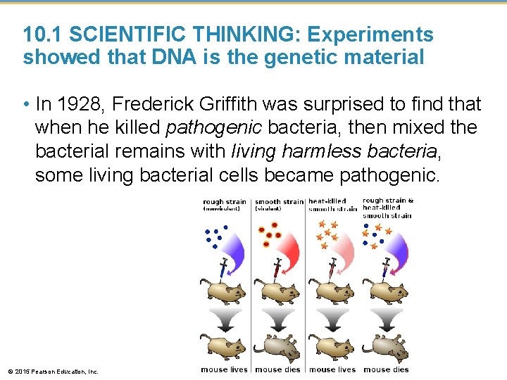
10. 1 SCIENTIFIC THINKING: Experiments showed that DNA is the genetic material • In 1928, Frederick Griffith was surprised to find that when he killed pathogenic bacteria, then mixed the bacterial remains with living harmless bacteria, some living bacterial cells became pathogenic. © 2015 Pearson Education, Inc.
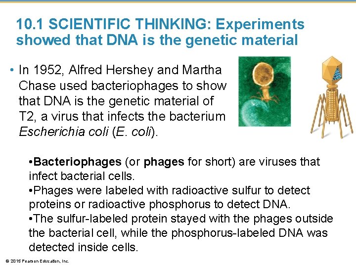
10. 1 SCIENTIFIC THINKING: Experiments showed that DNA is the genetic material • In 1952, Alfred Hershey and Martha Chase used bacteriophages to show that DNA is the genetic material of T 2, a virus that infects the bacterium Escherichia coli (E. coli). • Bacteriophages (or phages for short) are viruses that infect bacterial cells. • Phages were labeled with radioactive sulfur to detect proteins or radioactive phosphorus to detect DNA. • The sulfur-labeled protein stayed with the phages outside the bacterial cell, while the phosphorus-labeled DNA was detected inside cells. © 2015 Pearson Education, Inc.
Animation: Hershey-Chase Experiment © 2015 Pearson Education, Inc.
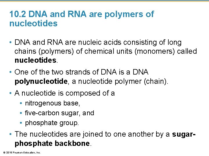
10. 2 DNA and RNA are polymers of nucleotides • DNA and RNA are nucleic acids consisting of long chains (polymers) of chemical units (monomers) called nucleotides. • One of the two strands of DNA is a DNA polynucleotide, a nucleotide polymer (chain). • A nucleotide is composed of a • nitrogenous base, • five-carbon sugar, and • phosphate group. • The nucleotides are joined to one another by a sugarphosphate backbone. © 2015 Pearson Education, Inc.
Figure 10. 2 a-0 A C T G A T C Sugar-phosphate backbone G T A C G C T A A T C Covalent bond joining nucleotides T A G A A G C Phosphate group Nitrogenous base Sugar Nitrogenous base (can be A, G, C, or T) C G T A A DNA double helix DNA nucleotide T G Phosphate group Thymine (T) Sugar (deoxyribose) DNA nucleotide G G Two representations of a DNA polynucleotide © 2015 Pearson Education, Inc.
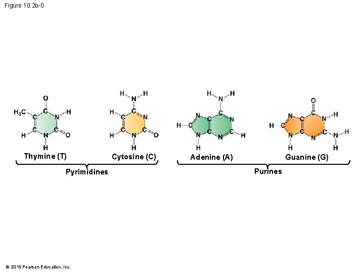
10. 2 DNA and RNA are polymers of nucleotides • Each type of DNA nucleotide has a different nitrogen-containing base: • • adenine (A), cytosine (C), thymine (T), and guanine (G). © 2015 Pearson Education, Inc.
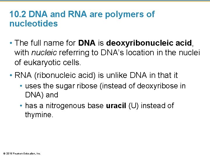
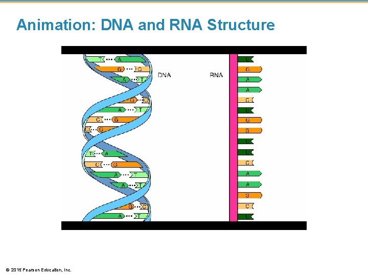
10. 2 DNA and RNA are polymers of nucleotides • The full name for DNA is deoxyribonucleic acid, with nucleic referring to DNA’s location in the nuclei of eukaryotic cells. • RNA (ribonucleic acid) is unlike DNA in that it • uses the sugar ribose (instead of deoxyribose in DNA) and • has a nitrogenous base uracil (U) instead of thymine. © 2015 Pearson Education, Inc.
Animation: DNA and RNA Structure © 2015 Pearson Education, Inc.
Figure 10. 2 b-0 Thymine (T) Pyrimidines © 2015 Pearson Education, Inc. Cytosine (C) Adenine (A) Guanine (G) Purines
10. 3 DNA is a double-stranded helix • After the 1952 Hershey-Chase experiment convinced most biologists that DNA was the material that stored genetic information, a race was on to determine how the structure of this molecule could account for its role in heredity. • Researchers focused on discovering the threedimensional shape of DNA. © 2015 Pearson Education, Inc.
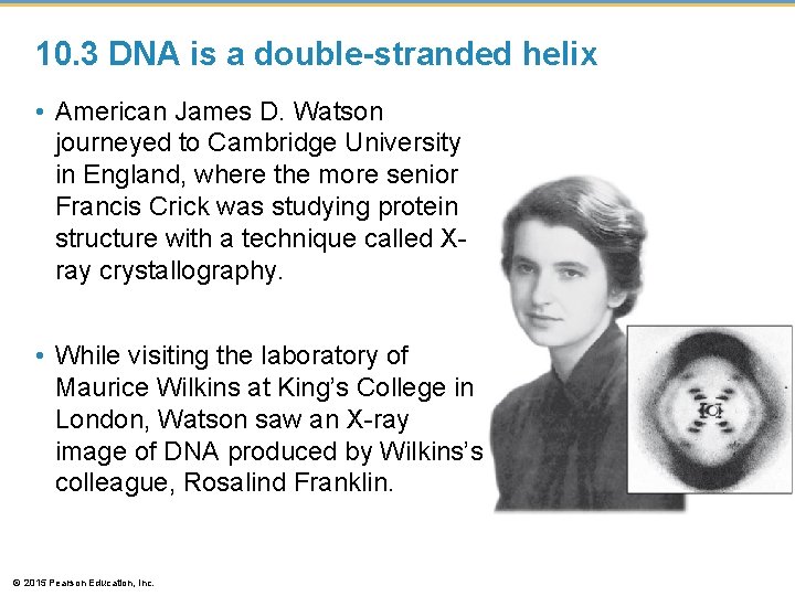
10. 3 DNA is a double-stranded helix • American James D. Watson journeyed to Cambridge University in England, where the more senior Francis Crick was studying protein structure with a technique called Xray crystallography. • While visiting the laboratory of Maurice Wilkins at King’s College in London, Watson saw an X-ray image of DNA produced by Wilkins’s colleague, Rosalind Franklin. © 2015 Pearson Education, Inc.
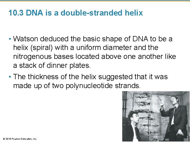
10. 3 DNA is a double-stranded helix • Watson deduced the basic shape of DNA to be a helix (spiral) with a uniform diameter and the nitrogenous bases located above one another like a stack of dinner plates. • The thickness of the helix suggested that it was made up of two polynucleotide strands. © 2015 Pearson Education, Inc.
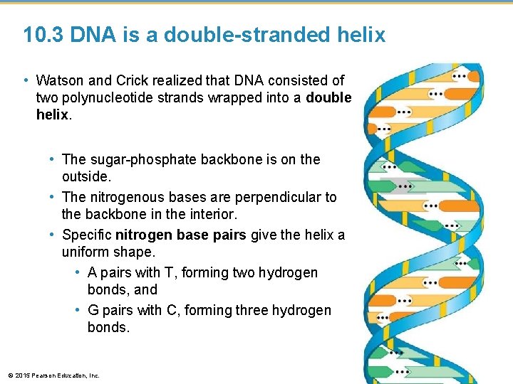
10. 3 DNA is a double-stranded helix • Watson and Crick realized that DNA consisted of two polynucleotide strands wrapped into a double helix. • The sugar-phosphate backbone is on the outside. • The nitrogenous bases are perpendicular to the backbone in the interior. • Specific nitrogen base pairs give the helix a uniform shape. • A pairs with T, forming two hydrogen bonds, and • G pairs with C, forming three hydrogen bonds. © 2015 Pearson Education, Inc.
10. 3 DNA is a double-stranded helix • In 1962, the Nobel Prize was awarded to James D. Watson, Francis Crick, and Maurice Wilkins. • Rosalind Franklin probably would have received the prize as well but for her death from cancer in 1958. • Nobel Prizes are never awarded posthumously. • The Watson-Crick model gave new meaning to the words genes and chromosomes. The genetic information in a chromosome is encoded in the nucleotide sequence of DNA. © 2015 Pearson Education, Inc.
DNA REPLICATION © 2015 Pearson Education, Inc.
I Can…. 1. EXPLAIN HOW THE STRUCTURE OF DNA FACILITATES ITS REPLICATION. 2. DESCRIBE THE PROCESS OF DNA REPLICATION. © 2015 Pearson Education, Inc.
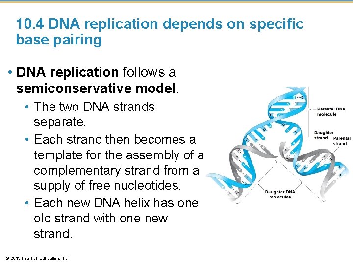
10. 4 DNA replication depends on specific base pairing • DNA replication follows a semiconservative model. • The two DNA strands separate. • Each strand then becomes a template for the assembly of a complementary strand from a supply of free nucleotides. • Each new DNA helix has one old strand with one new strand. © 2015 Pearson Education, Inc.
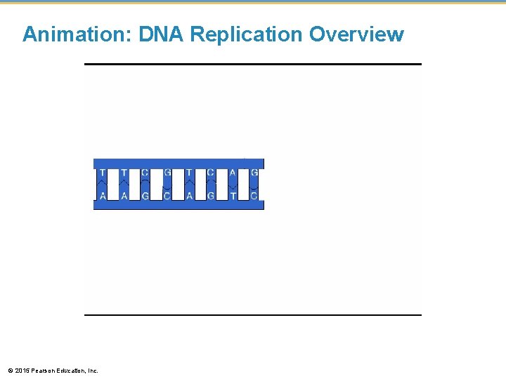
Animation: DNA Replication Overview © 2015 Pearson Education, Inc.
10. 5 DNA replication proceeds in two directions at many sites simultaneously • Replication of a DNA molecule begins at particular sites called origins of replication, short stretches of DNA having a specific sequence of nucleotides. • DNA Helicase, an enzyme/protein, initiates DNA replication • attach to the DNA at the origin of replication and • separate the two strands of the double helix. © 2015 Pearson Education, Inc.
Figure 10. 5 a Parental DNA molecule Origin of replication “Bubble” Two daughter DNA molecules © 2015 Pearson Education, Inc. Parental strand Daughter strand
10. 5 DNA replication proceeds in two directions at many sites simultaneously • DNA replication occurs in the 5 to 3 direction. • DNA polymerases add nucleotides only to the 3 end of the strand, never to the 5 end. • Replication is discontinuous on the 5 to 3 template, forming short Okazaki fragments. • An enzyme, called DNA ligase, links (or ligates) the pieces together into a single DNA strand. © 2015 Pearson Education, Inc.
Figure 10. 5 c DNA polymerase molecule 5′ 3′ Parental DNA Replication fork 5′ 3′ DNA ligase Overall direction of replication © 2015 Pearson Education, Inc. 3′ 5′ This daughter strand is synthesized continuously This daughter strand is 3′ synthesized 5′ in pieces
10. 5 DNA replication proceeds in two directions at many sites simultaneously • DNA polymerases and DNA ligase also repair DNA damaged by harmful radiation and toxic chemicals. • DNA replication ensures that all the somatic cells in a multicellular organism carry the same genetic information. © 2015 Pearson Education, Inc.
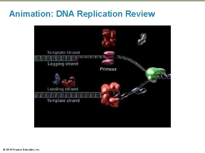
Animation: DNA Replication Review © 2015 Pearson Education, Inc.