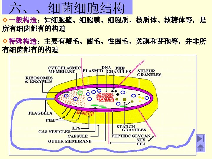
Figure 1 Schematic diagram of a typical procaryotic


- Slides: 13
Figure 1. Schematic diagram of a typical procaryotic cell. Minimally, a procaryote is composed of a cell wall and plasma membrane that surrounds its cytoplasm containing a chromosome, ribosomes, enzymes, several classes of RNA, and small molecules (precursors).
Figure 4 - Electron micrograph of a Gcell wall. Figure 3 - Electron micrograph of a G+ cell wall.
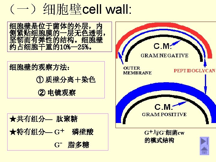
细胞壁cell wall:
Gram Positive
Gram Negative
Figure 5 - A comparison of L and D amino acids. Note that while the structure is identical, it is impossible to superimpose them.
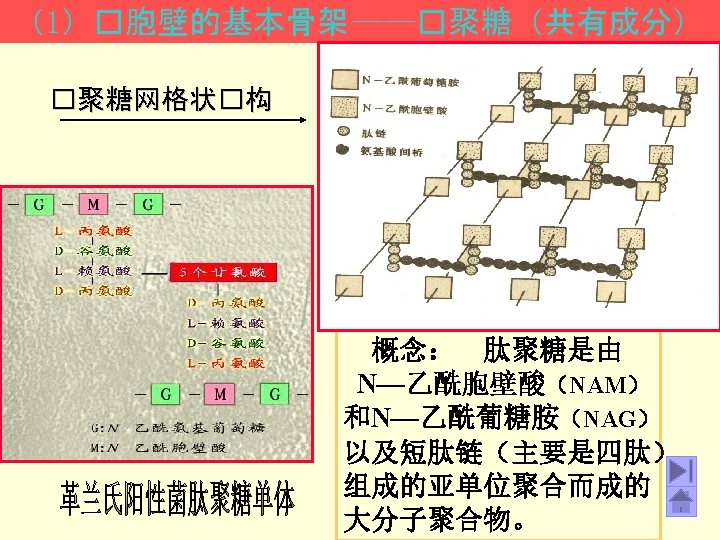
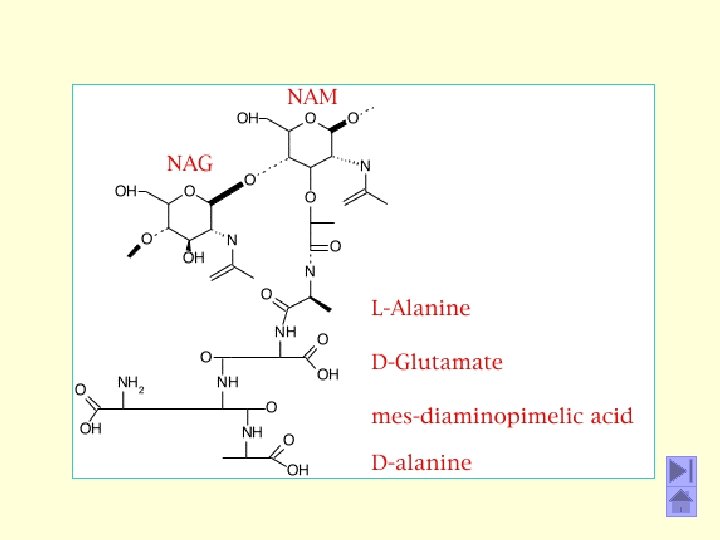
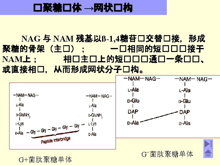
Figure 8. Schematic diagram of the peptidoglycan sheet of Staphylococcus aureus. G = N-acetyl-glucosamine; M = Nacetyl-muramic acid; L-ala = L-alanine; D-ala = D-alanine; Dglu = D-glutamic acid; L-lys = L-lysine. This is one type of murein found in Gram-positive bacteria. Compared to the E. coli peptidoglycan (Figure 7) there is L-lys in place of DAP (diaminopimelic acid) in the tetrapeptide. The free amino group of L-lys is substituted with a glycine pentapeptide (glygly-gly-) which then becomes an interpeptide bridge forming a link with a carboxy group from D-ala in an adjacent tetrapeptide side chain. Gram-positive peptidoglycans differ from species to species, mainly in regards to the amino acids in the third position of the tetrapeptide side chain and in the amino acid composition of the interpeptide bridge.