Fig 3 20 Reproduced from Biochemistry by T
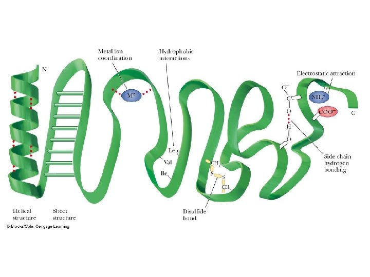

Fig 3. 20 Reproduced from: Biochemistry by T. A. Brown, ISBN: 9781907904288 © Scion Publishing Ltd, 2017

Fig 3. 27 Reproduced from: Biochemistry by T. A. Brown, ISBN: 9781907904288 © Scion Publishing Ltd, 2017

Fig 3. 28 Reproduced from: Biochemistry by T. A. Brown, ISBN: 9781907904288 © Scion Publishing Ltd, 2017

Fig 3. 29 Reproduced from: Biochemistry by T. A. Brown, ISBN: 9781907904288 © Scion Publishing Ltd, 2017

Fig 3. 30 Reproduced from: Biochemistry by T. A. Brown, ISBN: 9781907904288 © Scion Publishing Ltd, 2017

Quick Thoughts on Protein Folding • There are literally millions of possible ways a simple protein can fold, but only one conformation that works • Hydrophobic interactions help drive protein folding – Not so much the hydrophobic groups attracting each other (That only involves London Forces, right? ) – The dipole-dipole interactions between water molecules in solution are much stronger and push the hydrophobic side chains aside • The entropy of the universe must increase in a spontaneous process, and protein folding is a spontaneous process – When water molecules surround a nonpolar compound, they are restricted in the number of hydrogen bonds then can form which represents a lower entropy – By having the hydrophobic residues sequestered in the core of the folded protein, the water molecules are free to form up to 4 hydrogen bonds each. • This freedom represents greater entropy, thus helping drive folding of the protein
 Where are the side chains")
Protein Folding: Myoglobin Note Placement of hydrophobic residues (green) Where are the side chains pointing? Note Placement of polar residues (blue) Where are the side chains pointing?

Fig 7. 2 Reproduced from: Biochemistry by T. A. Brown, ISBN: 9781907904288 © Scion Publishing Ltd, 2017

Fig 7. 4 Reproduced from: Biochemistry by T. A. Brown, ISBN: 9781907904288 © Scion Publishing Ltd, 2017

Table 7. 1 Reproduced from: Biochemistry by T. A. Brown, ISBN: 9781907904288 © Scion Publishing Ltd, 2017

Fig 7. 7 Reproduced from: Biochemistry by T. A. Brown, ISBN: 9781907904288 © Scion Publishing Ltd, 2017

Fig 7. 10 Reproduced from: Biochemistry by T. A. Brown, ISBN: 9781907904288 © Scion Publishing Ltd, 2017

Fig 7. 11 Reproduced from: Biochemistry by T. A. Brown, ISBN: 9781907904288 © Scion Publishing Ltd, 2017

Fig 7. 12 Reproduced from: Biochemistry by T. A. Brown, ISBN: 9781907904288 © Scion Publishing Ltd, 2017

How Do Enzymes Work So Well? How do they bring about huge rate enhancements? What is the source of the energy necessary to decrease the activation energy? 1) Many enzyme catalyzed reactions involve the rearrangement of covalent bonds • The enzyme has prosthetic groups and amino acid side chain functional groups that can break and make bonds, thereby lowering G*

How Do Enzymes Work So Well? How do they bring about huge rate enhancements? What is the source of the energy necessary to decrease the activation energy? 2) Noncovalent or Intermolecular forces (Duh!) • By making A LOT of low energy interactions with the substrate, the enzyme can lower the activation energy • These interactions ALL contribute to the BINDING ENERGY, Gb, • This binding energy is the primary source of free energy used to lower the activation energy of reactions

How to Lower G - The Proximity Model Enzymes organize reactive groups into close proximity and proper orientation • Uncatalyzed bimolecular reactions two free reactants single restricted transition state conversion is entropically unfavorable • Uncatalyzed unimolecular reactions flexible reactant rigid transition state conversion is entropically unfavorable for flexible reactants • Catalyzed reactions Enzyme uses the binding energy of substrates to organize the reactants to a fairly rigid ES complex Entropy cost is paid during binding Rigid reactant complex transition state conversion is entropically OK

Support for the Proximity Model The rate of anhydride formation from esters and carboxylates shows a strong dependence on proximity of two reactive groups (work by Thomas C. Bruice’s group). The smaller the reactants for a given reaction, the fewer degrees of freedom and therefore the faster the reaction. Enzymes “hold” groups into place, minimizing the possibility of rotation and maximizing the possibility of encountering each other

How to Lower G Enzymes bind transition states best • The idea was proposed by Linus Pauling in 1946 – Enzyme active sites are complimentary to the transition state of the reaction – Enzymes bind transition states better than substrates – Stronger/additional interactions with the transition state as compared to the ground state lower the activation barrier

Transition State Stabilization: Imaginary Stickase
 Binding energy, Gb is the key player in")
Key Principles of Enzyme Function 1) Binding energy, Gb is the key player in catalysis – Gb determines Specificity and Catalytic Rate • If you don’t form as many low energy IMF interactions b/w enzyme and substrate, the G* is not decreased as much and the rate is decreased relative to another substrate with a higher Gb value 2) The weak interactions that comprise the Gb are optimized for the transition state NOT the substrate conformation - This helps “pull” the substrate over the hump and into product

Key Principles of Enzyme Function By combining these two ideas with everything we’ve learned to date, we can come up with a good thought model that describes how and enzyme functions: 1)Binding substrates restricts their degrees of rotational and translational freedom • • This places them in a mode that primes them for reaction It decreases their entropy (Uh oh) 2) Binding disrupts IMFs between solvent and substrate and even the active site residues • Desolvation of substrate Increases Solvent Entropy – Remember the hydrophobic effect and clathrate cages?

Key Principles of Enzyme Function By combining these two ideas with everything we’ve learned to date, we can come up with a good thought model that describes how and enzyme functions 3)The binding energy, Gb, is at its maximum during the transition state, so this helps pay the activation cost 4)The enzyme itself changes conformation when binding substrate • Remember the Induced Fit model? • Energetic Shock Absorber

Fig 7. 14 Reproduced from: Biochemistry by T. A. Brown, ISBN: 9781907904288 © Scion Publishing Ltd, 2017

Fig 7. 15 Reproduced from: Biochemistry by T. A. Brown, ISBN: 9781907904288 © Scion Publishing Ltd, 2017
- Slides: 26