
Fig 3 1 Copyright Mc GrawHill Education Permission
- Slides: 28
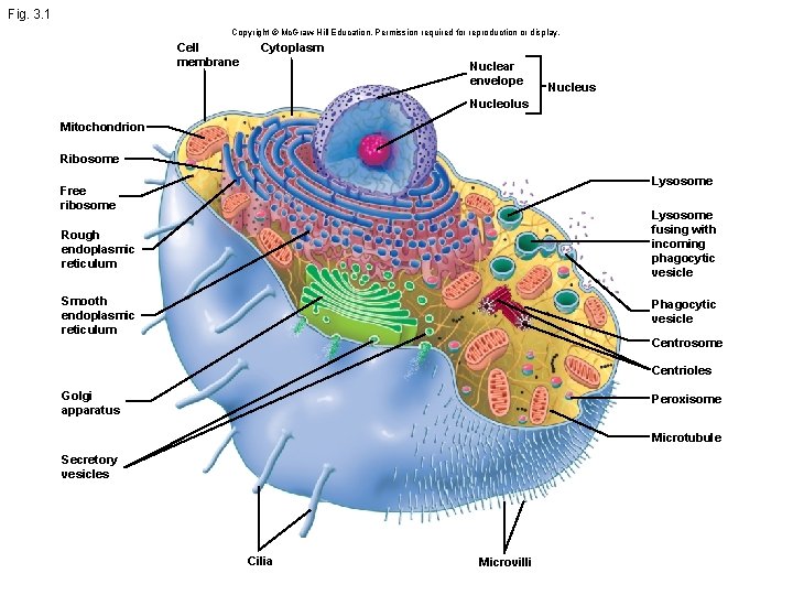
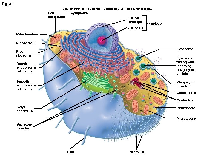
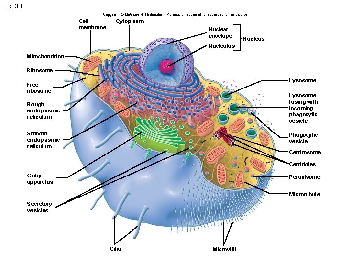
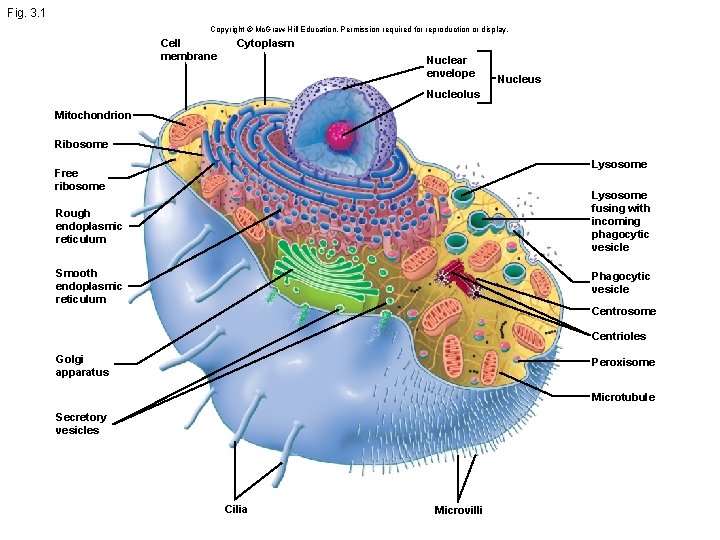
Fig. 3. 1 Copyright © Mc. Graw-Hill Education. Permission required for reproduction or display. Cell membrane Cytoplasm Nuclear envelope Nucleus Nucleolus Mitochondrion Ribosome Lysosome Free ribosome Rough endoplasmic reticulum Lysosome fusing with incoming phagocytic vesicle Smooth endoplasmic reticulum Phagocytic vesicle Centrosome Centrioles Golgi apparatus Peroxisome Microtubule Secretory vesicles Cilia Microvilli
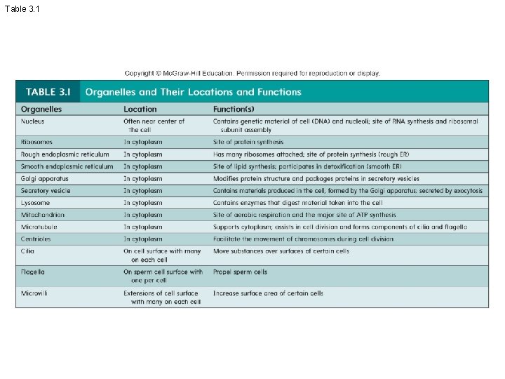
Table 3. 1
Fig. 3. 1 Copyright © Mc. Graw-Hill Education. Permission required for reproduction or display. Cell membrane Cytoplasm Nuclear envelope Nucleus Nucleolus Mitochondrion Ribosome Lysosome Free ribosome Rough endoplasmic reticulum Lysosome fusing with incoming phagocytic vesicle Smooth endoplasmic reticulum Phagocytic vesicle Centrosome Centrioles Golgi apparatus Peroxisome Microtubule Secretory vesicles Cilia Microvilli
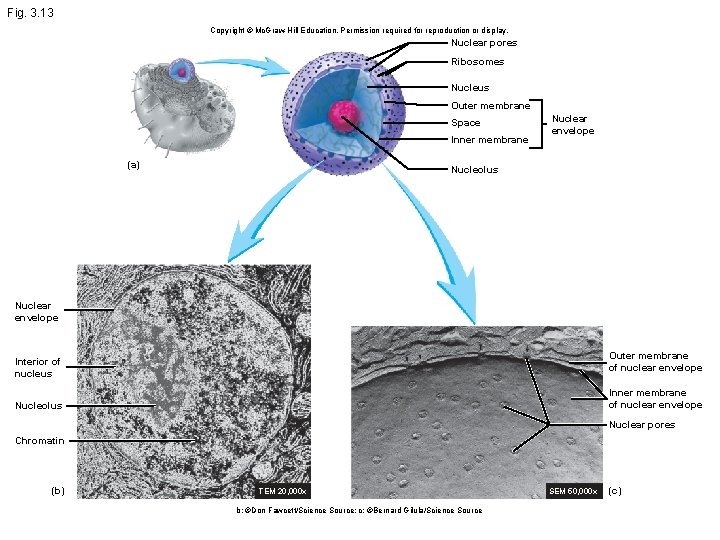
Fig. 3. 13 Copyright © Mc. Graw-Hill Education. Permission required for reproduction or display. Nuclear pores Ribosomes Nucleus Outer membrane Space Inner membrane (a) Nuclear envelope Nucleolus Nuclear envelope Outer membrane of nuclear envelope Interior of nucleus Inner membrane of nuclear envelope Nucleolus Nuclear pores Chromatin (b) TEM 20, 000 x b: ©Don Fawcett/Science Source; c: ©Bernard Gilula/Science Source SEM 50, 000 x (c)
Fig. 3. 1 Copyright © Mc. Graw-Hill Education. Permission required for reproduction or display. Cell membrane Cytoplasm Nuclear envelope Nucleus Nucleolus Mitochondrion Ribosome Lysosome Free ribosome Rough endoplasmic reticulum Lysosome fusing with incoming phagocytic vesicle Smooth endoplasmic reticulum Phagocytic vesicle Centrosome Centrioles Golgi apparatus Peroxisome Microtubule Secretory vesicles Cilia Microvilli
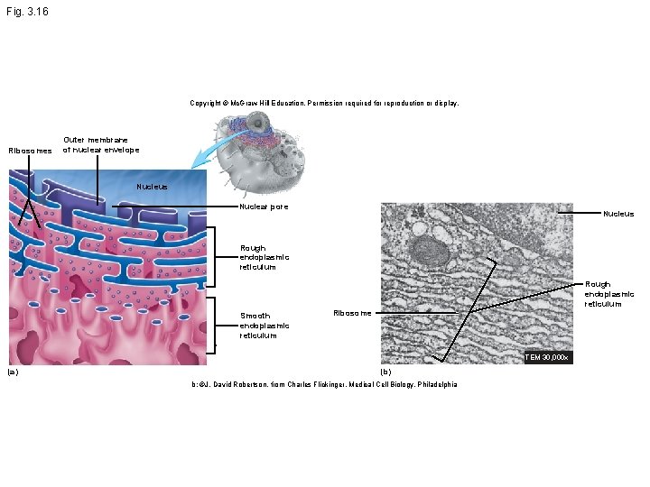
Fig. 3. 16 Copyright © Mc. Graw-Hill Education. Permission required for reproduction or display. Ribosomes Outer membrane of nuclear envelope Nucleus Nuclear pore Nucleus Rough endoplasmic reticulum Smooth endoplasmic reticulum Ribosome TEM 30, 000 x (a) (b) b: ©J. David Robertson, from Charles Flickinger, Medical Cell Biology, Philadelphia
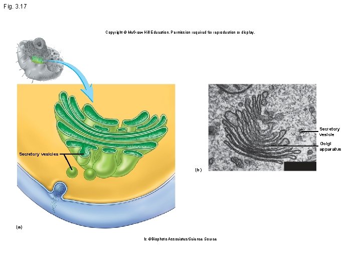
Fig. 3. 17 Copyright © Mc. Graw-Hill Education. Permission required for reproduction or display. Secretory vesicle Golgi apparatus Secretory vesicles (b) (a) b: ©Biophoto Associates/Science Source
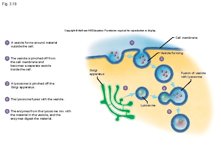
Fig. 3. 18 Copyright © Mc. Graw-Hill Education. Permission required for reproduction or display. Cell membrane 1 A vesicle forms around material outside the cell. 1 Vesicle forming 2 The vesicle is pinched off from the cell membrane and becomes a separate vesicle inside the cell. 3 A lysosome is pinched off the Golgi apparatus. 2 Golgi apparatus Fusion of vesicle with lysosome 3 4 4 The lysosome fuses with the vesicle. Lysosome 5 The enzymes from the lysosome mix with the material in the vesicle, and the enzymes digest the material. 5
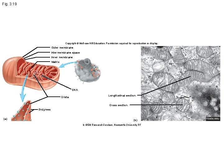
Fig. 3. 19 Copyright © Mc. Graw-Hill Education. Permission required for reproduction or display. Outer membrane Intermembrane space Inner membrane Matrix DNA Crista Longitudinal section Cross section Enzymes (a) (b) b: ©EM Research Services, Newcastle University RF TEM 34, 000 x
Fig. 3. 1 Copyright © Mc. Graw-Hill Education. Permission required for reproduction or display. Cell membrane Cytoplasm Nuclear envelope Nucleus Nucleolus Mitochondrion Ribosome Lysosome Free ribosome Rough endoplasmic reticulum Lysosome fusing with incoming phagocytic vesicle Smooth endoplasmic reticulum Phagocytic vesicle Centrosome Centrioles Golgi apparatus Peroxisome Microtubule Secretory vesicles Cilia Microvilli
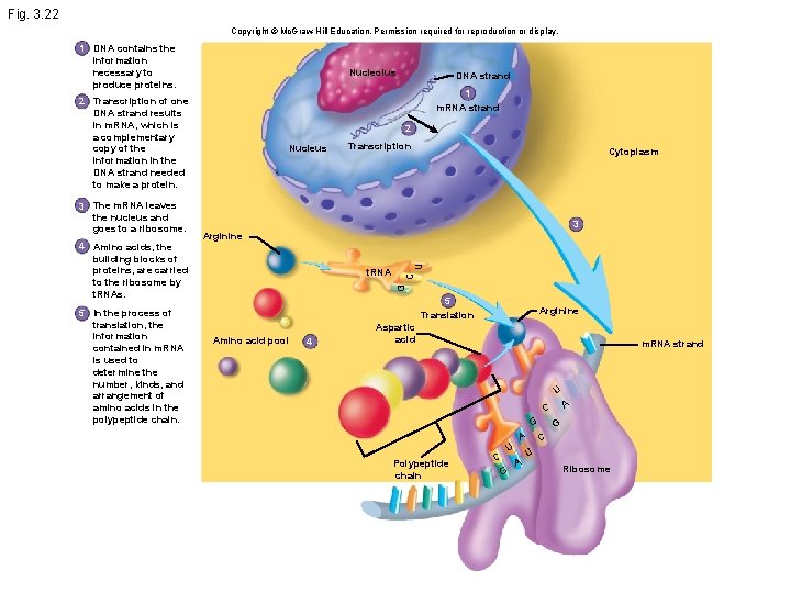
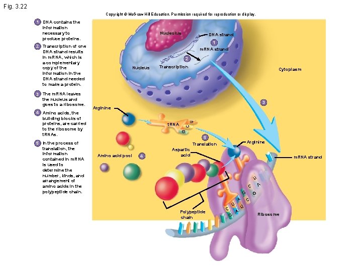
Fig. 3. 22 Copyright © Mc. Graw-Hill Education. Permission required for reproduction or display. 1 DNA contains the information necessary to produce proteins. Nucleolus 1 m. RNA strand 2 Transcription of one DNA strand results in m. RNA, which is a complementary copy of the information in the DNA strand needed to make a protein. 5 In the process of translation, the information contained in m. RNA is used to determine the number, kinds, and arrangement of amino acids in the polypeptide chain. Nucleus Transcription Cytoplasm 3 t. RNA U Arginine C 4 Amino acids, the building blocks of proteins, are carried to the ribosome by t. RNAs. 2 G 3 The m. RNA leaves the nucleus and goes to a ribosome. DNA strand 5 Translation Amino acid pool 4 Arginine Aspartic acid m. RNA strand U A C G A Polypeptide chain C U G A G C U Ribosome
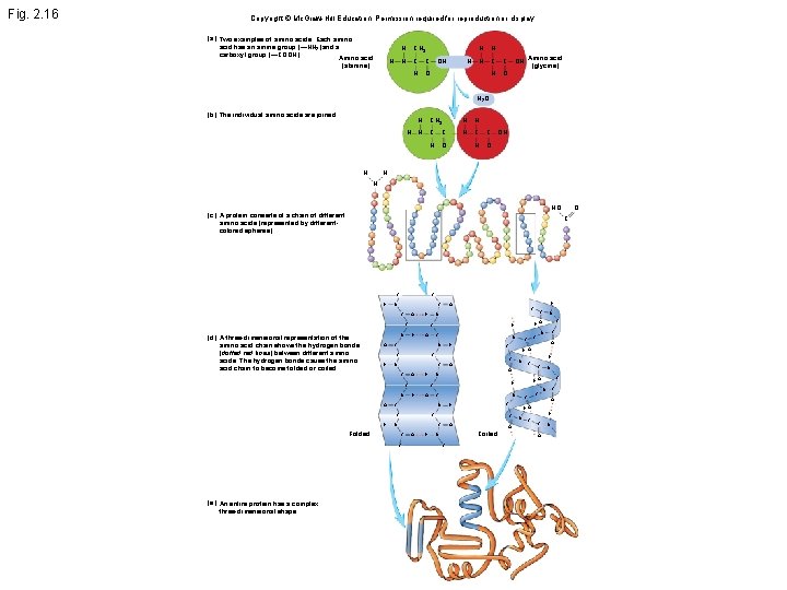
Fig. 2. 16 Copyright © Mc. Graw-Hill Education. Permission required for reproduction or display. (a) Two examples of amino acids. Each amino acid has an amine group (—NH 2) and a carboxyl group (—COOH). Amino acid H (alanine) H CH 3 N C C H OH H H N C C H O OH Amino acid (glycine) H 2 O (b) The individual amino acids are joined. H H H CH 3 N C C H O H H N C C H O OH H N HO (c) A protein consists of a chain of different amino acids (represented by differentcolored spheres). C H C N C C O H H O C (d) A three-dimensional representation of the amino acid chain shows the hydrogen bonds (dotted red lines) between different amino acids. The hydrogen bonds cause the amino acid chain to become folded or coiled. N O C O N C H N H H C O C C O H C C H C O O Coiled C C C N O H O C N N O C C C O H N N H C C N C C H N N H O O N O C Folded N N C H O H C N O C C H H O N C C (e) An entire protein has a complex three-dimensional shape. O C N H C C O N
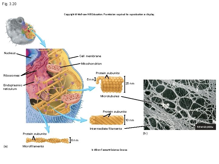
Fig. 3. 20 Copyright © Mc. Graw-Hill Education. Permission required for reproduction or display. Nucleus Cell membrane Mitochondrion Protein subunits Ribosomes 5 nm 25 nm Endoplasmic reticulum Microtubules Protein subunits 10 nm SEM 60, 000 x Intermediate filaments (b) Protein subunits 8 nm (a) Microfilaments b: ©Don Fawcett/Science Source
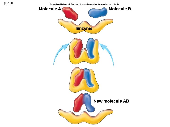
Fig. 2. 18 Copyright © Mc. Graw-Hill Education. Permission required for reproduction or display. Molecule A Molecule B Enzyme New molecule AB
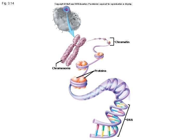
Fig. 3. 14 Copyright © Mc. Graw-Hill Education. Permission required for reproduction or display. Chromatin Chromosome Proteins DNA
Fig. 3. 22 Copyright © Mc. Graw-Hill Education. Permission required for reproduction or display. 1 DNA contains the information necessary to produce proteins. Nucleolus 1 m. RNA strand 2 Transcription of one DNA strand results in m. RNA, which is a complementary copy of the information in the DNA strand needed to make a protein. 5 In the process of translation, the information contained in m. RNA is used to determine the number, kinds, and arrangement of amino acids in the polypeptide chain. Nucleus Transcription Cytoplasm 3 t. RNA U Arginine C 4 Amino acids, the building blocks of proteins, are carried to the ribosome by t. RNAs. 2 G 3 The m. RNA leaves the nucleus and goes to a ribosome. DNA strand 5 Translation Amino acid pool 4 Arginine Aspartic acid m. RNA strand U A C G A Polypeptide chain C U G A G C U Ribosome
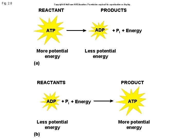
Fig. 2. 8 Copyright © Mc. Graw-Hill Education. Permission required for reproduction or display. REACTANT PRODUCTS ATP ADP More potential energy (a) Less potential energy + Pi + Energy REACTANTS PRODUCT ADP ATP + Pi + Energy Less potential energy (b) More potential energy
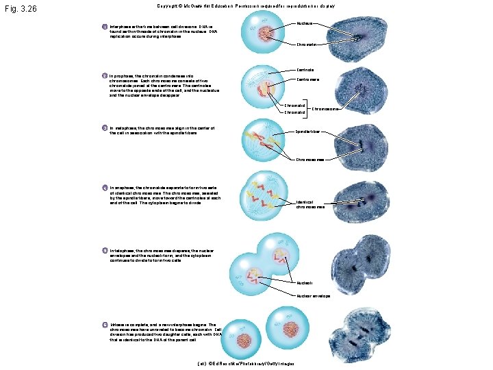
Fig. 3. 26 Copyright © Mc. Graw-Hill Education. Permission required for reproduction or display. Nucleus 1 Interphase is the time between cell divisions. DNA is found as thin threads of chromatin in the nucleus. DNA replication occurs during interphase. Chromatin Centriole 2 In prophase, the chromatin condenses into chromosomes. Each chromosome consists of two chromatids joined at the centromere. The centrioles move to the opposite ends of the cell, and the nucleolus and the nuclear envelope disappear. Centromere Chromatid 3 In metaphase, the chromosomes align in the center of the cell in association with the spindle fibers. Chromosome Spindle fiber Chromosomes 4 In anaphase, the chromatids separate to form two sets of identical chromosomes. The chromosomes, assisted by the spindle fibers, move toward the centrioles at each end of the cell. The cytoplasm begins to divide. Identical chromosomes 5 In telophase, the chromosomes disperse, the nuclear envelopes and the nucleoli form, and the cytoplasm continues to divide to form two cells. Nucleoli Nuclear envelope 6 Mitosis is complete, and a new interphase begins. The chromosomes have unraveled to become chromatin. Cell division has produced two daughter cells, each with DNA that is identical to the DNA of the parent cell. (all): ©Ed Reschke/Photolibrary/Getty Images
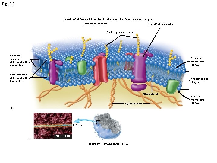
Fig. 3. 2 Copyright © Mc. Graw-Hill Education. Permission required for reproduction or display. Membrane channel Receptor molecule Carbohydrate chains Nonpolar regions of phospholipid molecules External membrane surface Polar regions of phospholipid molecules Phospholipid bilayer Cholesterol Cytoskeleton (a) 15 nm (b) TEM 1, 000 x b: ©Don W. Fawcett/Science Source Internal membrane surface
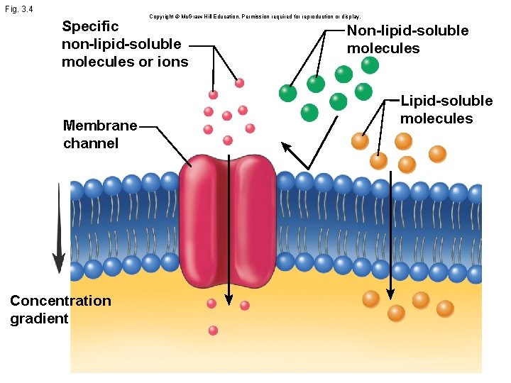
Fig. 3. 4 Copyright © Mc. Graw-Hill Education. Permission required for reproduction or display. Specific non-lipid-soluble molecules or ions Membrane channel Concentration gradient Non-lipid-soluble molecules Lipid-soluble molecules
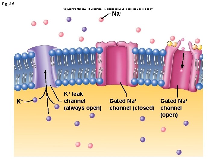
Fig. 3. 5 Copyright © Mc. Graw-Hill Education. Permission required for reproduction or display. Na+ K+ K+ leak channel (always open) Gated Na+ channel (closed) Gated Na+ channel (open)
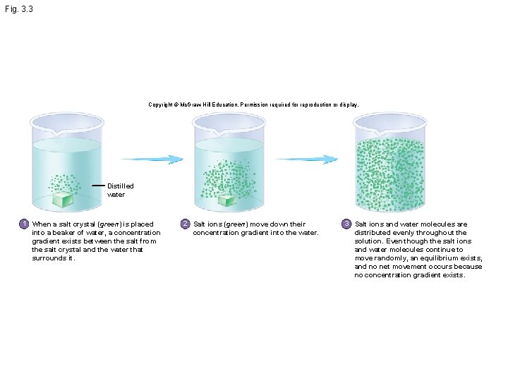
Fig. 3. 3 Copyright © Mc. Graw-Hill Education. Permission required for reproduction or display. Distilled water 1 When a salt crystal (green) is placed into a beaker of water, a concentration gradient exists between the salt from the salt crystal and the water that surrounds it. 2 Salt ions (green) move down their concentration gradient into the water. 3 Salt ions and water molecules are distributed evenly throughout the solution. Even though the salt ions and water molecules continue to move randomly, an equilibrium exists, and no net movement occurs because no concentration gradient exists.
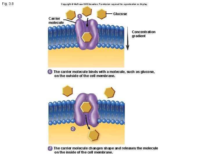
Fig. 3. 8 Copyright © Mc. Graw-Hill Education. Permission required for reproduction or display. 1 Carrier molecule Glucose Concentration gradient 1 The carrier molecule binds with a molecule, such as glucose, on the outside of the cell membrane. 2 2 The carrier molecule changes shape and releases the molecule on the inside of the cell membrane.
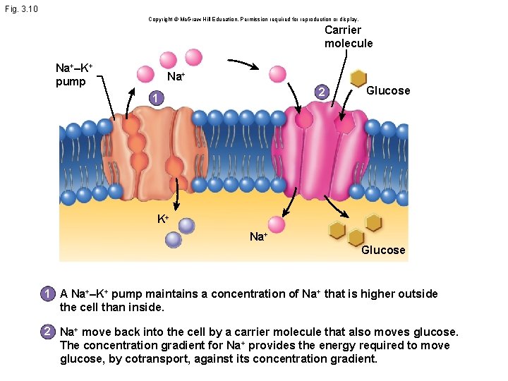
Fig. 3. 10 Copyright © Mc. Graw-Hill Education. Permission required for reproduction or display. Carrier molecule Na+–K+ pump Na+ 2 1 Glucose K+ Na+ Glucose 1 A Na+–K+ pump maintains a concentration of Na+ that is higher outside the cell than inside. 2 Na+ move back into the cell by a carrier molecule that also moves glucose. The concentration gradient for Na+ provides the energy required to move glucose, by cotransport, against its concentration gradient.
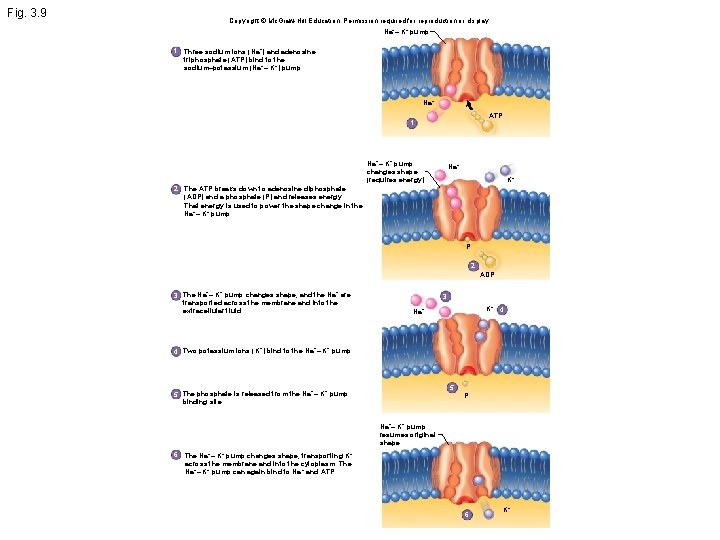
Fig. 3. 9 Copyright © Mc. Graw-Hill Education. Permission required for reproduction or display. Na+– K+ pump 1 Three sodium ions (Na+) and adenosine triphosphate (ATP) bind to the sodium–potassium (Na+– K+) pump. Na+ ATP 1 Na+– K+ pump changes shape (requires energy). Na+ K+ 2 The ATP breaks down to adenosine diphosphate (ADP) and a phosphate (P) and releases energy. That energy is used to power the shape change in the Na+– K+ pump. P 2 ADP 3 The Na+– K+ pump changes shape, and the Na+ are transported across the membrane and into the extracellular fluid. 3 K+ 4 Na+ 4 Two potassium ions (K +) bind to the Na +– K+ pump. 5 5 The phosphate is released from the Na +– K+ pump binding site. P Na+– K+ pump resumes original shape. 6 The Na+– K+ pump changes shape, transporting K+ across the membrane and into the cytoplasm. The Na+– K+ pump can again bind to Na + and ATP. 6 K+
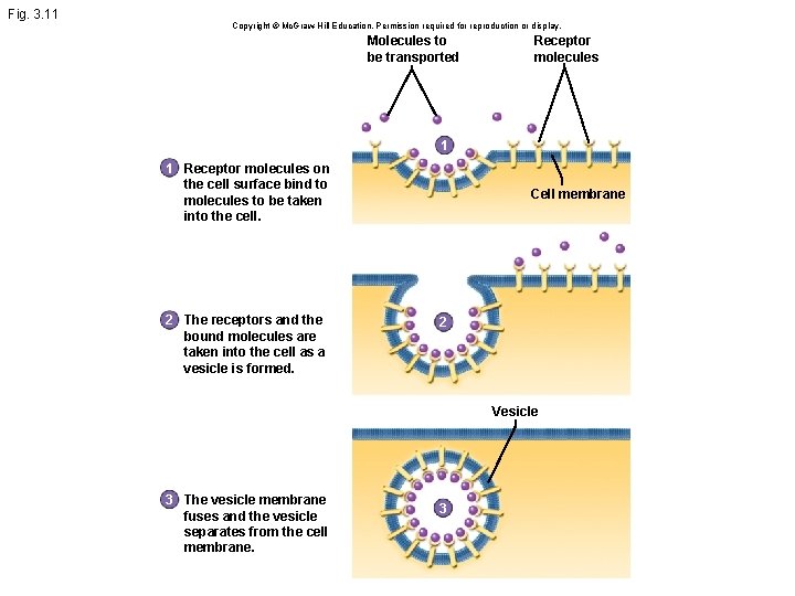
Fig. 3. 11 Copyright © Mc. Graw-Hill Education. Permission required for reproduction or display. Molecules to be transported Receptor molecules 1 1 Receptor molecules on the cell surface bind to molecules to be taken into the cell. 2 The receptors and the bound molecules are taken into the cell as a vesicle is formed. Cell membrane 2 Vesicle 3 The vesicle membrane fuses and the vesicle separates from the cell membrane. 3
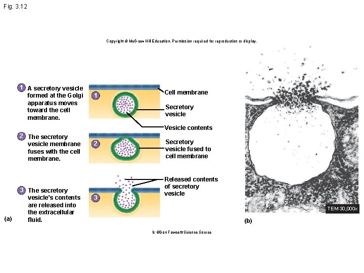
Fig. 3. 12 Copyright © Mc. Graw-Hill Education. Permission required for reproduction or display. 1 A secretory vesicle formed at the Golgi apparatus moves toward the cell membrane. 1 Cell membrane Secretory vesicle Vesicle contents 2 The secretory vesicle membrane fuses with the cell membrane. (a) 3 The secretory vesicle’s contents are released into the extracellular fluid. 2 3 Secretory vesicle fused to cell membrane Released contents of secretory vesicle TEM 30, 000 x (b) b: ©Don Fawcett/Science Source
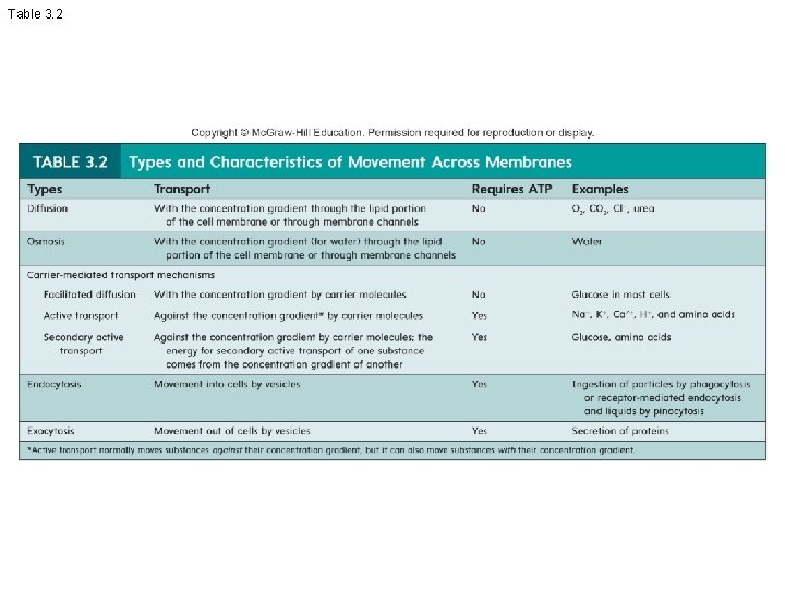
Table 3. 2