
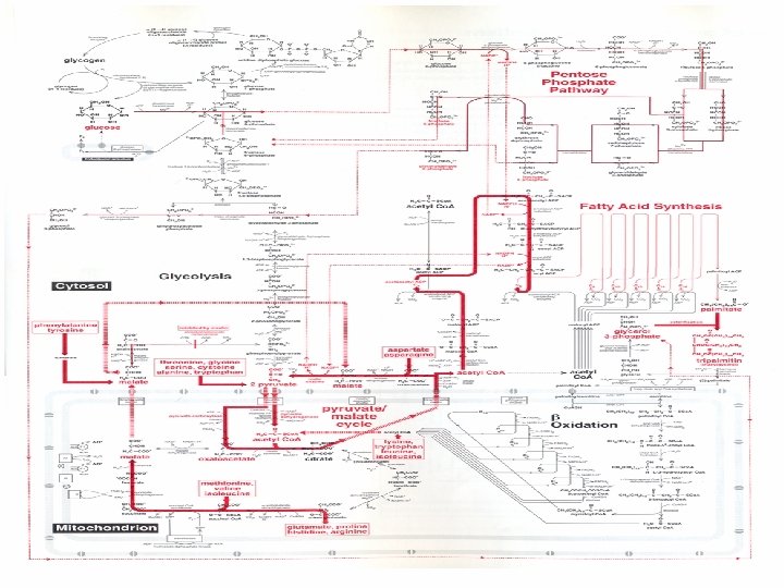
Fig 29 1 Amphibolic intermediates formed from the


Fig 29 -1 Amphibolic intermediates formed from the carbon skeletons of amino acids.
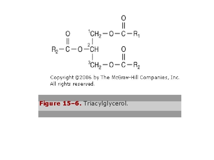
Figure 15– 6. Triacylglycerol.
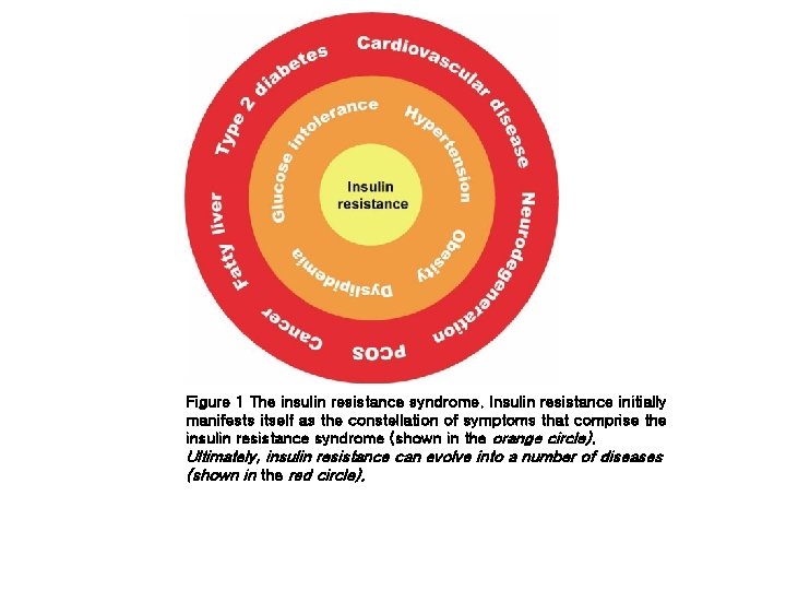
Figure 1 The insulin resistance syndrome. Insulin resistance initially manifests itself as the constellation of symptoms that comprise the insulin resistance syndrome (shown in the orange circle). Ultimately, insulin resistance can evolve into a number of diseases (shown in the red circle).
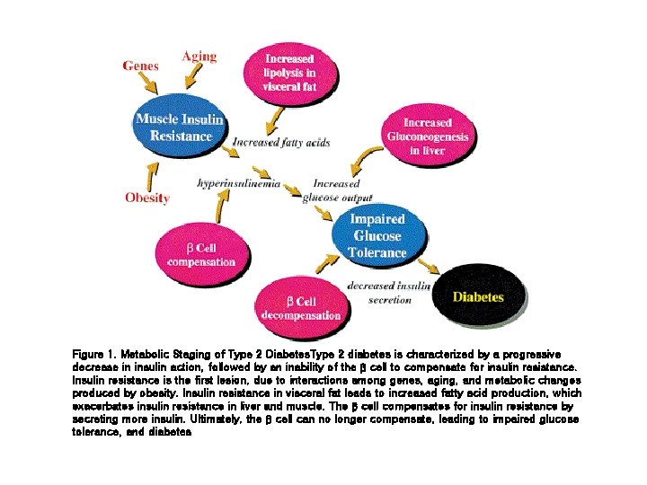
Figure 1. Metabolic Staging of Type 2 Diabetes. Type 2 diabetes is characterized by a progressive decrease in insulin action, followed by an inability of the β cell to compensate for insulin resistance. Insulin resistance is the first lesion, due to interactions among genes, aging, and metabolic changes produced by obesity. Insulin resistance in visceral fat leads to increased fatty acid production, which exacerbates insulin resistance in liver and muscle. The β cell compensates for insulin resistance by secreting more insulin. Ultimately, the β cell can no longer compensate, leading to impaired glucose tolerance, and diabetes
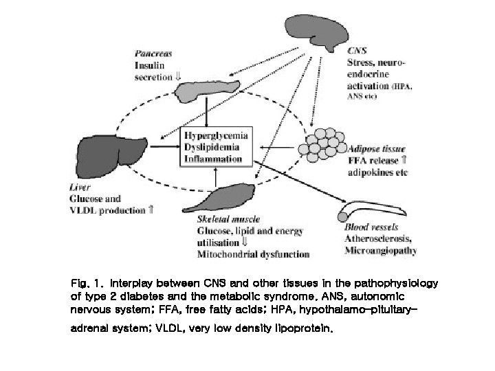
Fig. 1. Interplay between CNS and other tissues in the pathophysiology of type 2 diabetes and the metabolic syndrome. ANS, autonomic nervous system; FFA, free fatty acids; HPA, hypothalamo–pituitary– adrenal system; VLDL, very low density lipoprotein.
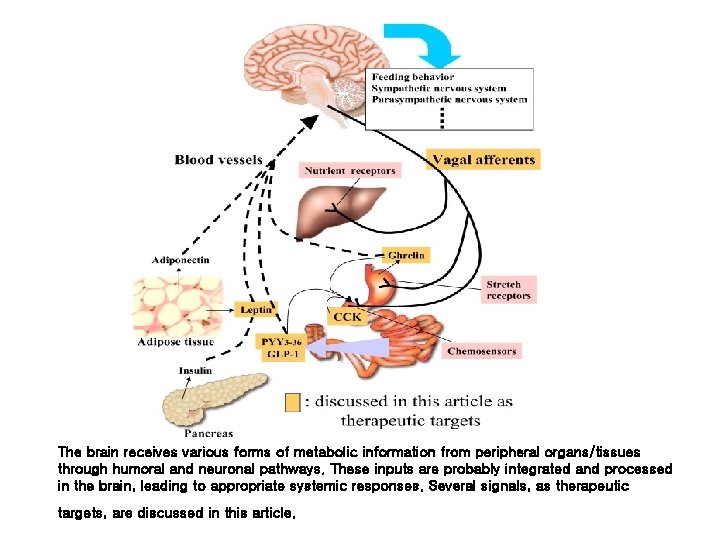
The brain receives various forms of metabolic information from peripheral organs/tissues through humoral and neuronal pathways. These inputs are probably integrated and processed in the brain, leading to appropriate systemic responses. Several signals, as therapeutic targets, are discussed in this article.
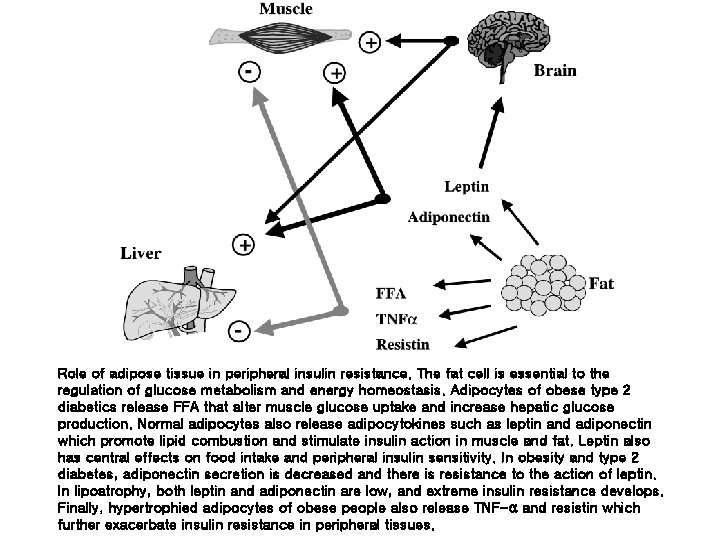
Role of adipose tissue in peripheral insulin resistance. The fat cell is essential to the regulation of glucose metabolism and energy homeostasis. Adipocytes of obese type 2 diabetics release FFA that alter muscle glucose uptake and increase hepatic glucose production. Normal adipocytes also release adipocytokines such as leptin and adiponectin which promote lipid combustion and stimulate insulin action in muscle and fat. Leptin also has central effects on food intake and peripheral insulin sensitivity. In obesity and type 2 diabetes, adiponectin secretion is decreased and there is resistance to the action of leptin. In lipoatrophy, both leptin and adiponectin are low, and extreme insulin resistance develops. Finally, hypertrophied adipocytes of obese people also release TNF-α and resistin which further exacerbate insulin resistance in peripheral tissues.
- Slides: 24