Fig 1 Defining features eight Puf repeats a
 The RNA-binding domain of PUF proteins.")


- Slides: 39
Fig. 1. Defining features: eight Puf repeats. (a) The RNA-binding domain of PUF proteins. The eight tandem repeats of approximately 40 amino acids are colored in boxes, and designated Puf repeats. These are flanked by two short sequences (anvil and oval) that form Puf repeat-like structures, named Csp 1 (anvil) and Csp 2 (oval). Supplementary information (including maps of other PUF proteins) is at http: //www. biochem. wisc. edu/wickens. (b) Alignments and conserved sequences. A simplified version of a Hidden Markov Model of the entire Puf repeat region is presented. At the top are three helical units (H 1¯H 3) present in each Puf repeat. Amino acids predicted to be present at a given position in at least 40% of all PUF proteins are shown; all other amino acids are indicated with dashes. Dots indicate spaces that are inserted for the alignment. The `core consensus' comprises helix 2; conserved amino acids in that helix are colored: green, aromatic; blue, basic; red, acidic; yellow, aliphatic. Supplementary information (a more complete Markov model) is at http: //www. biochem. wisc. edu/wickens. (c) Pumilio structure. Structure colored as in (a). The region and loop indicated with an * is required for binding of a protein collaborator, Nanos [6, 9 and 10]. RNA is thought to bind to the inside face of the crescent. Structure shown is that of Drosophila Pumilio, as presented in Ref. [9]. (d) The RNA-binding face of Pumilio. The orientation used in (c) is rotated 90 degrees, so that the conserved amino acids in helix 2 (the putative RNA-binding surface) face the reader. Conserved residues align in adjacent repeats, and are colored as in (b). Note the solvent exposed aromatic (green) and basic (blue) side chains, probably positioned to bind RNA. The structure shown is that of a human PUF, determined by Wang et al. [10]; we greatly appreciate their supplying pdb (protein database) coordinates before publication.
Fig. 4. Models for PUF protein-mediated repression of m. RNAs. PUF proteins convert an active m. RNA¯protein particle ¯ `active m. RNP' (messenger ribonucleoprotein), top ¯ into a `repressed m. RNP' (bottom). m. RNAs in the active complex are translated, stable and deadenylated only slowly; m. RNAs in the repressed complex are translationally silent, unstable and deadenylated rapidly. The transition from one form to another can occur through either of two paths, (a) and (b). Path (a): PUFs recruit the deadenylase. The number and nature of the interactions is not known, and recruitment of the deadenylase could simultaneously recruit components of the turnover machinery. Path (b): PUF proteins directly trigger a change in m. RNP organization. The key feature of this model is that PUF proteins inhibit the action of positive-acting components. Although the e. IF-4 E¯e. IF-4 G¯poly(A)binding protein (PAB) linkage (and associated translational components) is depicted (green), other candidates exist. Blue, PUF protein; green, positive-acting factors, including the translation initiation factors, e. IF-4 E and e. IF 4 G; red, negative-acting factors including the deadenylase (Ccr 4 p); black, components of the repressed m. RNP that are allowed access to the m. RNA after the transition.
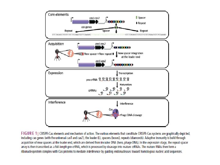
CRISPR/Cas 9 Clustered Regularly Interspaced Short Palindromic Repeats/ CRISPR-associated protein 9