
Fibrous dysplasia of bone It is a developmental
Fibrous dysplasia of bone It is a developmental, fibro osseous condition where normal medullary bone is gradually replaced by an abnormal fibrous tissue in which woven bone (newly mineralized product) is laid down resulting in an asymmetric distortion and expansion of bone. Types of fibrous dysplasia according to number of involved bone: a. Monostotic fibrous dysplasia: limited to single bone. b. Polyostotic fibrous dysplasia: several bones are affected.
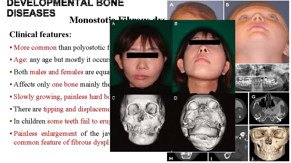
Monostotic Fibrous dysplasia of bone Clinical features: § More common than polyostotic fibrous dysplasia (up to 6 times). § Age: any age but mostly it occurs in the first and second decades (4 -18 years). § Both males and females are equally affected. § Affects only one bone mainly the jaws, specifically maxilla more than mandible. § Slowly growing, painless hard bony swelling, causing asymmetric facial disfigurement. § There are tipping and displacement of teeth. § In children some teeth fail to erupt. § Painless enlargement of the jaws reaching huge size leading to facial asymmetry is a common feature of fibrous dysplasia.
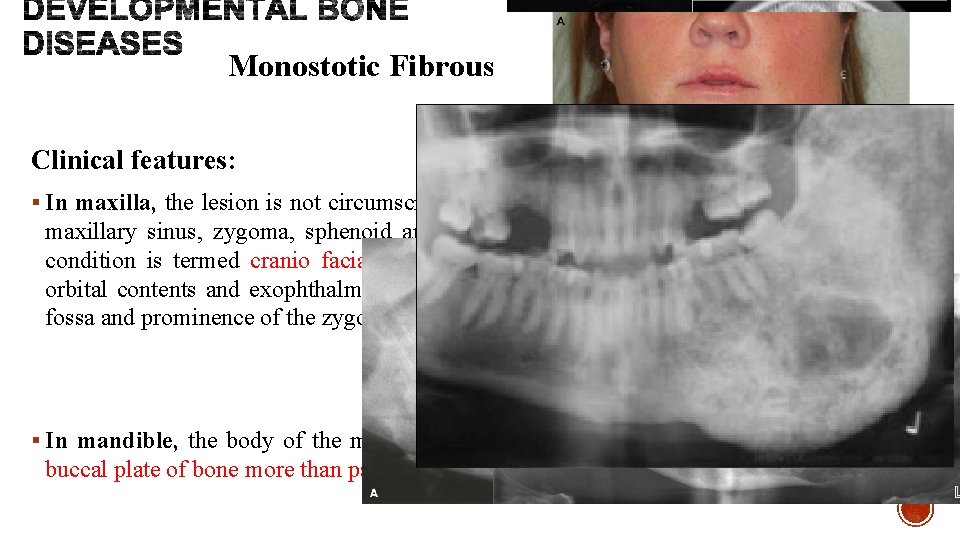
Monostotic Fibrous dysplasia of bone Clinical features: § In maxilla, the lesion is not circumscribed so spread to involve other adjacent bones as in maxillary sinus, zygoma, sphenoid and occipital, thus it is not strictly monostotic. This condition is termed cranio facial fibrous dysplasia. It is manifested by displacement of orbital contents and exophthalmos. Obliteration of maxillary sinus, bulging of the canine fossa and prominence of the zygomatic bone also can be seen. § In mandible, the body of the mandible is the most affected site leading to expansion of buccal plate of bone more than palatal or lingual plate.

Polyostotic Fibrous dysplasia of bone Clinical features: § It is less common than monostotic type. It is common in young individuals. § Female to male ratio is 3: 1. § Skull, long bones and jaws are the most affected bones. § In the jaws, the mandible is more affected than maxilla. § This type is painful. § When it affects long weight bearing bones, pathological fracture especially in upper head of femur is common. This leads to hockey stick deformity due to the replacement of bone by fibrous tissue and the weakened pathological nature of bone.
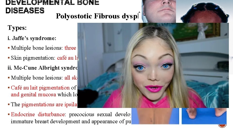
Polyostotic Fibrous dysplasia of bone Types: i. Jaffe’s syndrome: § Multiple bone lesions: three quarter of skeletal bones are affected. § Skin pigmentation: café au lait macules. ii. Mc-Cune Albright syndrome: § Multiple bone lesions: all skeletal bones are nearly affected. § Café au lait pigmentation of the skin of trunk and thighs is evident. Irregular macules on oral and genital mucosa which looks like a map with irregular outline. § The pigmentations are ipsilateral to the side of bony lesion. § Endocrine disturbance: precocious sexual development in females (menstrual bleeding, immature breast development and appearance of pubic hair), and hyperthyroidism in males.
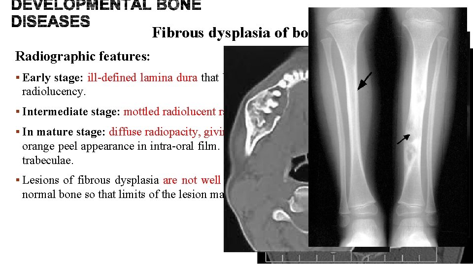
Fibrous dysplasia of bone Radiographic features: § Early stage: ill-defined lamina dura that blends with unilocular or multilocular ill-defined radiolucency. § Intermediate stage: mottled radiolucent radiopaque as the primitive bone is laid down. § In mature stage: diffuse radiopacity, giving ground glass appearance in extra-oral film and orange peel appearance in intra-oral film. This is due to disorganized poorly calcified bone trabeculae. § Lesions of fibrous dysplasia are not well defined and the margins blend into the adjacent normal bone so that limits of the lesion may be difficult to define.
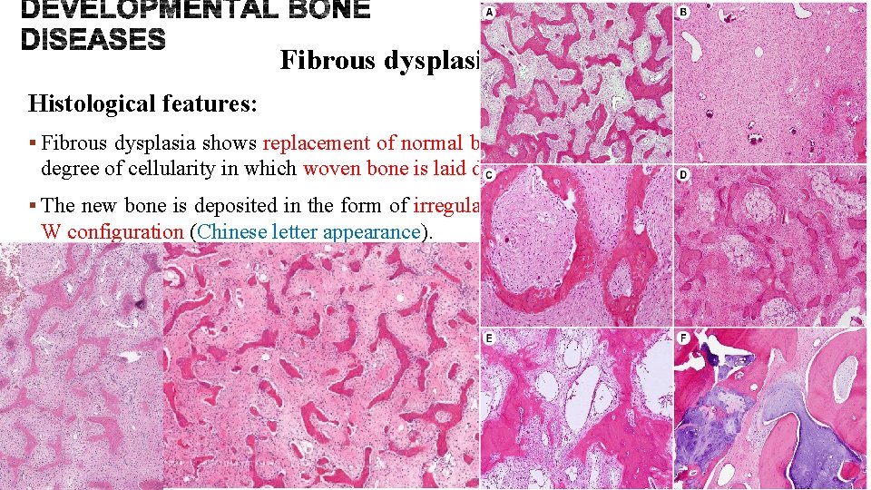
Fibrous dysplasia of bone Histological features: § Fibrous dysplasia shows replacement of normal bone by vascular fibrous tissue of varying degree of cellularity in which woven bone is laid down. § The new bone is deposited in the form of irregular, usually thin trabeculae often with V or W configuration (Chinese letter appearance). § The bone trabeculae are considered to arise by metaplasia and are not surrounded by osteoid seams or plump osteoblasts. At first the bony trabeculae are formed of osteoid, but they undergo calcification as they mature.
Fibrous dysplasia of bone Treatment: § Small lesions can be simply removed surgically. § Moderately to large sized lesions, surgical removal may cause regrowth and so a confirmation biopsy is only taken and the lesion is left until puberty, where it can be selfcorrected as bone remodeling occurs with skeletal maturation and only shaving down of the lesion can be done for functional and cosmetic reasons. § Radiotherapy is contraindicated because it may lead to post irradiation osteosarcoma. § Albright syndrome is associated with increased risk of death from heart arrhythmias which result from endocrinal disturbances. § Rarely malignant changes occur, however lesions of fibrosarcoma, osteosarcoma and chondrosarcoma have been reported.
- Slides: 9