FEVER WITH RASH WHAT IS THAT FEVER temporary


 Exposures - Ill contacts (home, day care…) -")
 Distribution pattern - symmetrical eruption - asymmetrical rashes")


 Maculopapular rash DESCRIPTION -macules – red/pink")
 Diffuse erythroderma - thickened, scaly skin on palms and soles BACT - Scarlet")
 vesicular, pustular, bullous -Vesicular – raised hemispherical lesions, < 0. 5 cm, contain")


 Host Human Invade")

 Incubation -IP = 8 to")
 Exanthematous (rash) - accompanied by high grade fever (40 -40. 5°C) -The rash")


 �Interstitial pneumonia (50 -75% pathological chest XR) �")


 Host Human")


 �Mild pharyngitis �Conjunctivitis �Anorexia �Headache �Low")


")

 �Causes: Varicella zoster virus (VZV, herpesvirus family) �Human are the only natural")

 �Characteristic")


, and �frequently a sore throat.")


 • transmission: person-to-person by respiratory droplets colonization")


 • transmission: direct contact through")
")


,")


 �Has particular tropism for B lymphocytes and")


- Slides: 74
FEVER WITH RASH
WHAT IS THAT? �FEVER -temporary ↑ in the body’s temperature in response to some disease or illness �RASH - temporary eruption of the skin - discrete red spots / generalized reddening - accompanied by itching
In HISTORY TAKING : a) Exposures - Ill contacts (home, day care…) - Travelling history -Pets, insects - Medications and drugs - Immunization a) Features of rash - Temporal association (onset relative to fever) - Progression and evolution - Location and distribution - Pain or pruritus
In PHYSICAL EXAMINATION : a) Distribution pattern - symmetrical eruption - asymmetrical rashes b) Morphology - monomorphic - pleomorphic c) Configuration - linear, annular, grouped, -Koebner phenomenon (eruption in an area local trauma) of
LINEAR RASH
ANNULAR RASH
KOEBNER PHENOMENON
DIFFERENTIAL DIAGNOSIS OF FEVER WITH RASH LESION a) Maculopapular rash DESCRIPTION -macules – red/pink discrete flat areas, blanch on pressure -papules – solid, raised hemispherical lesions, tiny, blanch on pressure PATHOGENS OR INFECTION VRS - Measles, rubella, roseola, erythema infectiosum, EBV, echovirus, HBV, HIV BACT - Erythema marginatum, scarlet fever, erysipelas, 2° syphilis, leptospirosis, Lyme dzs, RICK – Rocky Mountain Spotted fever, Typhus OTH – RA, Kawasaki dis, drug rxn
b) Diffuse erythroderma - thickened, scaly skin on palms and soles BACT - Scarlet fever, Toxic Shock Syndrome, Staph SSS FUNGI – Candida albicans OTH – Kawasaki syndrome c) Urticarial rash - swelling of the lesions - appear n resolve rapidly VRS – EBV, Hep B, HIV BACT – M. pneumonia, Grp A strep. OTH – Drug rxn
d) vesicular, pustular, bullous -Vesicular – raised hemispherical lesions, < 0. 5 cm, contain clear fluid -Pustular, bullous – raised hemispherical lesion, >0. 5 cm, contain clear/purulent fluid VRS - HSV, VZV, Coxsackievirus BACT - Staph. SSS, Staph. Bullous impetigo, Strep. crusted impetigo OTH - Toxic epidermal necrolysis, Steven. Johnson Syndrome. RICK – Rickettsial pox e) Petechial - purpuric - non-blanching red/purple spots VRS - Atypical measles, congenital rubella, CMV, enterovirus, HIV, HF viruses BACT - Sepsis (meningococcal, gonococcal, pneumococcal, Hib), IE
RICK – Rocky mountain spotted fvr FUNGI – Aspergillus, mucor OTH - Vasculitis, thrombocytopenia, Henoch-Schönlein purpura, malaria f) Erythema Nodosum - tender red nodules, due to exudation of blood and serum VRS - EBV, HBV BACT - Group A Streptococcus TB, yersinia, Cat. Scratch Dzs FUNGI Coccidiomycosis, histoplasmosis OTH - Sarcoidosis, Inf. Bowel dzs, OCP, SLE, Behçet dzs
CASE SCENARIO History: � 9 mo old girl, good general health condition �Progressive fever for 5 days (max. 39. 50 C) �Coryza, exudative conjunctivitis �Severe cough and irritability �No diarrhea, no vomiting �No recent travel, no pets Rashes - over trunk, abdomen and back - appear 4 days after onset of fever - not elevated and no itching - blanching on pressure
Confluent maculo-papular rash all over the body
MEASLES Characteristic EXPLAINATION Causative Agent Measles virus ( ss. RNA paramyxovirus) Host Human Invade Upper respiratory tract, regional LN Transmited by Large respiratory droplets with no fomites (close contact transm. ) Virus present Respiratory secretion, blood, urine Period of communicability Contagious from 5 days before to 4 days after the appearance of rash.
EPIDEMIOLOGY �Endemic in regions where measles vaccination is not available �Young infants - protected by transplacental antibody, but become more susceptible toward the end of the first year. �Passive immunity may interfere with effective vaccination until 12 to 15 months of age.
CLINICAL MANIFESTATION Divided into 4 phases : - a) Incubation -IP = 8 to 12 days from exposure to the onset of symptoms, 14 days from exposure to the onset of rash. b) Prodromal (catarrhal) -cough, coryza, conjunctivitis (Stimson line) Koplik spots (buccal mucosa)
a) Exanthematous (rash) - accompanied by high grade fever (40 -40. 5°C) -The rash starts behind the ears and on the forehead at the hair line spread down to the leg (descending) - show severity of the illness b) recovery
CONJUNCTIVITIS KOPLIK SPOTS MACULAR RASH
Other manifestations : �Cervical lymphadenitis �Spleenomegaly �Abdominal pain �Mesenteric lymphadenopathy �Otitis media �Pneumonia common in infants �Diarrhea Liver involvement – common in adult
INVESTIGATIONS �Serological test - Ig. M antibody - appear in 1 -2 days of rash - persist for 1 -2 month Chest X-ray - interstitial infiltration - -ve measle pneumonia vs bacterial superinfection
DIAGNOSIS �Clinical �Serology �Viral culture �PCR
COMPLICATIONS �Acute otitis media (10 -15%) �Interstitial pneumonia (50 -75% pathological chest XR) � Myocarditis and pericarditis �Encephalitis (1/1000 cases) 7 -10 days after rash �Subacute sclerosis panencephalitis �Mesenteric lymphadenitis
MANAGEMENT TREATMENT � Routine supportive care �maintain adequate hydration �antipyretics � IV ribavirin (severe infection) � High dose for vitamin A supplementation
PREVENTION MMR � Live attenuated measles vaccine � 1 st dose : 12 -15 month of life � 2 nd dose : 4 -6 yrs old * Contraindicated for severe immunosupression patient
RUBELLA Characteristic EXPLAINATION Causative Agent Rubella virus ( ss. RNA, togavirus family) Host Human Invade Respiratory epithelium Transmited by Susceptible contact Infection in utero (congenital rubella synd) during the 1 st trimester Virus present Nasopharyngeal secretion, urine Period of communicabilit y contagious from 2 days before until 5 to 7 days after the onset of rash
EPIDEMIOLOGY �Outbreak of rubella in nonvaccinated groups can occur in adults at their workplaces, prisons, colleges & healthcare centers �Transplacental antibody protection only during first 6 month of life
CLINICAL MANIFESTATION IP = 14 to 21 days Retroauricular, posterior cervical, posterior occipital lymphadenopathy Erythematous, maculopapular, discrete rashes Rashes - begins on the face, spreads down to the body and lasts far three days.
�Forschheimer spots (rose-colored spots on the soft palate) �Mild pharyngitis �Conjunctivitis �Anorexia �Headache �Low grade fever �Polyarthritis
Erythematous maculopapular discrete rash Forschheimer spots
INVESTIGATIONS �NON-SPECIFIC and do not aid in diagnosis �WBC – normal or low �Thrombocytopenia – rare �Serological test �Ig. M antibody �Fourfold rise in specific Ig. G antibodies in paired acute & convalescent sera
COMPLICATIONS �Rarely complicated compared to measles �pregnancy – congenital rubella syndrome - IUGR - cataracts - deafness - patent ductus arteriosus (PDA)
CONGENITAL RUBELLA SYNDROME
PRINCIPLE OF MANAGEMENT TREATMENT �No specific therapy �Routine supportive care �Congenital Rubella Syndrome baby should be isolated
PREVENTION Live attenuated MMR vaccine �Children at age 12 -15 months of life �Children at age 4 -6 yrs old �Pregnant woman should be immunized after delivery
DIFFERENCES BETWEEN MEASLES AND RUBELLA MEASLES RUBELLA RASH Yes FEVER Yes Mild or no fever CHARACTERISTIC Confluent macular rash, Koplik spots Maculopapular discrete rash, Forschheimer spots TRANSMISSION Droplet, airborne Droplet SYMPTOMS High fever, runny nose, Rash with minimal to cough and for 3 -5 days, no fever or toxicity; followed by rash infection possible without rash
SPREAD OF RASH Starts on head and upper body Spreads slowly down to feet over 7 days Starts on head and upper body Spreads quickly down to feet over 3 days COMPLICATION Viral or bacterial Congenital infection pneumonia, encephalitis, otitis media, blindness SEVERITY Moderate to severe Mild
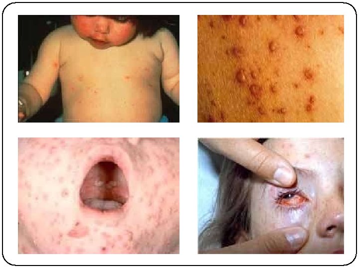
Varicella (chickenpox)
Clinical case �History: 5 y old boy, no special past medical history �Low grade fever (38. 30 C) for 48 h �Attends school �No travel history �No pets Vesicular rash on the trunk and face
Varicella (chickenpox) �Causes: Varicella zoster virus (VZV, herpesvirus family) �Human are the only natural host �Chickenpox (vericella) = manifestation of primary infection �Highly contagious among susceptible individuals; secondary attack rate is more than 90%) �Contagiosity: 2 days before to 7 days after the onset of the rash, when all lesions are crusted
�Peak age: 5 to 10 years old �Peak seasonal infection: late winter and spring �Transmission: direct contact, droplet, and air �Incubation period: 14 -16 days
Clinical manifestation �Prodromal symptoms: fever, malaise, anorexia (preceed the rash by 1 day) �Characteristic rash: small red papules> Erythematous papules> vesicular> vesicles ulcerate, crust and heal (new crops appear for 34 days) �Pattern of rash: beginning on the trunk followed by the head, face, and less commonly the extremities �Pruritus is universal and marked �Lesions may also present on mucosa membranes �Lymphadenopathy may be generalized
Complication � Common � More severe for neonates, adults, and immunocompromised persons. - Secondary infection of skin by streptococci pr staphylococci - Thrombocytopenia and haemorragic lesions or bleeding may occur (varicella gangrenosa) - Pneumonia (15 -20% 0 f healty adults and immunlcompromised persons, uncommon in healthy children) - Myocarditis, pericarditis, orchitis, hepatitis, ulcerative gastritis, glomerulonephritis and athritis may complicate - Reye syndrome may follow varicella (aspirin use is contraindicated) - Neurological complication: post infectious enencephaly, cerebellar ataxia, nystagmus and tremor.
�Congenital infection -characteristic: low birth weight, cortical atrophy, seizure, mental retardation, chorioretinitis, cataracts, microcephaly �Perinatal infection -severe form of noenatal varicella
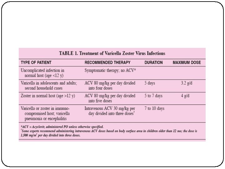
Treatment �Symptomatic therapy: Nonaspirin antipyretics, cool baths, careful hygiene �Antiviral treatment: acyclovir, famciclovir, valacyclovir
Prevention �Children with chickenpox should not return to school until all vesicle have crusted �Live attenuated varicella (primary prevention) �Passive immunity by VZIG (secondary prevention)
Must be administered by 96 h after exposure (or better if < 72 h)
Hand, foot and mouth disease
Hand, foot and mouth disease �most often occurs in children under 10 years old. �Causes: coxsackie virus A 16, enterovirus 71 (EV 71) and other enteroviruses. �The enterovirus group includes polioviruses, coxsackieviruses, echoviruses and other enteroviruses. � more frequent in summer and early autumn (in temperate countries)
�moderately contagious. �A person is most contagious during the first week of the illness. �transmitted from person to person via direct contact with nose and throat discharges, saliva, fluid from blisters, or the stool of infected persons. �(incubation period) is 3 to 7 days. Fever is often the first symptom of HFMD followed by blister/rash.
Clinical manifestation �mild fever, �poor appetite, �malaise ("feeling sick"), and �frequently a sore throat. �One or 2 days after the fever begins, painful sores develop in the mouth. They begin as small red spots that blister and then often become ulcers. �They are usually located on the tongue, gums, and inside of the cheeks. �The skin rash develops over 1 to 2 days with flat or raised red spots, some with blisters on the palms of the hand the soles of the feet.
Blister on the palms of the hands Blister then become ulcer on the inner gums Blister on the soles of the feet Blister on the dorsum of the feet
Complication HFMD caused by coxsackie virus A 16 infection is a mild disease and nearly all patients recover within 7 to 10 days. �Complications are uncommon. �HFMD caused by Enterovirus EV 71 may be associated with neurological complications such as aseptic meningitis and encephalitis �
Treatment �no specific effective antiviral drugs and vaccine available for the treatment of HFMD. �Symptomatic treatment is given to provide relief from fever, aches, or pain from the mouth ulcers. �Dehydration is a concern because the mouth sores may make it difficult and painful for children to eat and drink.
Prevention � good hygienic practices. Preventive measures include: � a. Frequent hand washing, especially after diaper changes, after using toilet and before preparing food, � b. Maintain cleanliness of house, child care center, kindergartens or schools and its surrounding, � c. Cleaning of contaminated surfaces and soiled items with soap and water, and then disinfecting them with diluted solution of chlorine-containing bleach (10% concentration), � d. Parents are advised not to bring young children to crowded public places such as shopping centers, cinemas, swimming pools, markets or bus stations, � e. Bring children to the nearest clinic if they show signs and symptoms. Refrain from sending them to child care centers, kindergartens or schools. � f. Avoidance of close contact (kissing, hugging, sharing utensils, etc. ) with children having HFMD illness to reduce of the risk of infection
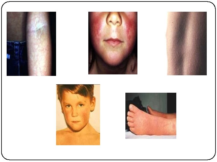
• caused by Neisseria meningitidis (meningococcus) • transmission: person-to-person by respiratory droplets colonization of URT penetrate into bloodstream go to CNS and causing meningitis (meningococcal meningitis) / infect the blood vessel (meningococcemia) • Meningococcemia / meningococcal septicaemia: cutaneous signs: . 1. Maculopapular – early, often on a painful joint or pressure point 2. Petechiae (50 -70%) – distribute at trunk and extremities (can be anywhere else) 3. Purpura (may start anywhere on the body and then spread) and necrotic area Non-cutaneous signs: altered mental status, neck
�Meningococcal septicemia can kill children in hours, therefore optimal outcome requires immediate recognition, prompt resuscitation and antibiotics. �Although there are now polysaccharide conjugate vaccines against groups A and C meningococcus, there is still no effective vaccines for group B meningococcus
CLINICAL CASE � History: 7 y. old boy, good general health condition �Sudden onset of sore throat since 24 hrs and fever at 39 o. C. Abdominal pain and 1 episode of vomiting � No conjuntivitis, � No rhinitis, � No hoarseness � No cough � Attends primary school, no recent travel
Scarlet Fever • caused by group A streptococcus (GAS) • transmission: direct contact through droplets • symptoms: rashes: develop 24 hours after the fever can begins at below ears , neck, chest and stomach then spread all over the body within 1 to 2 days look like sunburn and feel like sandpaper more apparent at skin fold of elbow, armpit and groin area last for about 2 -7 days • • • as the rash faded, skin at the tips of lips and fingers begin to peel flush face fever >38. 3°C swollen glands at the neck white or yellow spot coating on the throat and tonsil “strawberry tongue”
• In body folds, especially the armpits and elbows, fragile blood vessels (capillaries) can rupture and cause classic red streaks called Pastia lines. These may persist for 1 -2 days after the generalised rash has gone.
Diagnosis: 1. Throat culture remains the criterion standard for confirmation of group A streptococcal upper respiratory infection. 2. Complete blood count �White blood cell (WBC) count in scarlet fever may increase to 12, 000 -16, 000 per mm 3, with a differential of up to 95% polymorphonuclear lymphocytes. �During the second week, eosinophilia, as high as 20%, can develop. Treatment : Penicillin remains the drug of choice. Erythromycin can be considered as an alternative
KAWASAKI DISEASE • characterized by fever at least for 5 days together with 4 of the following 5 findings: conjungtival infection mucous membrane changes (pharyngeal red, dry, cracied lips, strawberry tongue) cervical lymphadenopathy rash redness or swelling of the hands and feet, generalized skin peeling • age: 4 month – 6 years • cause is unknown • complication: coronary artery aneurysm, sudden death
Complications �Coronary artery aneurysm �Prognosis � 75% no sequelae, 25% coronary abnormality (without treatment), � 1 -2% mortality in the acute phase
TYPHUS �A general name for various arthropod-borne rickettsial infections and that result in an acute febrile illness. �A Rickettsia- harboring louse bites a human to engage in a blood meal and causes a pruritic reaction on the host's skin. The louse defecates as it eats; when the host scratches the site, the lice are crushed, and the Rickettsia- laden excrement is inoculated into the bite wound. The Rickettsia travel to the bloodstream and rickettsemia develops.
Symptoms of Typhus �Severe headache �Chills �High fever �Stupor �Muscle aches �Swollen lymph nodes �Skin rash - macular, maculopapular, petechial or papulovesicular eruption �Forearm skin rash spreading to the body
� Diagnostic Tests � The list of diagnostic tests mentioned in various sources as used in the diagnosis of Typhus includes: � Blood tests for rickettsiae � Antibody blood tests (Ig. M, Ig. G)- Indirect Immunofluorescent Assay (IFA), rise indicate acute primary or secondary disease � Tests depend on the type of typhus � Treatment � Specific antimicrobial therapy effective against rickettsia should be used. Doxycycline and chloramphenicol are used as antirickettsial agents for the treatment of typhus.
INFECTIOUS MONONUCLEOSIS �Caused by Epstein-Barr virus (EBV) �Has particular tropism for B lymphocytes and epithelial cells of the pharynx �Transmission usually occurs by oral contact
Signs and symptoms �Fever �Malaise �Tonsillopharygitis – often severe, limiting oral ingestion of fluids and food, rarely breathing can be compromised �Lymphadenopathy – prominent cervical lymph nodes �Petechiae on the soft palate �Splenomegaly (50%), hepatomegaly (10%) �Maculopapular rash (5%)
�DIAGNOSIS �Patients with infectious mononucleosis in the differential diagnoses should have a CBC count with differential and an evaluation of the erythrocyte sedimentation rate (ESR) �Because the liver is uniformly involved in EBV infectious mononucleosis, mild elevation of the serum transaminases is a constant finding in early EBV infectious mononucleosis. �Heterophile antibody tests Patients with infectious mononucleosis should first be tested with a heterophile antibody test. The most commonly used is the latex
TREATMENT �Medical Care �Closely monitor patients with extreme tonsillar enlargement for airway obstruction. Steroids are indicated for impending or established airway obstruction in individuals with Epstein-Barr virus (EBV) infectious mononucleosis. Surgical Care �Surgery is necessary for spontaneous splenic rupture, which occurs in rare patients with EBV infectious mononucleosis and may be the initial manifestation of the condition.
THANK YOU