Fetal Membranes Dr Mujahid Khan The fetal part

Fetal Membranes Dr. Mujahid Khan

Ø The fetal part of the placenta and fetal membranes separate the fetus from the endometrium of the uterus Ø An interchange of substances such as nutrients and oxygen occurs between the maternal and fetal blood streams through the placenta

What constitute a Fetal Membrane Ø Decidua Ø Chorion Ø Amnion Ø Yolk sac Ø Allantois

Amnion Ø Thin but tough Ø Forms a fluid filled membranous amniotic sac that surrounds the embryo and fetus Ø Is attached to the margins of the embryonic disc Ø Its junction with embryo located on the ventral surface after the folding

Amniotic Fluid Ø Plays a major role in fetal growth and development Ø Most of it is derived from maternal tissue and by diffusion across the amniochorionic membrane from the decidua parietalis Ø Later there is a diffusion of fluid through the chorionic plate from blood in the intervillous space of the placenta

Amniotic Fluid Ø Amniotic fluid is similar to fetal tissue fluid Ø Before keratinization of the skin the pathway for passage of water and solutes in tissue fluid from the fetus to the amniotic cavity is through the skin Ø Fluid is also secreted by the fetal respiratory tract and enters the amniotic cavity

Amniotic Fluid Ø Daily contribution of fluid from respiratory tract is 300 -400 ml Ø Fetus contributes to the amniotic fluid by excreting urine into the amniotic cavity Ø Half a liter of urine is added daily during the late pregnancy Ø Amniotic fluid volume is 30 ml at 10 weeks, 350 ml at 20 weeks, 700 -1000 ml at 37 weeks

Circulation of Amniotic Fluid Ø Water content of amniotic fluid changes every 3 hours Ø It is been swallowed by the fetus and absorbed by respiratory & digestive tracts Ø Fetus swallows up to 400 ml of fluid per day during the end days of pregnancy

Circulation of Amniotic Fluid Ø Fluid passes into the fetal blood stream and the waste products in it cross the placental membrane and enter the maternal blood in the intervillous space Ø Excess water in the fetal blood is excreted by the fetal kidneys and returned to the amniotic sac as a urine

Disorders of Amniotic Fluid Volume Ø Oligohydromnios Ø Renal agenesis Ø Obstructive uropathy Ø Polyhydromnios Ø Esophageal atresia

Exchange of Amniotic Fluid Ø Large amount of amniotic fluid move in both directions between the fetal and maternal circulations mainly through the placental membrane Ø Most fluid passes into GIT but some passes into lungs Ø Fluid is absorbed in either case and enters the fetal circulation Ø It then passes into the maternal circulation through the placental membrane

Composition of Amniotic Fluid Ø 99 % is water Ø Desquamated fetal epithelial cells Ø Organic & inorganic salts Ø Protein, carbohydrates, fats, enzymes, hormones Ø Meconium & urine in the late stage Ø Amniocentesis can be performed to check the concentration of different compounds for diagnostic purpose
 in amniotic fluid usually")
Composition of Amniotic Fluid Ø High levels of alpha-phetoprotein (AFP) in amniotic fluid usually indicate the presence of a severe neural tube defect (meroanencephaly) Ø Low levels of AFP may indicate chromosomal aberrations such as trisomy 21

Significance of Amniotic Fluid Ø Permits symmetrical external growth of the embryo and fetus Ø Acts as a barrier to infection Ø Permits normal fetal lung development Ø Prevents adherence of amnion to fetus Ø Cushions & protects the embryo and fetus Ø Helps maintain the body temperature Ø Enables the fetus to move freely

Yolk Sac Ø It is large at 32 days Ø Shrinks to 5 mm pear shaped remnant by 10 th week & connected to the midgut by a narrow yolk stalk Ø Becomes very small at 20 weeks Ø Usually not visible thereafter
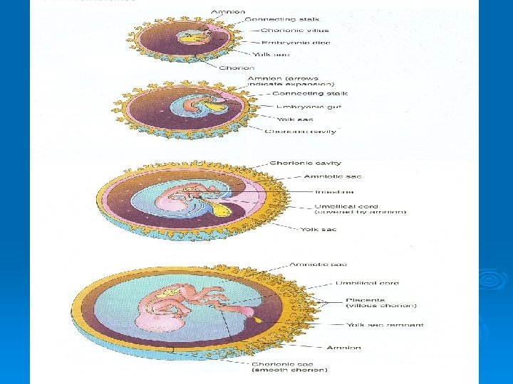

Significance of Yolk Sac Ø Has a role in transfer of nutrients during the 2 nd and 3 rd weeks Ø Blood development first occurs here Ø Incorporate into the endoderm of embryo as a primordial gut Ø Primordial germ cells appear in the endodermal lining of the wall of the yolk sac in the 3 rd week

Fate of Yolk Sac Ø At 10 weeks lies in the chorionic cavity between chorionic and amniotic sac Ø Atrophies as pregnancy advances Ø Sometimes it persists throughout the pregnancy but of no significance Ø In about 2% of adults the proximal intraabdominal part of yolk stalk persists as an ileal diverticulum or Meckel diverticulum

Allantois Ø In the 3 rd week it appears as a sausagelike diverticulum from the caudal wall of yolk sac that extends into the connecting stalk Ø During the 2 nd month, the extraembryonic part of the allantois degenerates
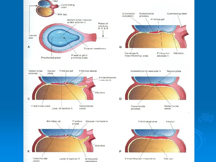

Functions of Allantois Ø Blood formation occurs in the wall during the 3 rd to 5 th week Ø Its blood vessels persist as the umbilical vein and arteries Ø Fluid from the amniotic cavity diffuses into the umbilical vein and enters the fetal circulation for transfer to maternal blood through placental membrane Ø Becomes Urachus and after birth is transformed into median umbilical ligament extends from the apex of the bladder to the umbilicus

Umbilical Cord Ø Is attached to the placenta usually near the center of the fetal surface of this organ Ø May attach to any other point Ø Is usually 1 -2 cm in diameter and 30 -90 cm in length Ø Long cord may cause prolapse or compression of the cord which may lead to fetal hypoxia Ø Short cord may cause premature separation of the placenta from the wall of the uterus during delivery
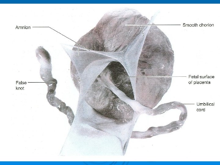

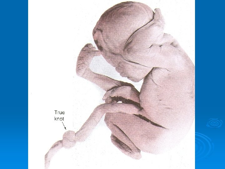
Umbilical Cord Ø Has two arteries and one vein surrounded by Wharton jelly Ø Umbilical vessels are longer than the cord, so twisting and bending of the vessels are common Ø They frequently form loops, producing false knots, that are of no significance Ø In about 1% of pregnancies, true knots form in the cord and cause fetal death

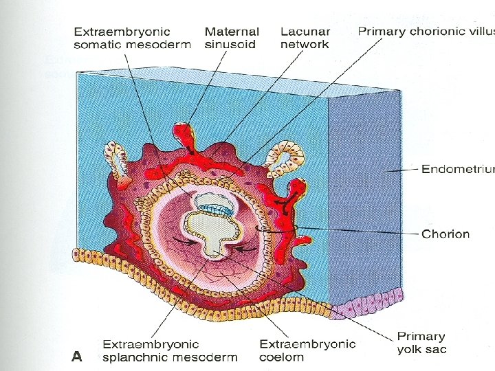
Chorion Ø Primary chorionic villi appear by the end of the 2 nd week Ø Growth of these extensions are caused by underlying extraembryonic somatic mesoderm Ø The cellular projections form primary chorionic villi

Chorion Ø The extraembryonic somatic mesoderm and the two layers of trophoblast form the chorion Ø Chorion forms the wall of chorionic sac Ø Embryo and its amniotic and yolk sacs are suspended into it by connecting stalk Ø The extraembryonic coelom is now called the chorionic cavity

Chorion Ø The amniotic sac with embryonic epiblast form its floor Ø The yolk sac with embryonic hypoblast form its roof Ø Are analogous to two balloons pressed together, suspended by a connecting stalk from the inside of a larger balloon (chorionic sac)

Chorion Ø Transvaginal ultrasound is used to measure the chorionic sac diameter Ø This measurement is valuable for evaluating the early embryonic development and pregnancy outcome

Chorion Ø Chorionic villi cover the entire chorionic sac until the beginning of 8 th week Ø As this sac grows, the villi associated with decidua capsularis are compressed, reducing the blood supply to them Ø These villi soon degenerates producing an avascular bare area smooth chorion (chorion laeve)
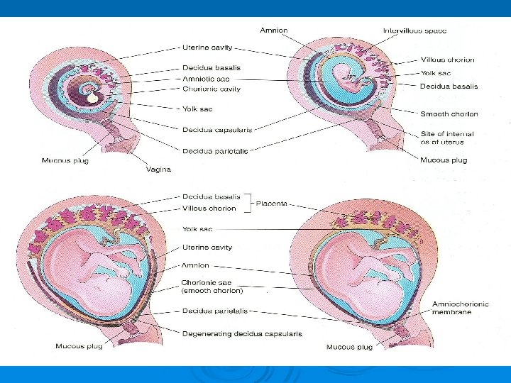

Chorion Ø As the villi disappear, those associated with the decidua basalis rapidly increase in number Ø Branch profusely and enlarge Ø This bushy part of the chorionic sac is villous chorion
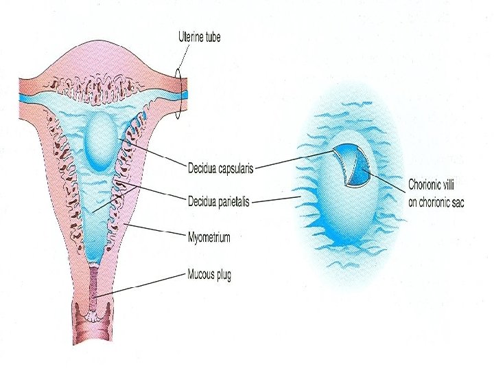

Decidua Ø The gravid endometrium is known as decidua Ø It is the functional layer of endometrium in a pregnant woman Ø This part of the endometrium separates from the rest of the uterus after parturition

Regions of Decidua 3 regions of decidua are: Ø Decidua basalis: lies deep to the conceptus that forms maternal part of the placenta Ø Decidua capsularis: superficial part that overlies the conceptus Ø Decidua parietalis: is all the remaining parts of the decidua


Decidua Ø In response to increasing progesterone levels in the maternal blood the connective tissue cells of the decidua enlarge to form decidual cells Ø These cells enlarge as glycogen and lipid accumulate in their cytoplasm

Decidua Ø The cellular and vascular changes occurring in the endometrium as the blastocyst implants constitute the decidual reaction Ø Many decidual cells degenerate near the chorionic sac in the region of the syncytiotrophoblast Ø Together with maternal blood the uterine secretions provide a rich source of nutrition for the embryo

Decidua Ø The full significance of decidual cells is not understood Ø They may protect the maternal tissue against uncontrolled invasion by the syncytiotrophoblast Ø They may be involved in hormonal production Ø Clearly recognizable during ultrasonography to diagnose early pregnancy
- Slides: 40