Fetal Diagnosis and Treatment of Cardiovascular Conditions Shanthi


 is measured at the")


- Slides: 44
"Fetal Diagnosis and Treatment of Cardiovascular Conditions" Shanthi Sivanandam, MD, FASE Medical Director, Fetal Cardiology Co- Director, Echocardiography Co-Director Fetal Diagnostic and Treatment Center University of Minnesota Medical School
I have no financial relationships to disclose.
Fetal Cardiology • Incidence of Congenital heart defects in general population 8 -10 per 1000 live births • Fetal echocardiography has given us an opportunity to diagnose CHD in utero, as well as to observe the natural history of many cardiac defects
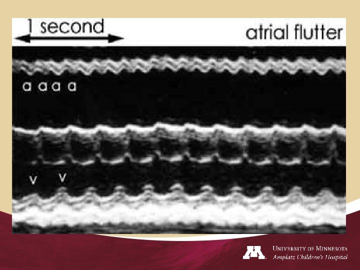
Fetal Cardiology • Fetal echocardiography is now paramount in making an early and accurate assessment of cardiovascular structure as well as diagnosing and treating fetal arrhythmias. • Complete fetal echocardiographic study includes the structural and rhythm analysis using a combination of two-dimensional imaging, Mmode scanning, pulsed- and continuous-wave Doppler measurements, and color-flow mapping
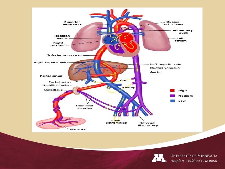
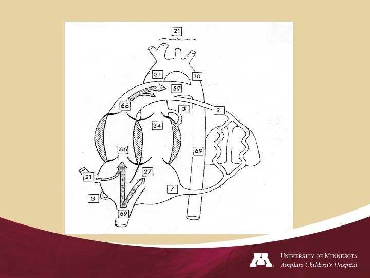
Fetal Cardiology • Fetal cardiovascular system differs from adult in many ways • Intrauterine ventricles work in parallel rather than in series • Three communications-ductus venosus, foramen ovale, ductus arteriosus • RV is the dominant ventricle-60 -65% combined cardiac output
Fetal Cardiology • The immature fetal myocardium has decreased contractility and compliance as well as slower contractility and relaxation rates than Neonatal and Adult myocardium.
Fetal Cardiology • Ultrasonic imaging -fetal heart began to appear in the literature as early as 1970’s • Ability to diagnose CHD and arrhythmias in the fetus utilizing M-mode and real time 2 D-1980’s • Transabdominal fetal echocardiography is performed at 18 to 24 weeks gestation
Indications • • • • Abnormal cardiac examination on routine ultrasound Parents with congenital heart disease Previous child with congenital heart disease Family Hx of left sided cardiac lesions (HLHS) Identification of other congenital malformation Identification of chromosomal abnormalities Abnormal fetal growth or evidence of fetal distress Exposure to a known Teratogen (Lithium, Alcohol, Anticovulsants, Paxil, Isotretinoin) Maternal Hx of Autoimmune disorder (Lupus, Sjogrens) Abnormal heart rate or rhythm Maternal Hx of Diabetes 2 vessel cord Heterotaxy TTTS
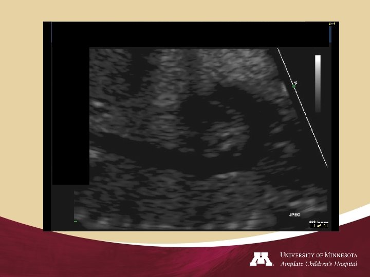
Fetal Cardiology • Fetus dictate the ease in which the images can be obtained • Determine position of the fetus, left and right side, position of the liver, stomach, and descending aorta • Heart occupy one third the volume of the thorax
Fetal Cardiology • • Right atrium- Eustachian valve, SVC, IVC Left atrium- pulmonary veins Four chamber Atrioventricular concordance Venticuloarterial concordance Aortic arch, Ductal arch, branch PA HR, IVS thickness, Cardiac function
Fetal Cardiology • When a prenatal diagnosis of structural or rhythm abnormalities is obtained, the health care team can outline a management strategy to optimize the care and support given to the fetus, mother, and family.
Fetal Cardiology • • Counseling Follow-up scans Planning Delivery Handling the Newborn Intervention after birth Quality of life Parent support group
Fetal Cardiology • 23% of infants with ductal-dependent circulation are d/c from hospital, only to return days later in shock from ductal closure. • True benefits prenatal diagnosis-improved longterm neurologic and functional result than mortality risk
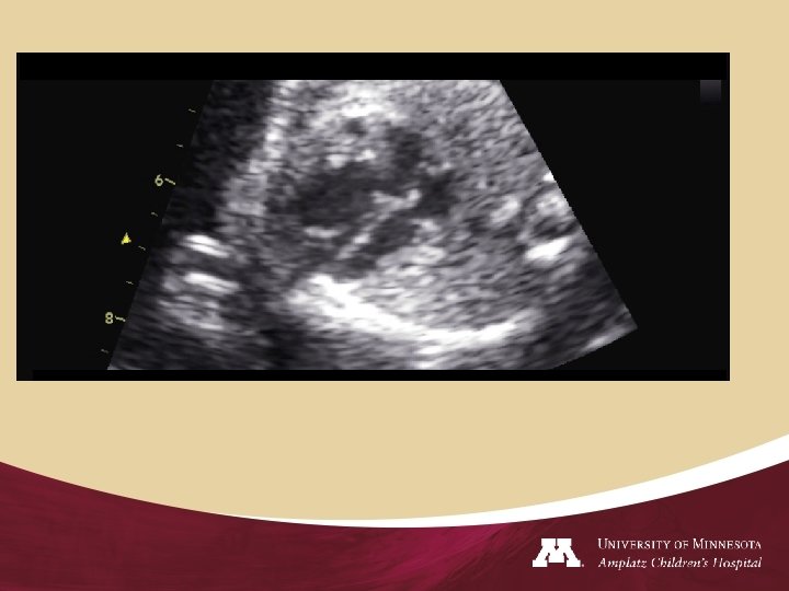
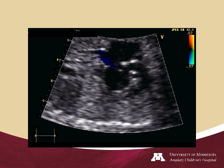
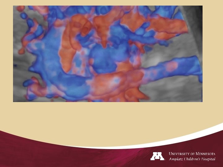
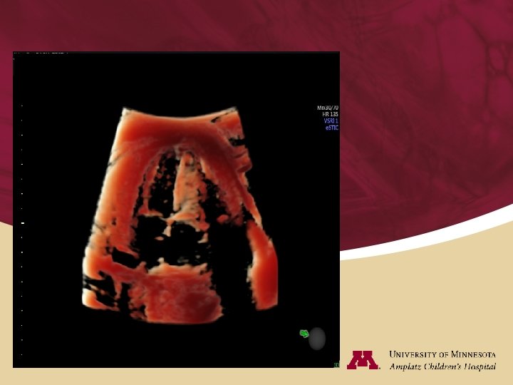
Fetal Echocardiography
Fetal Cardiology
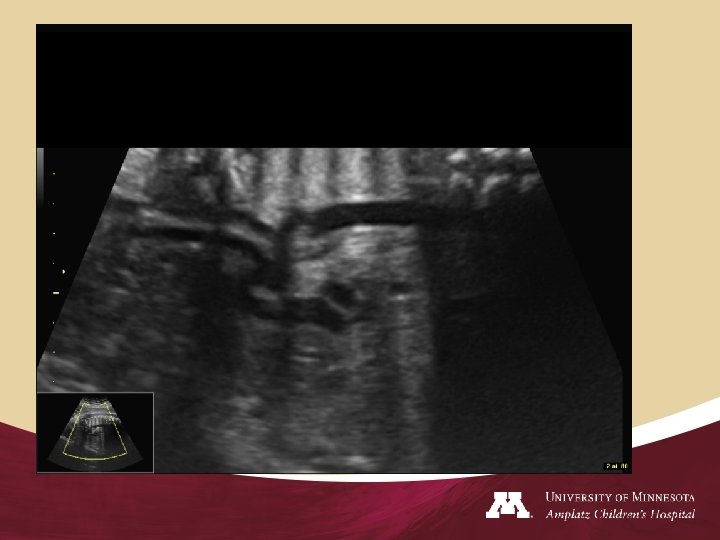
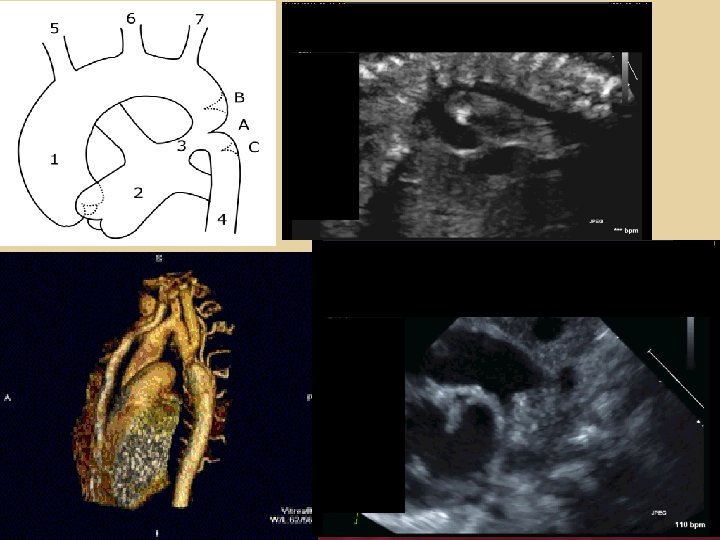
Fetal Aortic Arch • Aortic arch is a candy cane structures/ head and neck vessels, runs parallel to the spine
Fetal Aortic Arch • The ductal arch: Hockey stick-shaped structure that is continuation of the main pulmonary artery
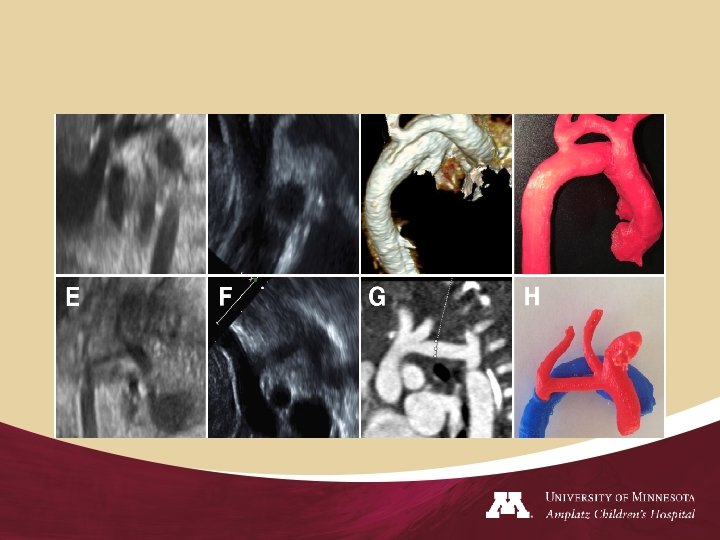
Fetal Aortic Arch • Carotid-subclavian artery index (d 1/d 2) is measured at the origin of the left subclavian artery (d 1) and the distance between the origin of the left carotid artery and the origin of the left subclavian artery (d 2).
Fetal Aortic Arch
Fetal Aortic Arch CS Index N- 1. 1/ Abnormal 0. 7
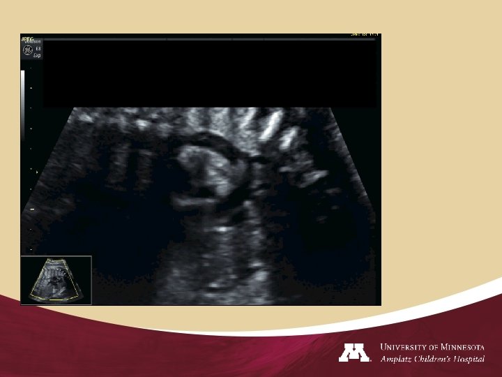
Fetal Cardiology Cardiomyopathy/coarctation
Fetal Cardiology
Fetal Cardiology
Fetal Aortic Arch
Fetal Hydrops Ascites
Fetal Cardiology
Fetal Cardiology
Fetal Cardiology • • Fetal Cardiac Intervention : Is feasible 1. Aortic stenosis evolving to HLHS 2. Pulmonary atresia evolving to Hypoplastic right heart 3. Creation of atrial communication
Fetal Cardiology • Fetal Diagnosis of Aortic Stenosis Patient selection for fetal intervention In midgestation fetuses with AS and normal LV length, reversed flow in the TAA and foramen ovale, monophasic mitral inflow, and LV dysfunction are predictive of progression to HLHS. These physiological features may help refine patient selection for fetal intervention to prevent the progression of AS to HLHS.
Fetal Cardiology • Suggested preoperative Echocardiographic Criteria with Threshold Z scores for Balloon Valvuloplasty • • Aortic valve Mitral Valve Left ventricular function Left ventricular measurements/ long axis/short axis
Fetal Cardiology • ASD • Pulmonary stenosis/Pulmonary atresia
THANK YOU QUESTIONS