Fetal Circulation Umbilical vein carries oxygenated blood from

Fetal Circulation • Umbilical vein carries oxygenated blood from placenta to right atrium • Liver and lungs mostly bypassed • Blood goes to left • atrium via foramen ovale Pulmonary artery connected to aorta via ductus arteriosus, another bypass

Fetal Circulation: Changes at birth • Pulmonary Circulation • Vasodilation of pulmonary vessels secondary to oxygen • Decreased resistance • Ductus Arteriosus • Closes functionally within 24 hours • Foramen ovale closes

Malformation Incidence per 1 Million Live Births % Ventricular septal defect 4482 42 Atrial septal defect 1043 10 Pulmonary stenosis 836 8 Patent ductus arteriosus 781 7 Tetralogy of Fallot 577 5 Coarctation of aorta 492 5 Atrioventricular septal defect 396 4 Aortic stenosis 388 4 Transposition of great arteries 388 4 Truncus arteriosus 136 1 Total anomalous pulmonary venous connection 120 1 Tricuspid atresia 118 1

Congenital Malformations: Causes

Congenital Malformations: Frequency

Congenital Heart Disease • abnormalities of heart or great vessels present at birth • 6 -8 per 1000 live born full term births • etiology unknown >60% • some clear genetic input, e. g. chromosomal --Turner's XO syndrome • some clear environmental, e. g. 1 st trimester maternal rubella • numerous other teratogens suspected

Congenital Heart Disease • Shunts • Obstructions

Congenital Heart Disease: Shunts • Shunt = abnormal communication between • chambers/vessels left-to-right (“pale” type): • gradual overload of right heart, pulmonary vascular bed • progressive pulmonary hypertension: difficulty breathing because the lungs are wet, congested, or fluid-filled • right ventricular hypertrophy • eventual increase of right sided pressures, reversal of flow » VSD, ASD, PDA

Congenital Heart Disease: Shunts • right-to-left shunt: • poorly oxygenated blood enters left side • blue skin, mucous membranes (cyanosis) • "cyanotic CHD type": tetralogy of Fallot, et al.

Congenital Heart Disease: Obstruction • Abnormal narrowing of chambers, valves, vessels • stenoses (partial narrowing) • atresias (complete obstruction) • Coarctation of aorta • Aortic valvular stenosis • Pulmonary valvular stenosis

Left-to-right shunt

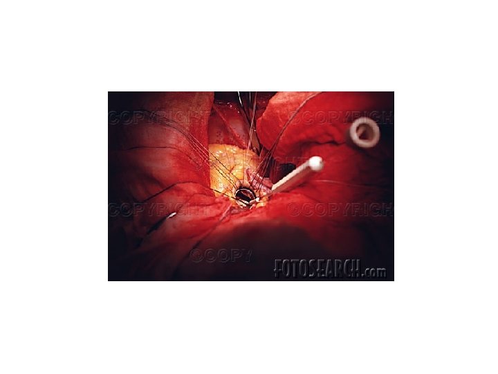
Ventricular Septal Defect • most common congenital cardiac anomaly • frequently associated with other anomalies • range of clinical manifestations (from selfclosure to problems at birth) • functional significance depends upon: • size • presence/absence of pulmonary stenosis

Ventricular Septal Defect
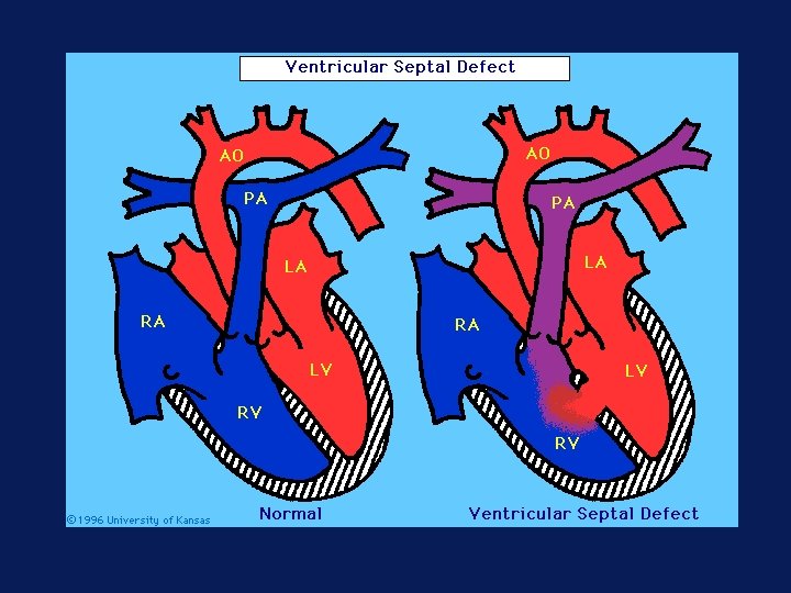

Ventricular Septal Defect • Consequences of large left-to-right shunt: • Right ventricular hypertrophy • Pulmonary hypertension » shunt reversal » cyanosis, polycythemia • congenital CHF • risk of infective endocarditis • Surgical repair in older patients before pulmonary lesions irreversible

Atrial Septal Defect • abnormal opening in wall between right and left atria • most common congenital cardiac anomaly first detected in adult life • normal: • fetal R atrial pressures > Left, flow follows through foramen ovale • at birth L > R, foramen ovale closes (2/3); patent 1/3, but little or no flow

Normal closure of foramen ovale at birth by increased left atrial pressure

Atrial septal defects Developmental defects in septum formation

• high L pressures shunt blood to R, increase • pulmonary blood flow 2 -4 X gradual volume hypertrophy of R atrium and ventricle • pulmonary hypertension in < 10% of cases • Surgical repair to prevent complications (emboli, pulmonary vascular disease)

Patent Ductus Arteriosus - persistent opening of ductus arteriosus • a normal aortopulmonary channel in fetal life • normally closes in first day or two of life » response to increased oxygen » prostaglandins • stays open in premature infants with hypoxemia • stays open in full-term infants with structural defect

PDA circulation

Patent Ductus Arteriosus Clinical problems in later childhood, early adult life: • Pulmonary hypertension (congestive heart failure) • problems with heart rate or rhythm (arrhythmias) • excessive work load on heart that interferes with breathing, feeding, or sleeping

PDA • detected by murmur early • ultimately complications of left-toright shunts • early closure accepted therapy • pharmacologic (block vasodilation by prostaglandin E) • surgical

PDA closure Type and timing of surgical repair depends on the child's condition and the type and severity of heart defects.

PDA Occlusion Gianturco Coil

Right-to-left

Tetralogy of Fallot • Obstruction to right ventricular outflow • Ventricular septal defect • Aorta overrides the VSD • Right ventricular hypertrophy derived from embryologic displacement of infundibular septum

Tetralogy of Fallot • Severity depends upon degree of right ventricular outflow obstruction • if mild, resembles VSD, left-to-right shunt • if severe, right-to-left shunt » cyanosis • Pulmonic origin does not grow with rest of heart • worsens as child gets older • Surgical repair possible in classic tetralogy

Tetralogy of Fallot

Congenital Heart Disease: Obstruction • Abnormal narrowing of chambers, valves, vessels • stenoses (partial narrowing) • atresias (complete obstruction) • Coarctation of aorta • Aortic valvular stenosis • Pulmonary valvular stenosis

Coarctation of aorta

Coarctation of Aorta • narrowing, constriction • • • of aorta M>F Turner's syndrome (XO) ~10% Preductal • manifests early (neonatal) • blood to lower body hypoxemic, upper normoxemic • surgical intervention needed

Coarctation of Aorta • Postductal • often asymptomatic until adulthood • hypertension in upper extremities, weak pulse & low pressure in lower extremities • collateral circulation from precoarctation to post- vessels develops • complications: CHF, intracranial hemorrhage, dissection (pressure related)

pda coarctation normal vsd

Transposition of great vessels

Complex Congenital Abnormality Double-outlet right ventricle with inversion of the ventricles, consecutive atrioventricular discordance, and malposition of aorta and pulmonary artery, pulmonic stenosis and a ventricular septum defect. Conduit graft to connect the anatomic left ventricle with the pulmonic bifurcation. The VSD was closed with a pericardial patch.

Robotic Surgery

International Public Health Initiatives in Heart Disease • Heart to Heart USA-Russia • International Outreach Program USA- Haiti, Brazil
- Slides: 39