Fertilization Spermatozoa maturation steps The maturation and activation


 •")


- Slides: 37
Fertilization
Spermatozoa maturation steps
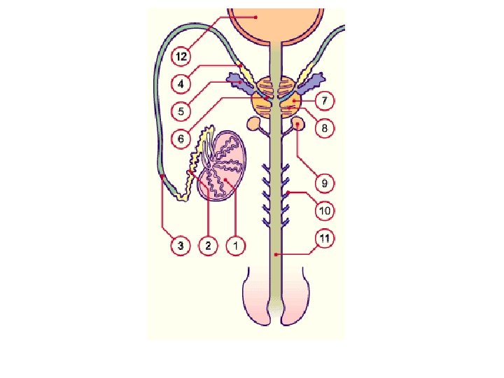
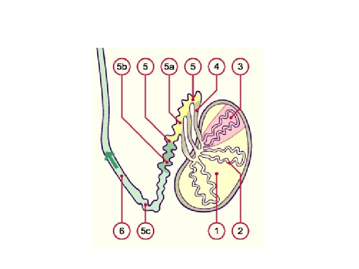
The maturation and activation of the spermatozoa occur in the following four steps: • • Storage in the epididymis Maturation Ejaculation Activation Ascension to the ovary Capacitation Near the oocyte Acrosome reaction
Composition of the seminal plasma Amount 2 -100 ml p. H value 7 -8 (light alkaline buffer) Seminal gland secretion 75% of the volume, alkaline fructose-rich secretion (1. 5 -6. 5 mg/ml fructose), phosphorylcholine, ascorbic acid Prostate secretion 20%-25% of the volume, biogenic amine (spermidine, spermine), citric acid, cholesterol, phospholipids, protease to fluidize the ejaculate (fibrinolysin, fibrinogenase) Further components Phosphate and bicarbonate as buffer, prostaglandin,
Human
COW
MARE SOW
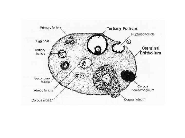
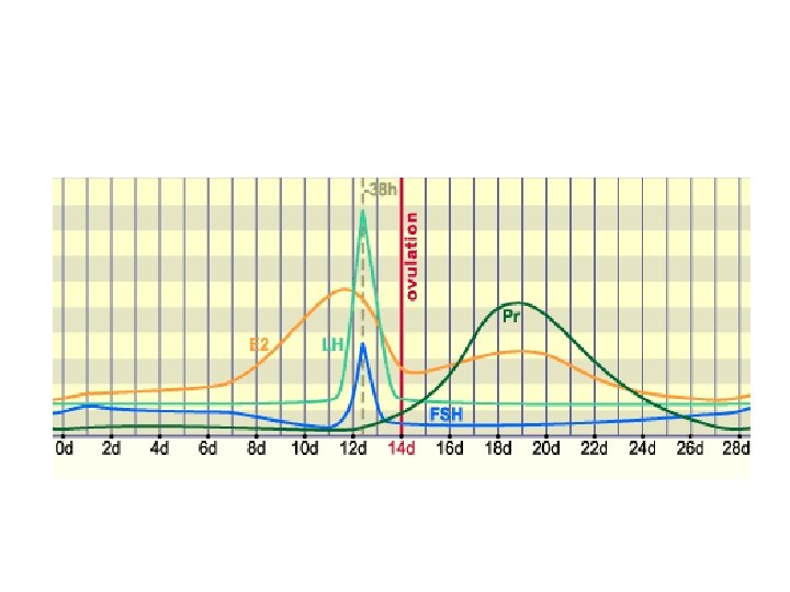
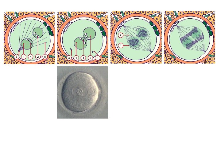
Maturation of the oocyte in the dominant follicle shortly before ovulation
Termination of the first meiosis
The secondary oocyte
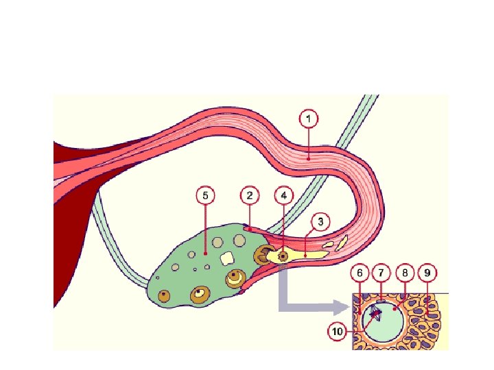
The follicle that is about to rupture
Secondary oocyte in metaphase 2
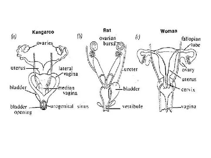
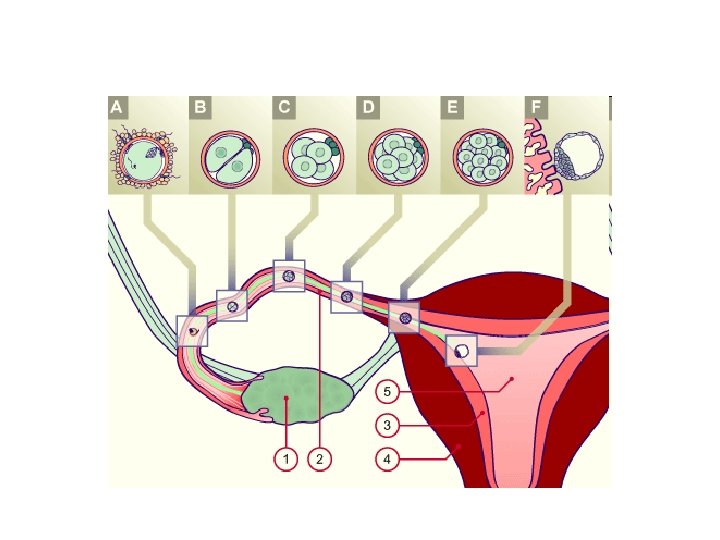
The acquisition of the oocyte by the fallopian tube
Consequences of multiple ovulation • Twin pregnancy • from one mating male • Superfecundation: from two males, after synchrone ovulation • Superfoetation: from two males, after asynchrone ovulation
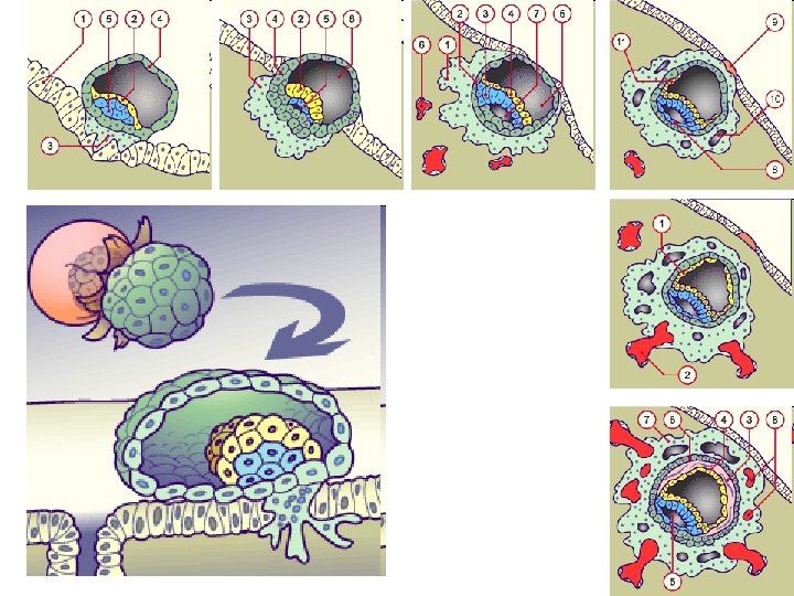
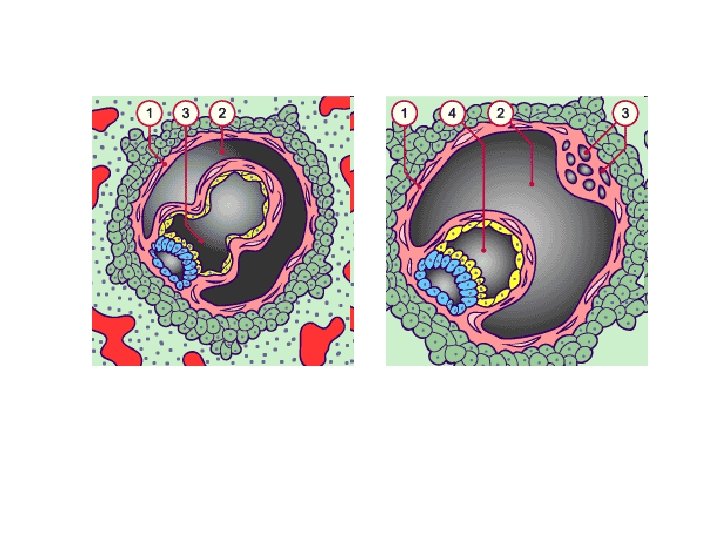
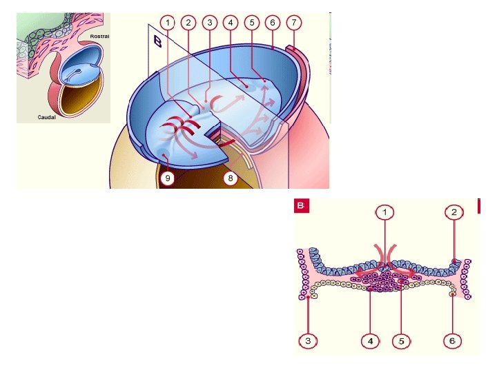
Formation of a bilaminar germ disk • In the bilaminar primordium of the embryo (hypoblast or primary endoderm and epiblast) one recognizes in the epithelium of the epiblast a fluid-filled space, the first primordium of the amniotic cavity. • Ventrally, the roof of the still incompletely uncovered primary umbilical vesicle (previously the blastocyst cavity) is formed by the hypoblast.
Develomental differences in early embriogenesis • A: Folding (Carnivores, Rodents, Ungulate (hoofed animal) • B: Exocoel formation (Primates, Human, Insectivores)
1. Extra-embryonic mesoblast 2. Extra-embryonic reticulum 3. Primary umbilical vesicle 4. Cytotrophoblast 5. Lacunae in the reticulum 6. Hypoblast 7. Heuser´s membrane between hypoblast and mesoblast cells
Formation of the ‘Trilaminar germ disc’ AKA epithelio-mesenchymal transition • Immigrated cells form a third germinal layer: the intraembryonic mesoblast • The mesoblast cells wander in all directions: laterally, cranially and caudally. • This middle germinal layer lies between the definitive endoblast and epiblast. • Exceptions are the cloacal membrane as well as the pharyngeal membrane, where the ectoderm and endoderm lie directly opposite each other!