Fertilization and Cleavage Dr Nandor Nagy Semmelweis University


")

, ZP")
 ZP 2 (120 k. Da)")


 - Sodium/potassium ATPase pumps sodium interior of")


- Slides: 32
Fertilization and Cleavage Dr. Nandor Nagy Semmelweis University
Process of fertilization -maturation of sperms: capacitation -The chemoattraction of sperm to the egg -attachment to and penetration of the zona pellucida The exocytosis of acrosomal vesicle to release enzymes The binding of the sperm to the extracellular envelope The passage of the sperm through the envelope The membrane fusion between sperm and egg -the process end with the intermingling of maternal and paternal chromosomes
Spermatozoa Undergo a Terminal Step of Functional Maturation Called Capacitation -freshly ejaculated sperm are unable to fertilize oocytes
-capacitated sperm is metabolically more active and beats its flagellum rapidly -Thermotaxis: capacitated sperm can sense thermal gradient between isthmus of oviduct and ampulla (about 2 o. C). Acrosome (Proacrosin-GFP) Nucleus/mitochondria/mictotubule Increased membran fluidity and Increased motility albumin and lipid transfer protein (LTP-1; both protein present in female genital organ) decrease the level of cholesterol more fluid spermatozoa cell membrane
Chemotaxis: secreted molecules from cumulus cells and oocyte. obstacles… First obstacle: -granulosa cells embedded in a loose extracellular matrix (hyaluronic acid) Before the encounter
second obstacle: Zona pellucida (13 µm)
Recognition of egg and sperm: 1. Chemoattraction of the sperm to the egg by soluble molecules 2. The binding of the sperm to the extracellular envelop of the egg 3. The exocytosis of the acrosomal vesicle to release its enzyme 4. The passage of the sperm through this envelop 5. Fusion of egg and sperm cell membrane Events Leading to the Fusion of Egg and Sperm Plasma Membranes
zona pellicida 3 protein of zona pellucida : ZP 1 (200 k. Da), ZP 2 (120 k. Da), ZP 3 (83 k. Da). When a spermatozoon reaches the tough zona pellucida surrounding the oocyte, it binds in a species(that is, human-) specific interaction with a glycoprotein sperm receptor molecule in the zona (ZP 3, one of three glycoproteins composing the zona pellucida).
A zona pellucida fehérjéi: ZP 1 (200 k. Da) ZP 2 (120 k. Da) ZP 3 (83 k. Da, ebből csak 44 k. Da a polipeptidváz, a többi a piros „dobverőként” illusztrált oligoszaccharid oldallácok révén) Binding to ZP 3 is mediated by a sperm surface protein called SED 1 galactosyltransferase-I -binds to ZP-3 –activate G protein— open Ca channel—acrsomal rxn. Release proteases that lyse the ZPmake a hole in egg
The acrosome reaction -The exocytosis of the acrosomal vesicle to release its enzyme -Enzymes are released from the acrosome [esterases, collagenase, acrosin and neuraminidase] -Enzymes cause lysis of the zona pellucida
As a result of this binding, the acrosome is induced to release degradative enzymes that allow the sperm to penetrate the zona pellucida.
The cell membrane of the two cells fuse. Molecules required for this event: -CD 9 -Fertilin -tetraspanin -Izumo
Robert Edwards
ART – assisted reproductive technology ZIFT - Zygote Intra-Fallopian Transfer But: laparascopy IUI - Intra-Uterine Insemination IVF – In Vitro Fertilization Nobel prize 2010 Edwards Ovarian hyperstimulation GIFT – Gamete Intra-Fallopian Transfer But: laparascopy ICSI Intra-Cytoplasmatic Sperm Injection
Infertility Statistics on conception in normal fertile couples: within a month: 20– 25%. within a year: 90% Number of months attempting to get pregnant Figure from www. ivf. indiana. com website Definition of infertility: the inability of a couple to conceive after a year of regular intercourse without contraception. Data from HFEA (human fertilization embryology authority) 2
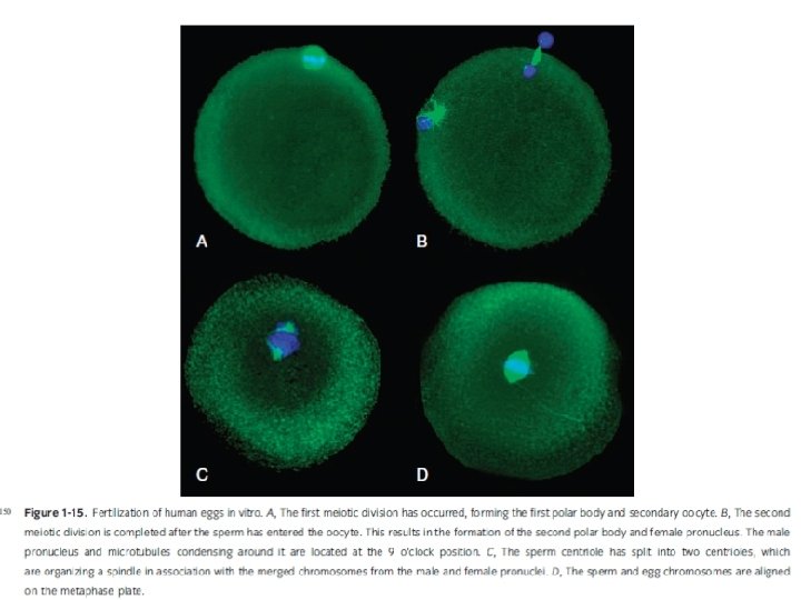
Binding and fusion of sperm cell and egg -in normal monospermy: only one sperm enters a haploid sperm nucleus + a haploid egg nuleus combine: diploid nucleus of the fertilized egg (zygote) http: //www. advancedfertility. com/triploid. htm Restoration of the chromosome number Polyspermy leads to disastrous consequences in most organism -two sperm entry results in triploid nucleus -each sperm’centriole divides to form the two poles of mitotic apparatus -triploid chromosomes -such cells either die or develop abnormally (triploid embryos nearly always abort) -amorphous, nodular embryos 3 pronuclei are seen in the center of the cell Each pronucleus contains 23 chromosomes This embryo has 69 chromosomes instead of the normal 46 Zona pellucida (shell) is visible as a halo around periphery Sperm are visible at 1 and 8 o'clock - outside of the zona
polyspermy block Membrane fusion immediately causes two events to occur: formation of a calcium wave that radiates over the surface of the egg from the point of sperm contact; and release of the contents of thousands of small cortical granules, located just beneath the oocyte cell membrane, into the perivitelline space between the oocyte and the zona pellucida. These two events alter the sperm receptor molecules, causing the zona to become impenetrable by additional spermatozoa. – - These changes prevent polyspermy or the fertilization of the oocyte by more than one spermatozoon.
Wave of Ca 2+ release across a sea urchin egg during fertilization induces cortical granule reaction polyspermy block: step 1
Cortical granule exocytosis polyspermy block: step 2
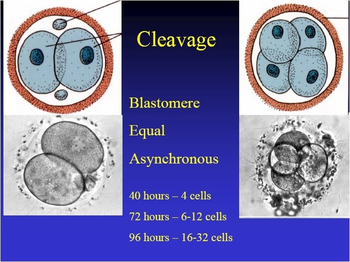
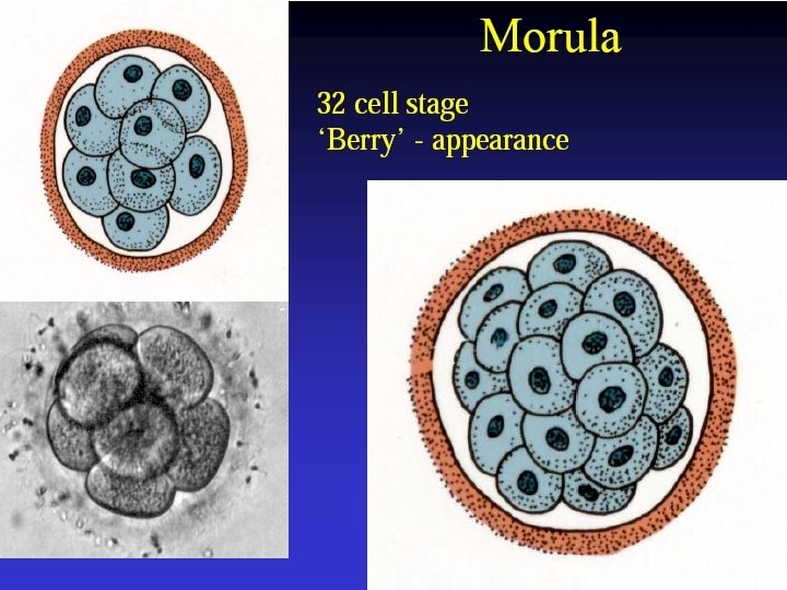
Early blastomeres lack well developed intercellular junctions. Cells are in contact through microvilli and other cellular projections Morula microvilli
Compaction E-cadherin E-cadherin=uvomorulun: 54, 000 D cell adhesion molecule
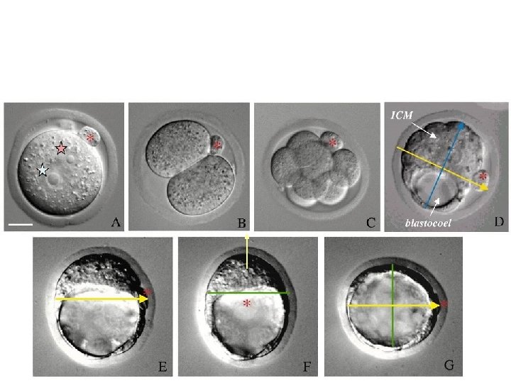
Compacting Human Morula Biology of Reproduction Nikas et al. 1996, Biol. Reprod. 55: 32 -37
compaction Before and after
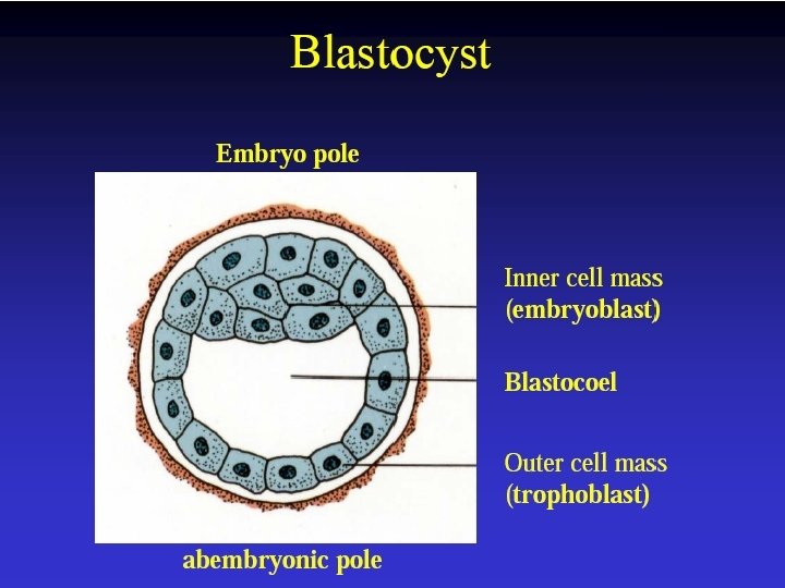
Blastocyst cavity forms within the morula (cavitation) - Sodium/potassium ATPase pumps sodium interior of the morula and water follows through osmosis
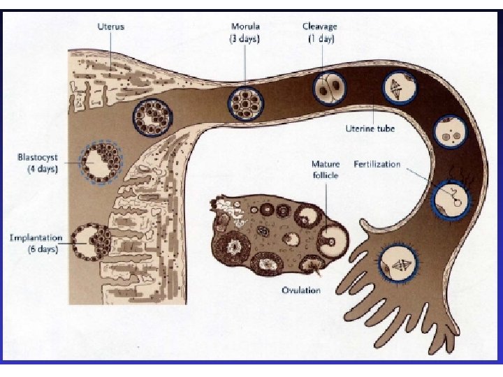
Early Embryonic Development Location All cell divisions are mitotic Smaller cell size with each cleavage division Day Development Ampullary- 0 -2 Isthmic Junction 1 -3 Ampullary. Isthmic Junction One cell Isthmus 2 -3 Four cell Isthmus 3 -5 Eight cell Uterus 4 -5 Sixteen cell Uterus 5 -8 Morula Two cell
Day Development 6 -7 Tight Morula 7 -8 Early Blastocyst 7 -9 Blastocyst Inner Cell Mass Forms Embryo Compact Morula form tight cell contacts so that Blastocyst formation is possible Fills with fluid in center - Cells have established polarity Blastocoel Trophoblast - Forms Placenta