FEMORAL SHAFT FRACTUTES The femoral shaft is well


, shock, fat embolism and")

young adults, usually")


 CLASSIFICATION")


- Slides: 24
FEMORAL SHAFT FRACTUTES The femoral shaft is well padded with musclesan advantage in protecting the bone from all but the most powerful forces but a disadvantages in that fractures are often severely displaced by muscle pull making reduction difficult. Mechanism of injury: This is essentially a fracture of young adults and usually results from a high energy injury. Diaphyseal fractures in elderly patients should be considered pathological until proved otherwise. In children under 4 years of age, the possibility of physical abuse must kept in mind.
Clinical features: There is swelling and deformity of limb with painful movement with sign of blood loss, its important to exclude neurovascular problems and other limb or pelvic fractures with high risk of multisystem injury in the patient. x-ray: It may be difficult to obtain adequate views in accident and emergency room setting which can postponed until better positioning are possible, but never forget to x-ray of the hip and knee also chest x-ray in ARDS.
Femoral shaft # with hip dislocation
EMERGENCY TREATMENT: At the site of the accident, shock should be treated by iv fluids and blood and the fracture splinted before the patient is moved by Thoma’s splint with skin traction until the patient transported to the hospital. DEFINITIVE TREATMENT: 1 -TRACTION AND BRACING: indications: a-fractures in children, b-contraindications to anaesthesia, and c-lack of suitable skill or facilities for internal fixation. it is apoor choice for elderly patients, for pathological fractures and those with multiple injuries. the traction take long time(10 -14 weeks)for adults.
Skeletal traction
2 -OPEN REDUCTION AND PLATING: indications: a-the combination of shaft and femoral neck fractures and b-a shaft fracture with an associated vascular injury. c-shaft # in growing children 3 -INTRAMEDULLARY NAILING: It is a method of choice for most femoral shaft fractures, this operation done closed reduction of fracture under screen(fluoroscopy) apply suitable size with or without reaming, with locking screws can be inserted transversly at the proximal and distal ends; this controls rotation with good stability even for subtrochanteric and distal-third fractures. 4 -EXTERNAL FIXATION: indications: a-the treatment of severe open injuries, b-management of patients with multiple injuries where there is need to reduce operating time, c-dealing with severe bone loss by the technique of bone transport, d- treating femoral fractures in adolescents.
Plating of femur
Locked intramedullary nailling
External fixation
OPEN FRACTURES Open femoral fractures should be carefully assessed for 1 -skin loss 2 -wound contamination. 3 muscle ischemia and 4 - vessels and nerves. the immediate treatment is similar to that of closed fractures. antibiotics and wound debridment should done with little delay, the major decision is how to stabilize the fracture. with small, clean wounds and little delay from time of injury, the fracture can be treated as for aclosed injury with addition of prophylactic antibiotics BUT, with large wounds, contaminated wounds, skin loss or tissue destruction, internal fixation should be avoided. after debridment, the wound should be left open and the fracture stabilized by applying an external fixation.
FEMORAL RACTURES IN CHILDREN Infants need no more than 1 or 2 weeks in balanced traction followed by a spica for another 3 or 4 weeks. children up to 10 years can be treated in asimilar manner, allowing twice as long in traction and then 6 weeks in a spica. teenager may require even longer in traction before changing to a spica, however, if satisfactory reduction cannot be obtained or held, internal fixation(plate and secrews or flexible intra medullary nails)or by external fixation.
COMPLICATIONS: EARLY GENERAL: such as blood loss(1 -2 liters), shock, fat embolism and acute respiratory distress are common in high energy injuries. VASCULAR INJURY: it is takes the priority and the vessel must be repaired or grafted without delay. at the same operation, the fracture is secured by interal fixation. THROMBOEMBOLISM : prolonged traction in bed predisposed to thrombosis, movement and exercise are important in preventing this; they can be supplemented by foot compression devices or prophylactic doses of anticoagulants. INFECTION : in open injuries and following internal fixation, there’s always risk of infection, prophylactic antibiotics and careful attention to the principles of fracture surgery kept the incidence below 2%.
LATE: DELAYED UNION AND NON-UNION: It’s said that a fractured femur should unite in 100 days plus or minus 20. if union is delayed beyond this time, an exchange nailing is performed using aslightly larger nail; in addition, the fracture may need bone grafting. MALUNION: Fractures treated by traction and bracing often develop some deformity; no more than 15 degrees of angulation should be accepted. JOINT STIFFNESS: The knee is often affected after a femoral shaft fracture either due to injury of knee at the same time or stiffeness because of soft tissue adhesions during treatment; hence the importance of exercise and knee movements. REFRACTURE AND IMPLANT FAILURE.
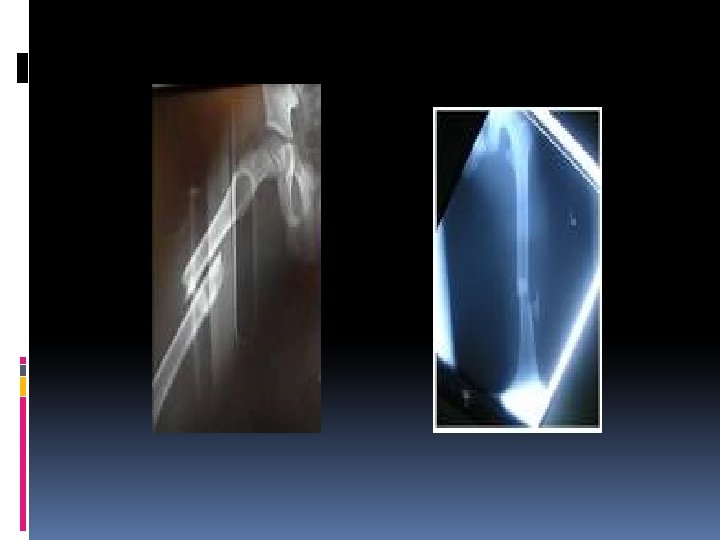
SUPRACONDYLAR FRACTURES OF THE FEMUR These types of fractures are seen in(a)young adults, usually as aresult of high-energy trauma and (b)elderly, osteoporotic individuals. direct violence is the usual cause. the fracture line is just above the condyles, but it may be branch off distally between them. the pull of the gastrocnemius attachments may tilt the distal fragment backwards. Clinical features: the knee is swollen and deformed; movement is too painful to be attempted. the tibial pulses should always be palpated. X-ray: the fracture is just above the femoral condyles and is transverse or comminuted. the distal fragment is often tilted backwards. the entire femur must be x-rayed so not to miss a proximal fracture or dislocated hip.
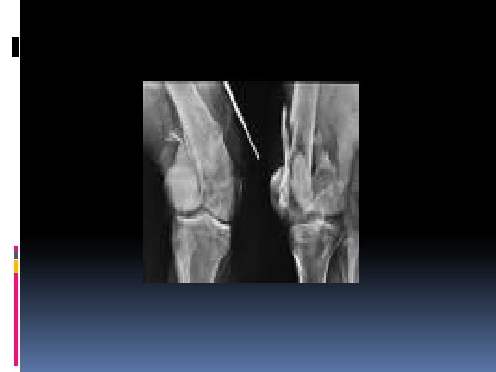
TREATMENT 1 -conservative: if the fracture is only slightly displaced and extra-articular or if it reduces easily with knee in flexion, it can be treated easily by skeletal traction through the proximal tibia and the limb is cradled on a Thomas’ splint with knee in flexion piece and the movements are encouraged. the fracture may need vertical traction by apply second pin above knee. at 4 -6 weeks when the fracture is beginning to unite the traction can be replaced by a cast brace and partial weight bearing with crutches. 2 -operative: if closed reduction fail, open reduction and internal fixation with an angled compression device(dynamic condylar compression secrew and plate)or blade condylar plate and secrews. unprotected weight-bearing is not permitted until the fracture has consolidated(usually around 12 weeks). Locked intramedullary nails which are introduced retrograde through the intercondylar notch are also used for these fractures.
Conservative treatment
Operative treatment
COMPLICATIONS: EARLY: -ARTERIAL DAMAGE: there’s a small but definite risk of arterial damage and distal ischemia. careful assessment of the leg and peripheral pulses is essential. LATE: -JOINT SYIFFNESS: knee stiffness is almost inevitable, along period of exercise is necessary but full movement is rarely regained. NON-UNION: knee increases the like of nonunion. This combination is difficult to treat and unless great care is exercised, the ultimate range of movement at the knee may be less than that at the fracture. OSTEOARTHRITIS(OA): supracondylar fractures often extend into the joint surface; anatomical reduction is necessary to reduce the risk of OA.
FEMORAL CONDYLE FRACTURES Condylar fractures are often associated with supracondylar fractures where a distal extension into the knee joint may cause one or both condyles to be split apart, they also occur in isolation: a direct injury or a fall from a hieght may drive the tibia upwards into the intercondylar fossa. Pathological anatomy: This fractures are classified by AO (Muller ) classification into three groups of fractures: A-Purely extra-articular, supracondylar fractures. B-Intra-articular fracture of one condyle-ysually the lateral one. C-Inta-articular bicondylar fractures, which are effectively also ‘supracondylar’.
AO (MULLER) CLASSIFICATION
Clinical features: The knee is swellen and may be deformed, there’s a tender”doughy” feel characteristic of a haemoarthrosis, the joint is too painful to move but the foot should be examined to exclude nerve and arterial damage. X-ray: One femoral condyle may be fractured obliquely and shifted upwards or both condyles may split apart so that the fracture line is T or Y shaped.
Treatment : The haemoarthrosis must be aspirated as soon as possible. because the articular surface is involved, accurate anatomical reduction is important, so open reduction and internal fixation are therefore often employed by cannulated secrews that fixed the articular fragments with blade plate or dynamic condylar screw and plate. The patient can begin knee exercises as soon possible to prevent the stiffness. Complications: 1 -stiffness of the knee: -this is a common complication. it usually responds to prolonged physiotherapy, although movement may not be fully restored. 2 -osteoarthritis: -as with other intra-articular fractures, secondary osteoarthritis is a late complication.