Female Reproductive System Kristine Krafts M D Female


 • Used to call the dysplastic precursor lesions “cervical intraepithelial")


 •")


")


 • Benign tumor of smooth muscle • Very common! • Stimulated by")


 • Frequently arises in")


 • Lobular carcinoma")


- Slides: 57
Female Reproductive System Kristine Krafts, M. D.
Female Reproductive System Outline • Cervix • Uterus • Ovaries • Breast
Female Reproductive System Outline • Cervix • Cervical carcinoma
Cervical Carcinoma • Most are squamous cell carcinomas; a few are adenocarcinomas • Caused by high-risk HPV (mostly 16, 18) • Low-risk HPV (6, 11) causes condylomas • Preceded by dysplastic epithelial changes • Pap smears catch most cases before they become invasive
Squamous Intraepithelial Lesion (SIL) • Used to call the dysplastic precursor lesions “cervical intraepithelial neoplasia” (CIN) • Current term is “SIL” • Low-grade SIL (LSIL): mild dysplasia • High-grade SIL (HSIL): mod-severe dysplasia • LSIL usually reverts to normal, but HSIL usually progresses to carcinoma
Cervical carcinoma usually arises in the transformation zone Transformation zone
Tissue biopsy Pap smear Normal LSIL HSIL
Invasive cervical carcinoma
Invasive Cervical Carcinoma • Peak age: 45 (10 -15 years after SIL develops) • Spreads slowly • Staging is super important for prognosis • Localized disease: 90% 5 -year survival • Regional mets: 50% 5 -year survival • Distant mets: 15% 5 -year survival
Female Reproductive System Outline • Cervix • Uterus • Endometriosis • Endometrial hyperplasia • Tumors
Endometriosis • Presence of endometrial glands outside uterus • Endometrium undergoes cyclic bleeding • Can cause pain, scarring, infertility
Endometriosis in ovary (“chocolate cyst”)
Endometrial Hyperplasia • Proliferation of endometrium • Risk factors involve estrogen excess (obesity, nulliparity, estrogen replacement therapy) • The more severe the hyperplasia, the greater the chance that it will evolve into carcinoma
Normal endometrium
Simple Complex Endometrial hyperplasia Atypical
Leiomyoma (“Fibroid”) • Benign tumor of smooth muscle • Very common! • Stimulated by estrogen • Can cause pain and abnormal bleeding
Leiomyosarcoma • Malignant tumor of smooth muscle • Necrotic, with atypical cells and lots of mitoses • Metastasizes early, often to lungs • 5 -year survival = 40%
Leiomyoma Leiomyosarcoma
Leiomyoma Leiomyosarcoma
Endometrial Carcinoma • Peak age: 55 -65 (not before 40) • Frequently arises in endometrial hyperplasia • Risk factors involve estrogen excess • Symptoms: leukorrhea, irregular bleeding • Metastasizes late
Female Reproductive System Outline • Cervix • Uterus • Ovaries • Tumors
Origin of Ovarian Tumors Most common! Surface epithelial tumors Germ cell tumors Sex cord-stromal tumors • Cystadenoma • Cystadenocarcinoma • Granulosa-theca cell tumor • Sertoli-Leydig cell tumor • • Teratoma Dysgerminoma Yolk sac tumor Choriocarcinoma
Cystadenoma • Benign tumor derived from surface epithelium of the ovary • Contains cystic spaces filled with fluid • Typically large, occasionally bilateral
Patient with ovarian cystadenoma
Ovarian cystadenoma
Ovarian cystadenoma
Teratoma • Benign tumor arising from germ cells • Usually cystic and lined with skin (“dermoid cyst”) • Often contains many types of tissue such as cartilage, bone, neural tissue • Rare cases become malignant
Teratoma
Ovarian Cancer • 2019 estimate: 22, 500 new cases / 14, 000 deaths • Common and deadly • Often noticed until metastatic • Peak age: 50 • Most are cystadenocarcinomas
Papillary cystadenocarcinoma
Papillary cystadenocarcinoma
Ovarian Cancer Symptoms • Bloating • Pelvic/abdominal pain • Early satiety • Urinary frequency/urgency Risk factors • Estrogen excess (eg, nulliparity) • Family history (BRCA gene mutation) • NOT using oral contraceptives!
Ovarian Cancer • Treatment: surgery, radiation, chemotherapy • Prognosis depends on stage • Cancer inside ovary: 5 y survival 70% • Cancer outside ovary: 5 y survival 13%
Female Reproductive System Outline • Cervix • Uterus • Ovaries • Breast • Fibrocystic change • Tumors
Breast • Many breast diseases present as lumps • Most lumps represent benign things… • …but a lump always needs to be evaluated • Ultrasound, mammography, fine needle aspiration, and biopsy are the usual methods
Most breast lumps are benign
Fibrocystic Change • “Lumpy bumpy” breast tissue containing cysts and fibrous tissue • Very common • Not associated with increased cancer risk
Fibrocystic change
Fibrocystic change
Fibroadenoma • Most common benign breast tumor • Stimulated by estrogen • Peak incidence in 20 s • Solitary, discrete, moveable mass • Fibrous tissue with compressed ducts and lobules
Fibroadenoma
Fibroadenoma
Breast Carcinoma • 2019 estimate: 268, 000 new cases / 42, 000 deaths • Most common, 2 nd deadliest cancer in women • Lifetime risk: 1 in 8 • 75% of patients are >50
Breast Carcinoma Risk Factors • Age • Family history • Increased estrogen exposure • Alcohol consumption • High-fat diet
Breast Carcinoma Family History • 5 -10% of all cases are hereditary • Worry if first degree relative with breast cancer • Most have BRCA-1 or BRCA-2 mutations • Tumor suppressor genes; help repair DNA • Genetic testing difficult • Most carriers get cancer by age 70
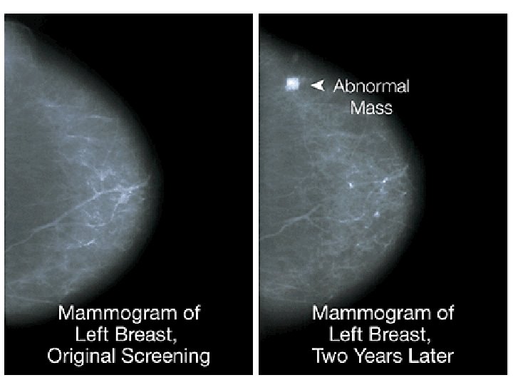
Breast Carcinoma Clinical Findings If discovered by palpation: • Solitary, painless, moveable mass • 2 -3 cm in diameter • Axillary nodes positive in 50% of patients If discovered by mammography: • 1 cm in diameter • Axillary nodes positive in 15% of patients As disease progresses: • Fixation to chest wall • Adherence to overlying skin • Peau d’orange
Advanced breast carcinoma: fixation to skin
Peau d’orange
Breast Carcinoma Histologic Types Non-invasive • Ductal carcinoma in situ (DCIS) • Lobular carcinoma in situ (LCIS) Invasive • Ductal • Lobular • Inflammatory • Others
Lobular carcinoma in situ
Low-grade invasive ductal carcinoma
High-grade invasive ductal carcinoma
Inflammatory breast carcinoma
Breast Carcinoma Prognostic Factors • Size of tumor • Lymph node involvement • Distant metastases • Grade of tumor • Histologic type of tumor
Sentinel node biopsy
TNM* staging system for breast cancer Stage T N M 5 -year survival Stage 0 DCIS 0 M 0 92% Stage I <2 cm 0 M 0 87% Stage II <5 cm >5 cm <3 0 M 0 75% Stage III <5 cm >5 cm Any T 4+ 1+ 10+ Any N M 0 46% M 0 skin or chest wall Stage IV Any T Any N M 1 13% * Tumor (size), nodes (# positive), metastases