
FEMALE REPRODUCTIVE SYSTEM By Prof Dr Indu K
FEMALE REPRODUCTIVE SYSTEM By Prof. (Dr. ) Indu K Pisharody Department of Physiology CIMS&H
Female Reproductive System Objectives 1. FEMALE REPRODUCTIVE ORGANS ØPrimary Sex Organs Ø� Accessory Sex Organs Ø� Functional Anatomy of Accessory Sex Organs 2. SEXUAL LIFE IN FEMALES Ø�First Period Ø�Second Period ØThird Period
FEMALE REPRODUCTIVE ORGANS Female reproductive system comprises of primary sex organs and accessory sex organs. PRIMARY SEX ORGANS Primary sex organs are a pair of ovaries, which produce eggs or ova and secrete female sex hormones, the estrogen and progesterone. ACCESSORY SEX ORGANS Accessory sex organs in females are: 1. A system of genital ducts: Ø Fallopian tubes, uterus, cervix and vagina 2. External genitalia: ØLabia majora, labia minora and clitoris.
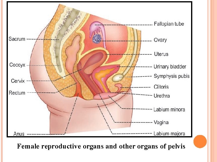
Female reproductive organs and other organs of pelvis
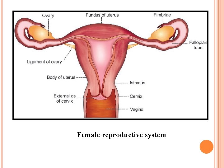
Female reproductive system
�PRIMARY SEX ORGANSOVARIES ØOvary is the gonad or primary sex organs in females. ØA woman has two ovaries. ØOvaries have two functions: I. Gametogenic II. Endocrine functions. ØGametogenic function is the production and release of ovum or female gamete ØEndocrine function of ovaries is the secretion of female sex hormones-estrogen and progesterone.
ØOvaries are flattened ovoid bodies, with dimensions of 4 cm in length, 2 cm in width and 1 cm in thickness. ØEach ovary is attached at hilum to the broad ligament. ØEach ovary has two portions: 1. Medulla 2. Cortex MEDULLA ØMedulla or zona vasculosa is the central deeper portion of the ovary. It has the stroma of loose connective tissues. ØIt contains blood vessels, lymphatics, nerve fibers and bundles of smooth muscle fibers near the hilum.
CORTEX ØCortex is the outer broader portion and has compact cellular layers. ØCortex is lined by the germinal epithelium underneath a fibrous layer known as ‘tunica albuginea’. ØThe Cortex consists of ovarian follicles at different stages, connective tissue and interstitial. ØIn the intrauterine life, outer part of cortex contains the germinal epithelium, which is derived from the germinal ridges. When fetus develops, the germinal epithelium gives rise to a number of primordial ova.
ØThe primordial ova move towards the inner substance of cortex. A layer of spindle cells called granulose cells. ØPrimordial ovum along with granulosa cells is called the primordial follicle ØAt 7 th or 8 th month of intrauterine life, about 6 million primordial follicles are found in the ovary. ØBut at the time of birth, only 1 million primordial follicles are seen in both the ovaries and the rest of the follicles degenerate. ØAt the time of puberty, the number decreases further to about 300, 000 to 400, 000. ØAfter menarche during every menstrual cycle, one of the follicles matures.
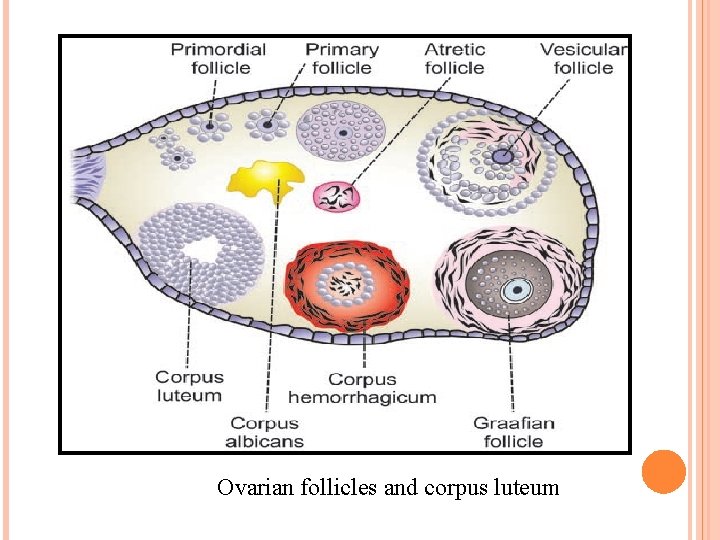
Ovarian follicles and corpus luteum
Functions of Ovaries ØOvaries are the primary sex organs in females. ØFunctions of ovaries are: 1. Secretion of female sex hormones 2. Oogenesis 3. Menstrual cycle.
ACCESSORY SEX ORGANS Accessory sex organs in females are: 1. A system of genital ducts: Fallopian tubes, uterus, cervix and vagina. 2. External genitalia: Labia majora, labia minora and clitoris.
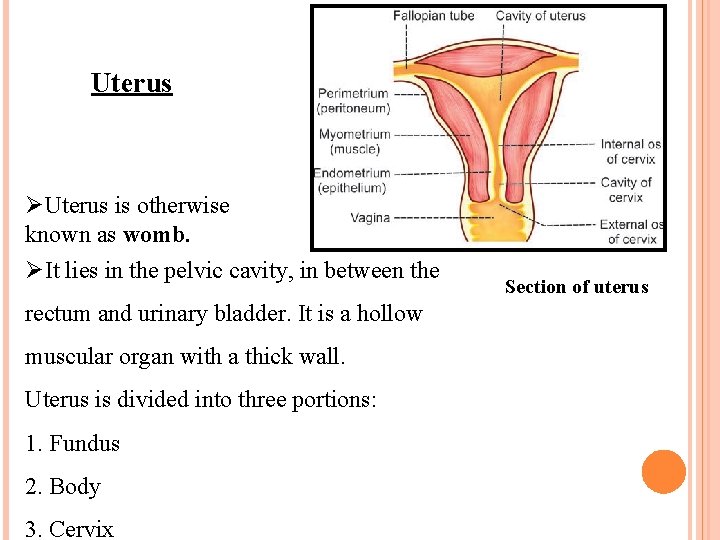
Uterus ØUterus is otherwise known as womb. ØIt lies in the pelvic cavity, in between the rectum and urinary bladder. It is a hollow muscular organ with a thick wall. Uterus is divided into three portions: 1. Fundus 2. Body 3. Cervix Section of uterus
Structure of uterus ØUterus is made up of three layers: 1. Outer Serous layer derived from peritoneum. 2. Middle muscular layer or myometrium made up of smooth muscle fibers. 3. Inner mucus layer or endometrium made up of ciliated columnar epithelial cells, connective and uterine glands.
Cervix is the lower constricted part of uterus. It is divided into two portions: 1. Upper supravaginal portion, which communicates with body of uterus through internal os (orifice) of cervix. 2. Lower vaginal portion, which projects into the anterior wall of the vagina and it communicates with vagina through external os. Vagina Ø Vagina is a short tubular organ. Ø It is lined by mucus membrane, which is formed by stratified epithelial cells.
OVARIAN HORMONES-ESTROGEN ØIn a normal non-pregnant woman, estrogen is secreted in large quantity by theca interna cells of ovarian follicles and in small quantity by corpus luteum of the ovaries. ØA small quantity of estrogen is also secreted by adrenal cortex. ØIn pregnant woman, a large amount of estrogen is secreted by the placenta. ØEstrogen is a C 18 steroid and is present in 3 forms in plasma: 1. β-estradiol 2. Estrone 3. Estriol. ØAll the three forms of estrogen are present in significant quantities in plasma. ØThe quantity and potency of β-estradiol are more than those of estrone and estriol.
FUNCTIONS OF ESTROGEN The major function of estrogen is to promote cellular proliferation and tissue growth in the sexual organs and in other tissues, related to reproduction. ØEstrogen promotes the growth of ovarian follicles by increasing the proliferation of the follicular cells. It also increases the secretory activity of theca cells ØIt prepare uterus for pregnancy by ØEnlargement of uterus to about double of its childhood size due to the proliferation of endometrial cells ØProliferation and dilatation of blood vessels of endometrium. ØProliferation and dilatation of the endometrial glands, which become more tortuous with increased blood flow ØIncrease in the spontaneous activity of the uterine muscles and their sensitivity to oxytocin. ØIncrease in the contractility of the uterine muscles.
ØEstrogen is responsible for the development of secondary sexual characters in females. i. Hair distribution: ii. Skin-Skin becomes soft and smooth. iii. Body shape ØIt also effect on breast for feeding baby. ØEstrogen increases osteoblastic activity therefore it aslo effect on development of bones.
PROGESTERONE ØA small quantity of progesterone is secreted by theca interna cells of ovaries during the first half of menstrual cycle. ØBut, a large quantity of progesterone is secreted during the latter half of each menstrual cycle ØSmall amount of progesterone is secreted from adrenal cortex also. ØIn pregnant woman, large amount of progesterone is secreted by the corpus luteum in the first trimester. In the second trimester, corpus luteum degenerates. ØPlacenta secretes large quantity of progesterone in second and third trimesters.
Chemistry of Progesterone It is a C 21 steroid. Plasma level of progesterone in females at normal reproductive age varies during different phases of menstrual cycle. In follicular phase, it is about 0. 9 ng/m. L.
Functions of Progesterone is concerned mainly with the final preparation of the uterus for pregnancy and the breasts for lactation. The effects of progesterone are: 1. Effect on Fallopian Tubes Progesterone promotes the secretory activities of mucosal lining of the fallopian tubes. Secretions of fallopian tubes are necessary for nutrition of the fertilized ovum, while it is in fallopian tube before implantation.
2. Effect on the Uterus Progesterone promotes the secretory activities of uterine endometrium during the secretory phase of the menstrual cycle. Thus, the uterus is prepared for implantation of the fertilized ovum. 3. Effect on Cervix Progesterone increases the thickness of cervical mucosa and thereby inhibits the transport of sperm into uterus. 4. Effect on the Mammary Glands Progesterone promotes the development of lobules and alveoli of mammary glands by proliferating and enlarging the alveolar cells. It makes the breasts to swell by increasing the secretory activity and fluid accumulation in the subcutaneous tissue.
5. Effect on Hypothalamus Progesterone inhibits the release of LH from hypothalamus through feedback effect. This effect is utilized for its contraceptive action. 6. Thermogenic Effect Progesterone increases the body temperature after ovulation. The mechanism of thermogenic action is not known. It is suggested that progesterone increases the body temperature by acting on hypothalamic centers for temperature regulation. 7. Effect on Respiration During luteal phase of menstrual cycle and during pregnancy, progesterone increases the ventilation via respiratory center. This decreases the partial pressure of carbon dioxide in the alveoli.
Regulation of Progesterone Secretion ØLH from anterior pituitary activates the corpus luteum to secrete progesterone. ØSecretion of LH is influenced by the gonadotropin-releasing hormone secreted in hypothalamus. ØProgesterone inhibits the release of LH from anterior pituitary by negative feedback.

Thank You
- Slides: 25