Female Genital System of birds BY DR MAHMOUD


Female Genital System of birds BY DR/ MAHMOUD ABDELGHAFFAR

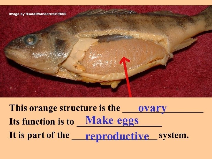
Ovary • The ovary is a cluster of developing ova, and is located midway between the neck and the tail of the bird and attached at the back. • Birds has only LEFT ovary
 epithelium A thin layer")
The ovary is covered by simple cuboidal epithelium (germinal epithelium) epithelium A thin layer of CT lies under the epithelium (Tunica albuginea) albuginea The ovary consists of an outer cortex and an inner medulla The medulla contains Bl. Vs, lymph vessels and nerves which are connected together by loose CT.

• Each ovum starts as a single cell surrounded by a vitelline membrane • As the ovum develops, additional yolk forms. • The color of the yolk comes from fatsoluble pigments, called xanthophylls, contained in the hen's diet.
 or allowed to range")
• Hens fed diets with yellow maize (field corn) or allowed to range on grass typically produce eggs with dark yellow yolks. • Hens fed diets with white maize, maize millet, millet or wheat typically produce eggs with pale yolks.

1 - Stage of oocytes Oocytes are embedded in the cortex. They are small cells with spherical, central nuclei. oocytes are not surrounded by follicular cells but by cortical stroma

2 - Stage of growing follicles • Under the effect of FSH, oocytes grow and are transformed into follicles. as follows, oocytes are increased in size and the cytoplasm accumulates yolk droplets. • Yolk condenses around the cytoplasm to form a perivitelline membrane.

• Oocytes are surrounded by granulosa cells (s. cub then becomes ps. st. col. epithelium). • Stroma around the follicles form theca folliculi (theca interna and theca externa). • Further growth of the follicles causes them to hanged from the ovary by stalks The presence of numerous follicles suspended from the ovary by stalks causes the ovary to resemble a bunch of grape
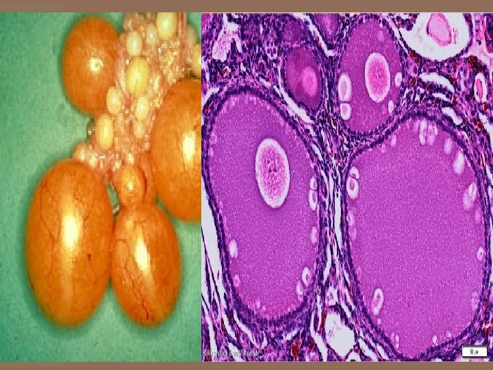

3 - Stage of mature follicles A mature follicle is formed of the following layers: 1. Primary oocyte: surrounded by a perivitelline membrane. 2. Zona radiata: thin zone secreted by granulosa cells. It contains processes extending radially from the oocyte and the granulosa cells 3. Granulosa cells: s. cuboidal epithelium 4. Theca folliculi: theca interna and theca externa

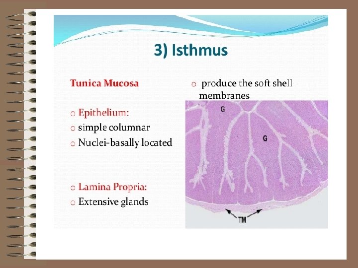
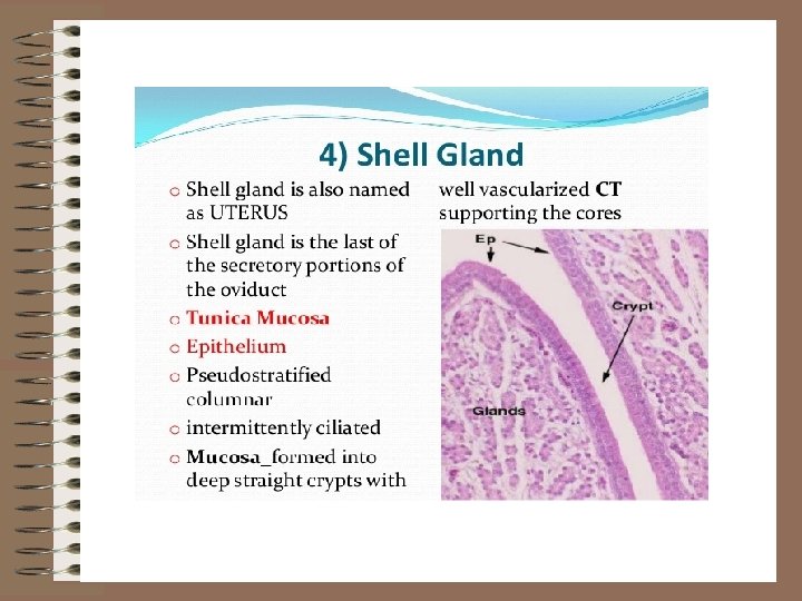
Oviduct • It is a long tortuous tube extends from close to the left ovary till it opens into the cloaca. • The functions of the oviduct is to receive the ovulated oocyte, allows fertilization and to add albumen, shell membrane and shell • The oviduct is divided into 5 regions. All regions possess the same basic structure.

1 - Tunica mucosa Mucosa project into the lumen in the form of primary folds carrying secondary folds. L. Ep --Simple columnar ciliated epith. L. Prop --Contains branched tubular glands. 2 - Tunica musculosa of 2 layers of SMF (inner thick circular & outer thin longitudinal) 3 -Tunica serosa part of the peritoneum. • Although the 5 regions have the same basic structure, each region possesses special modifications to adapt its function.
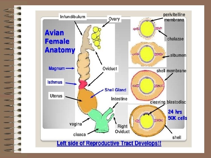

Infundibulum Magnum Isthmus Uterus Anat Formed of 2 parts: funnel neck part Longest & thickest region Mucosa * folds are spiral More folded * propria contains glands Muscul Scattered osa bundles of SMF Serosa +ve functio Receives the n ovum +ve Thick layer Vagina Shorter & smaller in diameter Folded Expanded Short & thin narrow walled & coiled Folds are leaf like Folds are long. +ve -ve Thicker than magnum +ve egg white shell memb. Well developed develop ed +ve T. Adv shell ovipositi on
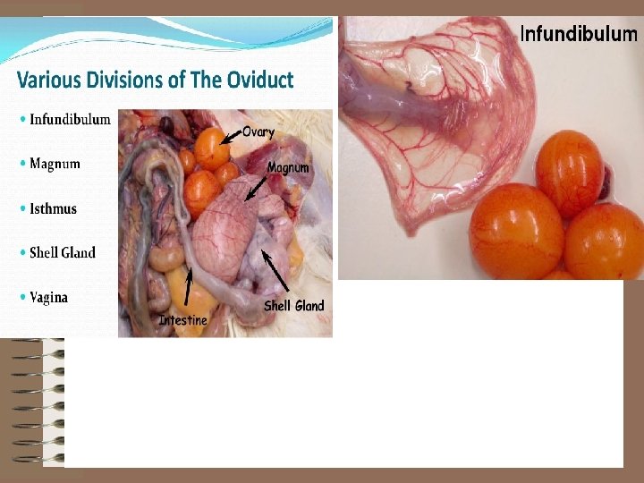

Infundibulum

Magnum

Female genital System of Fish BY DR/ MAHMOUD ABDELGHAFFAR

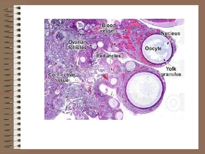
Ovary • Most of fish species in Egypt have ovaries of cyst-ovarian type. • This ovary has a lumen continuous with the oviduct. • Ova are shed unto the lumen and go through the oviduct.

• Histologically, the ovary of the fish is consisted of 1. stroma 2. parenchyma.

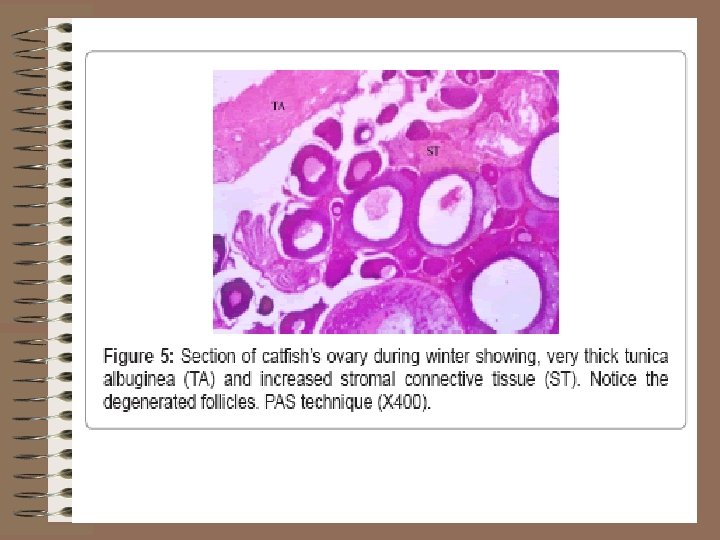
a. Stroma • The ovary of the fish is surrounded by tunica albuginea from which dense fine collagen fibers (ovigerous lamellae) that divide the ovary into compartments.

• Tunica albuginea consists of dense irregular connective tissue containing smooth muscle layer which help in squeezing the ovary to evacuate the eggs. • The smooth muscles in the tunica albuginea of some fish like catfish, bagrus species are differentiated into 2 distinct layers; outer longitudinal and inner circular layers.

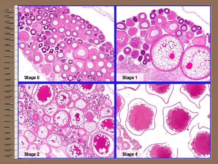
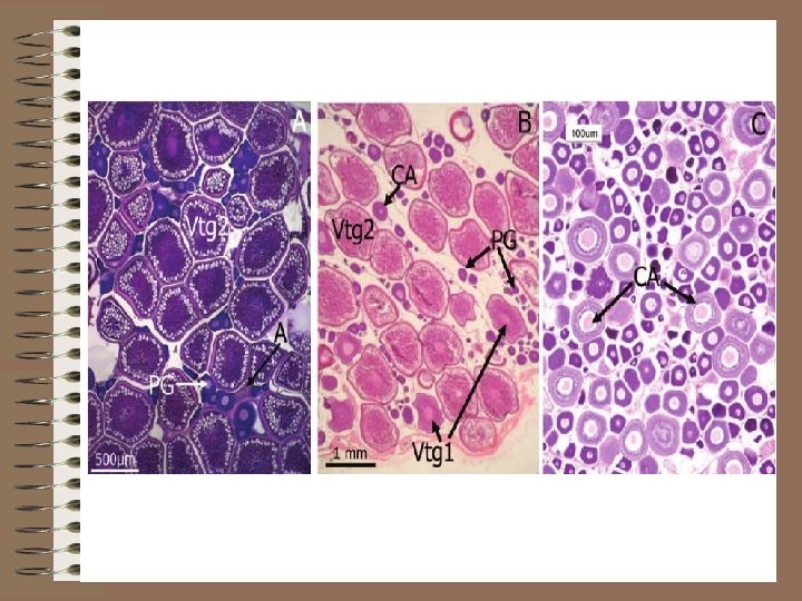
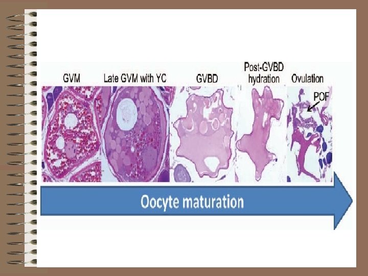
b. Parenchyma Ovigerous lamellae divide the ovary into compartments that contain oogonia and follicles in different developmental stages. These stages are; pre-vitellogenic, post-vitellogenic and atretic stages.
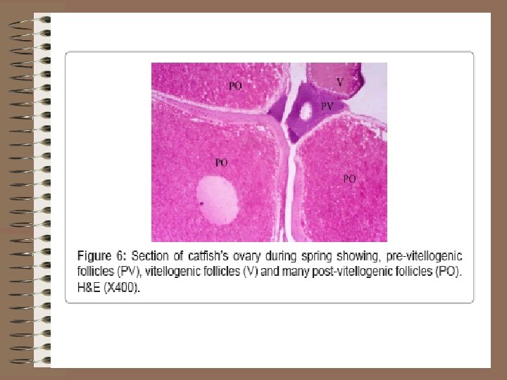

• The oogonia are the primordial cells that appear small spherical cells arranged in small groups around and in between the other developmental stages. • The pre-vitellogenic stage includes early and late oocytes. Both of them have basophilic cytoplasm due to that they have no yolk yet.

• The vitellogenic stage in which vitellogenesis begins to occur. • It includes early and late vacuolated follicles which are characterized by acidophilic cytoplasm due to the appearance of the yolk granules and droplets within the cytoplasm.

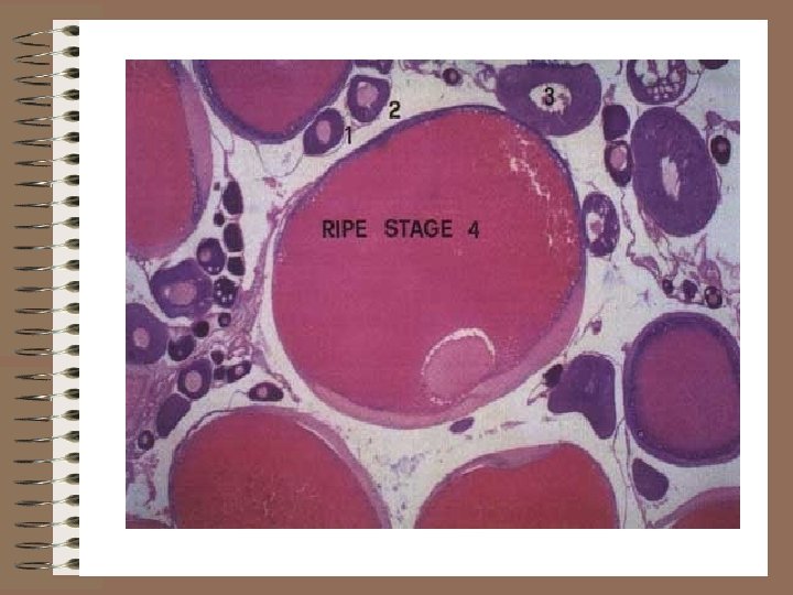
• They named vacuolated due to appearance of empty vacuoles of fat within their cytoplasm. • The pos-tvitellogenic stage includes the mature (ripe) follicles which are characterized by the migration of their nuclei from the center and the cytoplasm being full of large yolk globules.

• The atretic stage is that in which atresia (death of follicles before being mature) occur. • Atretic follicles are characterized by hypertrophy of the follicular epithelium with degeneration of the yolk beneath it. • Later on, macrophages are aggregates around the atretic follicles to phagocytize them.

Seasonal changes

Thank You
- Slides: 40