
Fedfast cycle Dr Sadia Haroon Git module lecture
- Slides: 59
Fed/fast cycle Dr Sadia Haroon Git module lecture 2019
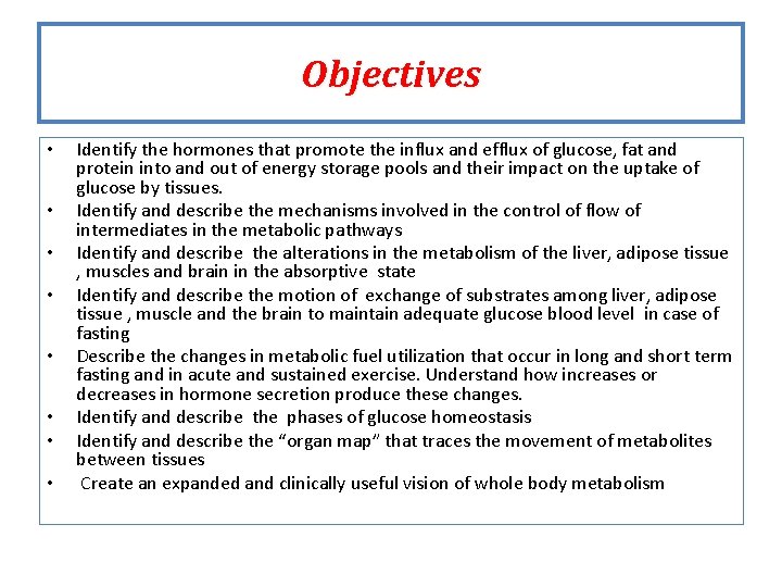
Objectives • • Identify the hormones that promote the influx and efflux of glucose, fat and protein into and out of energy storage pools and their impact on the uptake of glucose by tissues. Identify and describe the mechanisms involved in the control of flow of intermediates in the metabolic pathways Identify and describe the alterations in the metabolism of the liver, adipose tissue , muscles and brain in the absorptive state Identify and describe the motion of exchange of substrates among liver, adipose tissue , muscle and the brain to maintain adequate glucose blood level in case of fasting Describe the changes in metabolic fuel utilization that occur in long and short term fasting and in acute and sustained exercise. Understand how increases or decreases in hormone secretion produce these changes. Identify and describe the phases of glucose homeostasis Identify and describe the “organ map” that traces the movement of metabolites between tissues Create an expanded and clinically useful vision of whole body metabolism
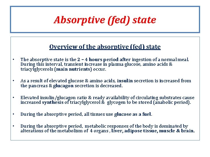
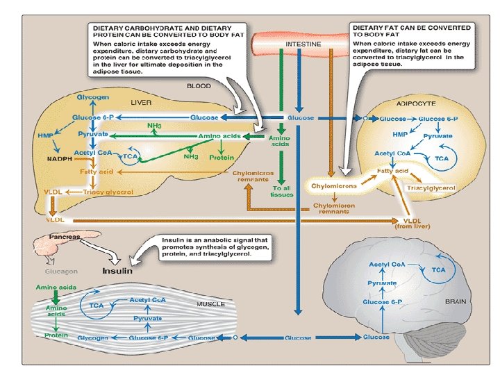
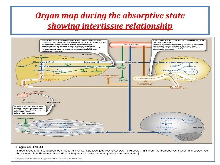
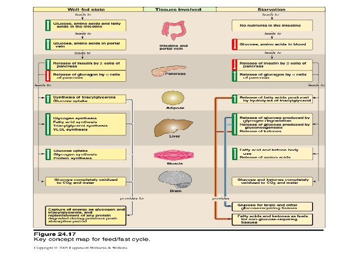
Absorptive (fed) state Overview of the absorptive (fed) state • The absorptive state is the 2 – 4 hours period after ingestion of a normal meal. During this interval, transient increase in plasma glucose, amino acids & triacylglycerols (main nutrients) nutrients occur. • As a result of elevated glucose & amino acids, insulin secretion is increased from the pancreas & glucagon secretion is decreased. • Elevated insulin/glucagon ratio & ready availability of circulating substrates cause increased synthesis of triacylglycerol & glycogen to be stored (anabolic period). • During the absorptive period, all tissues use glucose as a fuel • During the absorptive period, metabolic responses of the body is dominated by alterations of the metabolism of 4 organs , liver, adipose tissue, muscle & brain.
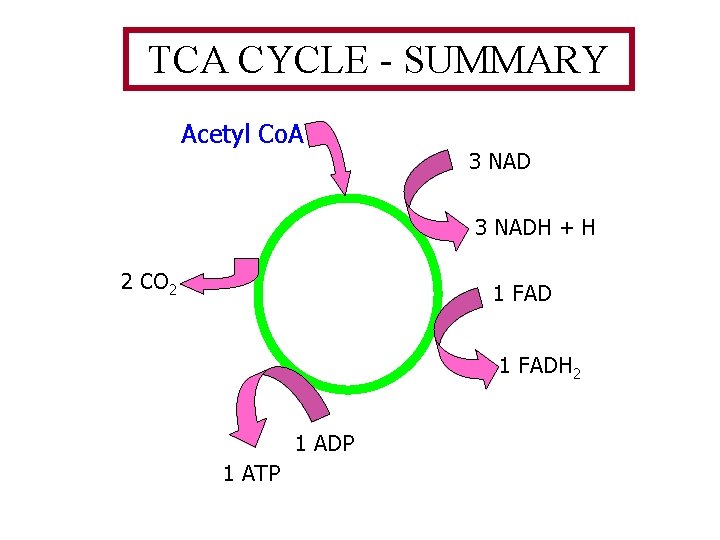
TCA CYCLE - SUMMARY Acetyl Co. A 3 NADH + H 2 CO 2 1 FADH 2 1 ADP 1 ATP
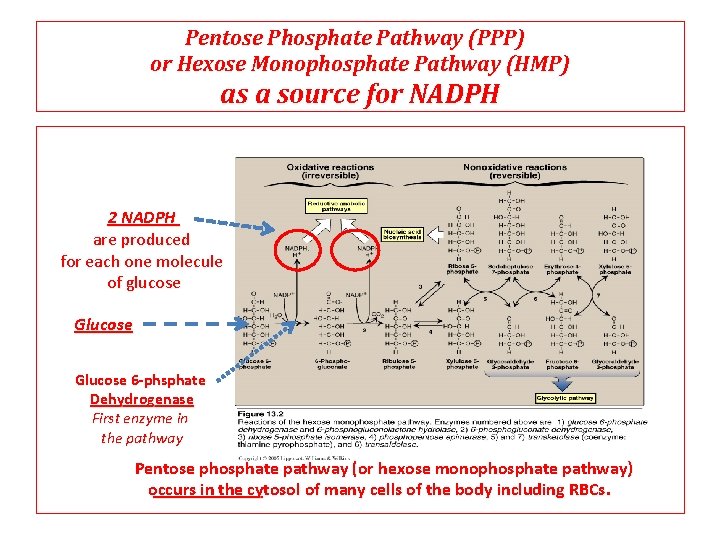
Pentose Phosphate Pathway (PPP) or Hexose Monophosphate Pathway (HMP) as a source for NADPH 2 NADPH are produced for each one molecule of glucose Glucose 6 -phsphate Dehydrogenase First enzyme in the pathway Pentose phosphate pathway (or hexose monophosphate pathway) occurs in the cytosol of many cells of the body including RBCs.
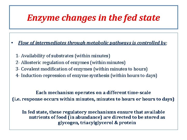
Enzyme changes in the fed state • Flow of intermediates through metabolic pathways is controlled by: by 1 - Availability of substrates (within minutes) 2 - Allosteric regulation of enzymes (within minutes) 3 - Covalent modification of enzymes (within minutes to hours) 4 - Induction-repression of enzyme synthesis (within hours to days) Each mechanism operates on a different time-scale (i. e. response occurs within minutes, minutes to hours or hours to days) In fed state, these regulatory mechanisms ensure that available nutrients of food (in abundance) are directed to be stored as glycogen, triacylglycerol & protein
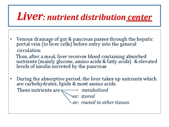
Liver: nutrient distribution center • Venous drainage of gut & pancreas passes through the hepatic portal vein (to liver cells) before entry into the general circulation Thus, after a meal, liver receives blood containing absorbed nutrients (mainly glucose, amino acids & fatty acids) & elevated levels of insulin secreted by the pancreas • During the absorptive period, the liver takes up nutrients which are carbohydrates, lipids & most amino acids. These nutrients are metabolized or: stored or: routed to other tissues
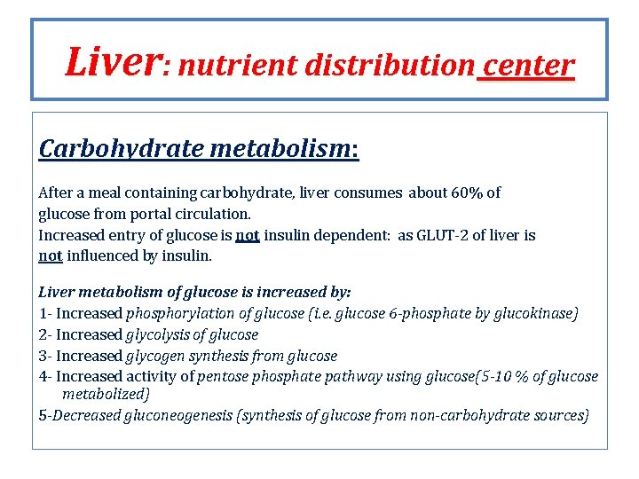
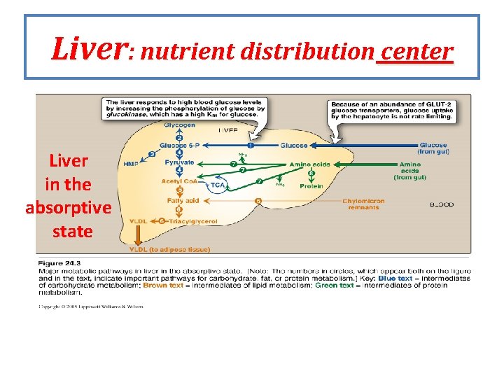
Liver: nutrient distribution center Carbohydrate metabolism: After a meal containing carbohydrate, liver consumes about 60% of glucose from portal circulation. Increased entry of glucose is not insulin dependent: as GLUT-2 of liver is not influenced by insulin. Liver metabolism of glucose is increased by: 1 - Increased phosphorylation of glucose (i. e. glucose 6 -phosphate by glucokinase) 2 - Increased glycolysis of glucose 3 - Increased glycogen synthesis from glucose 4 - Increased activity of pentose phosphate pathway using glucose(5 -10 % of glucose metabolized) 5 -Decreased gluconeogenesis (synthesis of glucose from non-carbohydrate sources)
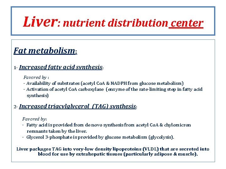
Liver: nutrient distribution center Fat metabolism: 1 - Increased fatty acid synthesis: Favored by : - Availability of substrates (acetyl Co. A & NADPH from glucose metabolism) - Activation of acetyl Co. A carboxylase (enzyme of the rate-limiting step in fatty acid synthesis) 2 - Increased triacylglycerol (TAG) synthesis: Favored by: by - Fatty acid is provided from de novo synthesis from acetyl Co. A & chylomicron remnants taken by the liver. - Glycerol 3 -phosphate is provided by glucose metabolism (glycolysis). Liver packages TAG into very-low density lipoproteins (VLDL) that are secreted into blood for use by extrahepatic tissues (particularly adipose & muscle).
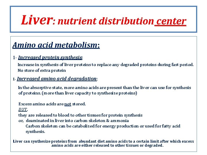
Liver: nutrient distribution center Amino acid metabolism: 1 - Increased protein synthesis: Increase in synthesis of liver proteins to replace any degraded proteins during fast period. No store of extra protein 1 - Increased amino acid degradation: degradation In the absorptive state, more amino acids are present than the liver can use for synthesis of proteins. (more than liver capacity to synthesize proteins) Excess amino acids are not stored. BUT, they are released to blood to other tissues for protein synthesis or, deaminated in liver into carbon skeleton & ammonia Carbon skeleton can be catabolized for energy production or used for fatty acid synthesis. Liver can synthesize proteins from abundant diet amino acids to a certain limit after which excess amino acids are either released to other tissues or degraded.
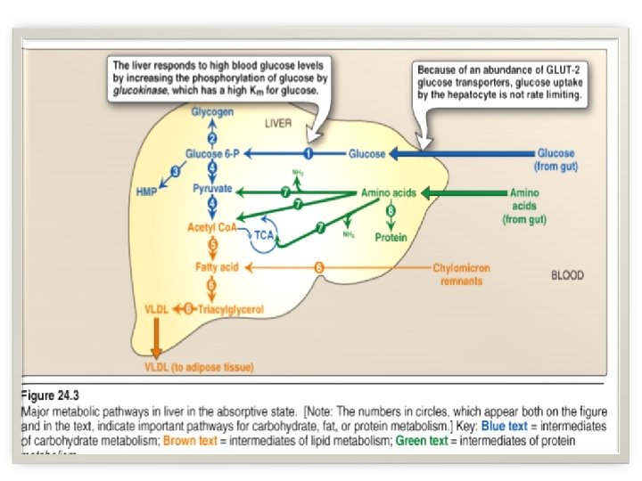
Liver: nutrient distribution center Liver in the absorptive state
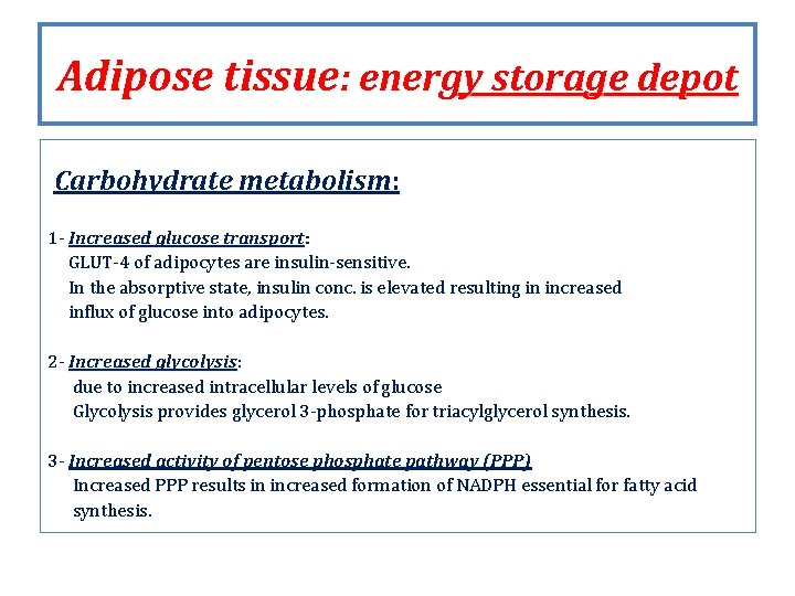
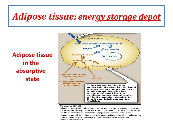
Adipose tissue: energy storage depot Carbohydrate metabolism: 1 - Increased glucose transport: transport GLUT-4 of adipocytes are insulin-sensitive. In the absorptive state, insulin conc. is elevated resulting in increased influx of glucose into adipocytes. 2 - Increased glycolysis: glycolysis due to increased intracellular levels of glucose Glycolysis provides glycerol 3 -phosphate for triacylglycerol synthesis. 3 - Increased activity of pentose phosphate pathway (PPP) Increased PPP results in increased formation of NADPH essential for fatty acid synthesis.
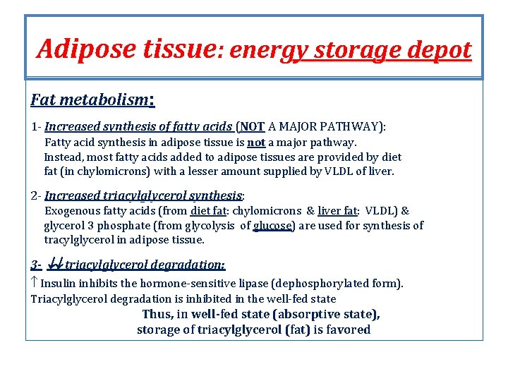
Adipose tissue: energy storage depot Fat metabolism: 1 - Increased synthesis of fatty acids (NOT A MAJOR PATHWAY): Fatty acid synthesis in adipose tissue is not a major pathway. Instead, most fatty acids added to adipose tissues are provided by diet fat (in chylomicrons) with a lesser amount supplied by VLDL of liver. 2 - Increased triacylglycerol synthesis: synthesis Exogenous fatty acids (from diet fat: chylomicrons & liver fat: VLDL) & glycerol 3 phosphate (from glycolysis of glucose) are used for synthesis of tracylglycerol in adipose tissue. 3 - triacylglycerol degradation: Insulin inhibits the hormone-sensitive lipase (dephosphorylated form). Triacylglycerol degradation is inhibited in the well-fed state Thus, in well-fed state (absorptive state), storage of triacylglycerol (fat) is favored
Adipose tissue: energy storage depot Adipose tissue in the absorptive state
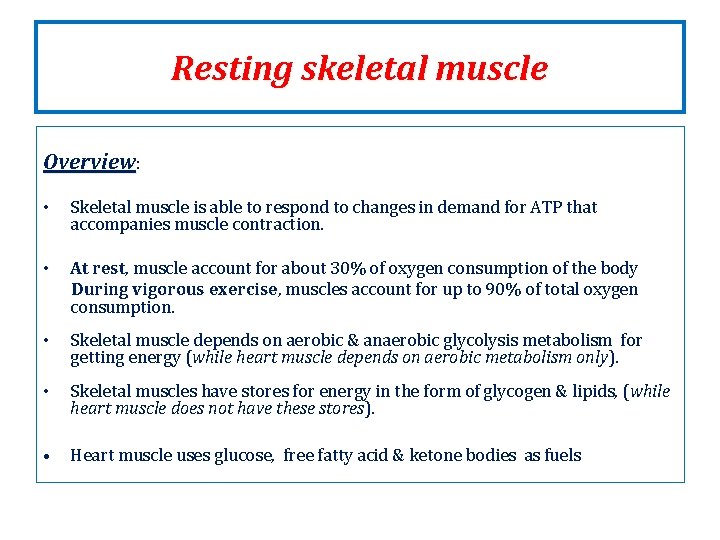
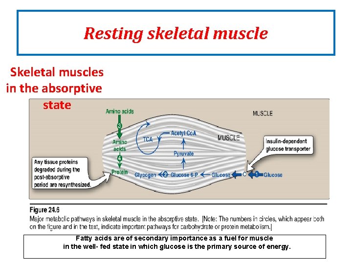
Resting skeletal muscle Overview: • Skeletal muscle is able to respond to changes in demand for ATP that accompanies muscle contraction. • At rest, rest muscle account for about 30% of oxygen consumption of the body During vigorous exercise, exercise muscles account for up to 90% of total oxygen consumption. • Skeletal muscle depends on aerobic & anaerobic glycolysis metabolism for getting energy (while heart muscle depends on aerobic metabolism only). • Skeletal muscles have stores for energy in the form of glycogen & lipids, (while heart muscle does not have these stores). • Heart muscle uses glucose, free fatty acid & ketone bodies as fuels
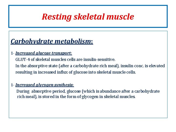
Resting skeletal muscle Carbohydrate metabolism: 1 - Increased glucose transport: GLUT-4 of skeletal muscles cells are insulin-sensitive. In the absorptive state (after a carbohydrate rich meal), insulin conc. is elevated resulting in increased influx of glucose into skeletal muscle cells. 1 - Increased glycogen synthesis: During absorptive period, glucose (which is abundance after a carbohydrate rich meal), is stored in the form of glycogen in skeletal muscles.
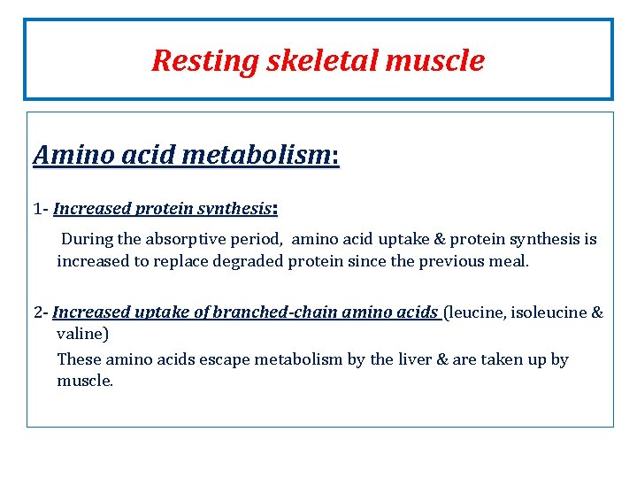
Resting skeletal muscle Amino acid metabolism: 1 - Increased protein synthesis: During the absorptive period, amino acid uptake & protein synthesis is increased to replace degraded protein since the previous meal. 2 - Increased uptake of branched-chain amino acids (leucine, isoleucine & valine) These amino acids escape metabolism by the liver & are taken up by muscle.
Resting skeletal muscle Skeletal muscles in the absorptive state Fatty acids are of secondary importance as a fuel for muscle in the well- fed state in which glucose is the primary source of energy.
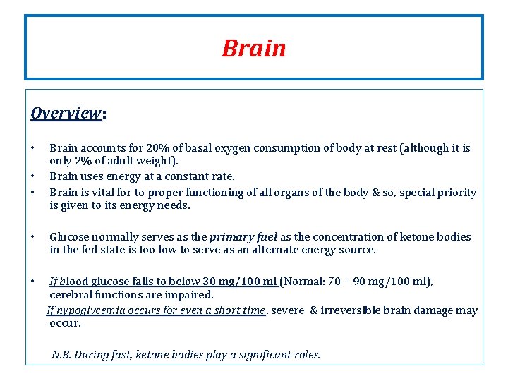
Brain Overview: • • • Brain accounts for 20% of basal oxygen consumption of body at rest (although it is only 2% of adult weight). Brain uses energy at a constant rate. Brain is vital for to proper functioning of all organs of the body & so, special priority is given to its energy needs. • Glucose normally serves as the primary fuel as the concentration of ketone bodies in the fed state is too low to serve as an alternate energy source. • If blood glucose falls to below 30 mg/100 ml (Normal: 70 – 90 mg/100 ml), cerebral functions are impaired. If hypoglycemia occurs for even a short time, time severe & irreversible brain damage may occur. N. B. During fast, ketone bodies play a significant roles.
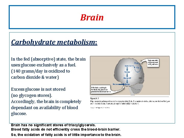
Brain Carbohydrate metabolism: In the fed (absorptive) state, the brain uses glucose exclusively as a fuel. (140 grams/day is oxidized to carbon dioxide & water) Excess glucose is not stored (no glycogen stores). Accordingly, the brain is completely dependant on availability of blood glucose. Brain has no significant stores of triacylglycerols. Blood fatty acids do not efficiently cross the blood-brain barrier. So, the oxidation of fatty acids is of little importance to the brain.
Organ map during the absorptive state showing intertissue relationship
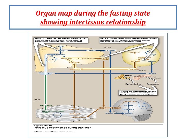
Fasting Overview of fasting Fasting may result from: from - Ramadan fasting for Muslims - Inability to obtain food - Desire to lose weight rapidly - Clinical situations in which an individual cannot eat (trauma, surgery , etc. . ) In absence of food, plasma levels of glucose, amino acids & triacylglycerol (main nutrients) fall with a resulting decline in insulin secretion & increase in glucagon release. The decreased insulin/glucagon ratio & decreased availability of circulating substrates, favors a catabolic period in which degradation of triacylglycerol, glycogen & protein is characteristic. Exchange of substrates between liver, adipose tissue, muscle & brain is guided by two priorities: priorities 1 - Need to maintain adequate plasma levels of glucose to secure energy metabolism to brain, RBCs & other tissues utilizing glucose as sole fuel. 2 - Need to mobilize fatty acids from adipose tissue, synthesis & release of ketone bodies to supply energy to other tissues.
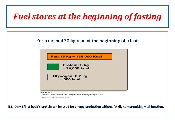
Fuel stores at the beginning of fasting For a normal 70 kg man at the beginning of a fast: N. B. Only 1/3 of body`s protein can be used for energy production without fatally compromising vital function.
Enzymatic changes in fasting • Flow of intermediates through metabolic pathways is controlled by: by 1 - Availability of substrates (within minutes) 2 - Allosteric regulation of enzymes (within minutes) 3 - Covalent modification of enzymes (within minutes to hours) 4 - Induction-repression of enzyme synthesis (within hours to days) The metabolic changes in fasting are generally opposite to those in fed state. In fasting, substrates are not provided by diet, but are available from the breakdown of tissues stores (e. g. lipolysis: lipolysis with release of fatty acids & glycerol from adipose tissue & proteolysis with release of amino acids from muscles.
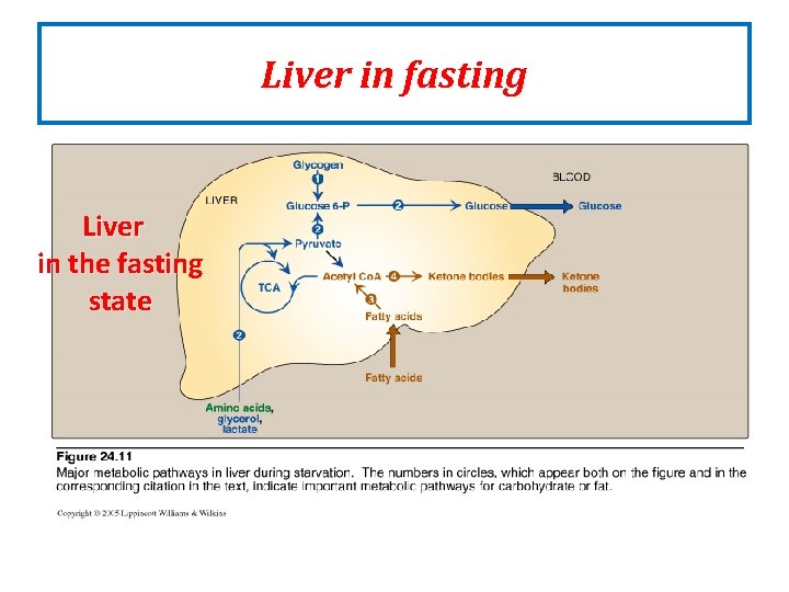
Liver in fasting Overview The primary role of liver in energy metabolism during fasting is maintaining of blood glucose through production & release of energy molecules for use by other organs.
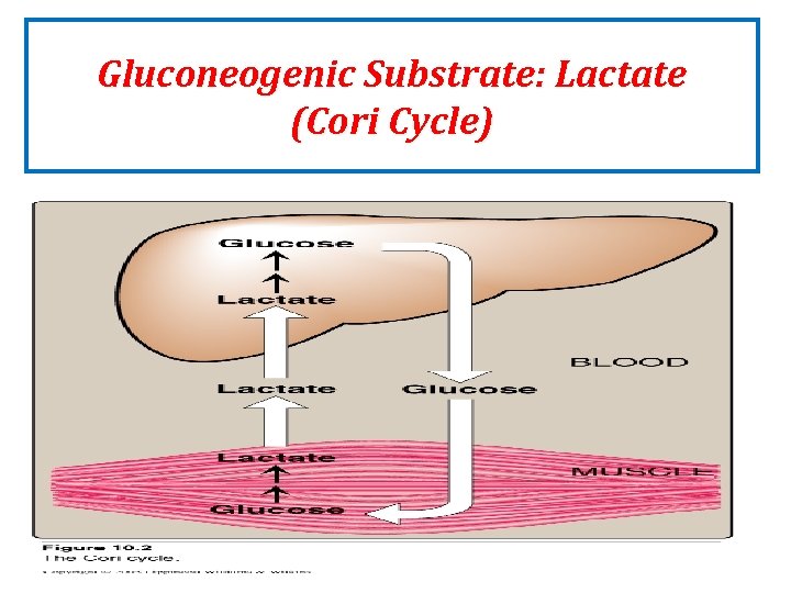
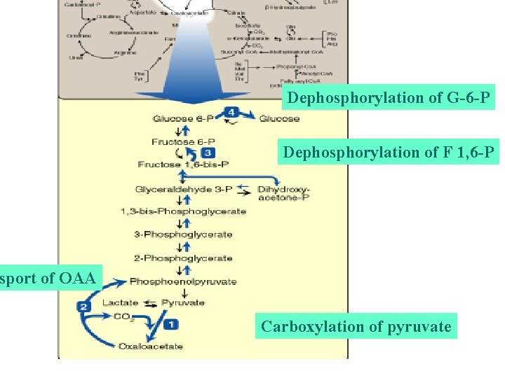
Liver in fasting Carbohydrate metabolism: In liver during fasting, glycogen is degraded first (10 -18 hrs of fasting) & then gluconeogenesis (after 18 hrs to secure glucose to brain & other tissues utilizing glucose as a sole fuel. 1 - Increased glycogen degradation (glycogenlysis) to produce glucose to blood : exhausted after 10 – 18 hours of fasting (early fasting). 2 - Increased gluconeogenesis: gluconeogenesis Gluconeogenesis is the synthesis of glucose from non-carbohydrate sources: amino acids & lactate from muscles & glycerol from adipose, plays an essential role during overnight & prolonged fasting. Gluconeogenesis begins 4 - 6 hours after the last meal & becomes fully active when stores of glycogen are depleted.
Gluconeogenic Substrate: Lactate (Cori Cycle)
Liver in fasting Fat metabolism: 1 - Increased fatty acid oxidation: Fatty acids obtained from adipose tissue is the major source of energy to liver during the fasting state. 2 - Increased synthesis of ketone bodies: bodies The liver can synthesize & release ketone bodies (only tissue) for use as a fuel by peripheral tissues (BUT: liver cannot use ketone bodies as a fuel). Ketone bodies formation is favored by the availability of fatty acids obtained from adipose tissue (fatty acids are degraded to acetyl Co. A, the precursor of ketone bodies). In this case, acetyl Co. A produced from fatty acids exceeds the capacity of citric acid cycle. Significant synthesis of ketone bodies starts during the first days of fasting. Ketone bodies are water-soluble & appears in blood & urine by the second day of a fast. Ketone bodies in blood during fasting is important as they can be used as fuel for most tissues including the brain tissue. This reduces the need for gluconeogenesis from amino acids & thus slowing the loss of essential protein.
Liver in fasting Liver in the fasting state
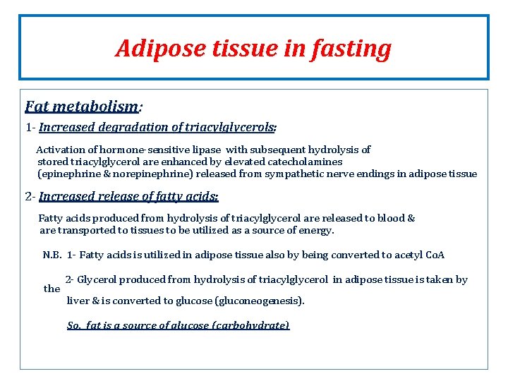
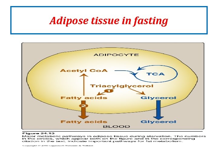
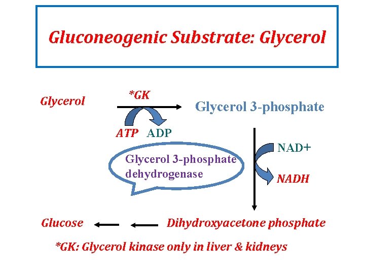
Adipose tissue in fasting Fat metabolism: 1 - Increased degradation of triacylglycerols: Activation of hormone-sensitive lipase with subsequent hydrolysis of stored triacylglycerol are enhanced by elevated catecholamines (epinephrine & norepinephrine) released from sympathetic nerve endings in adipose tissue 2 - Increased release of fatty acids: Fatty acids produced from hydrolysis of triacylglycerol are released to blood & are transported to tissues to be utilized as a source of energy. N. B. 1 - Fatty acids is utilized in adipose tissue also by being converted to acetyl Co. A the 2 - Glycerol produced from hydrolysis of triacylglycerol in adipose tissue is taken by liver & is converted to glucose (gluconeogenesis). So, fat is a source of glucose (carbohydrate)
Adipose tissue in fasting
Gluconeogenic Substrate: Glycerol *GK Glycerol 3 -phosphate ATP ADP Glycerol 3 -phosphate dehydrogenase Glucose NAD+ NADH Dihydroxyacetone phosphate *GK: Glycerol kinase only in liver & kidneys
Dephosphorylation of G-6 -P Dephosphorylation of F 1, 6 -P sport of OAA Carboxylation of pyruvate
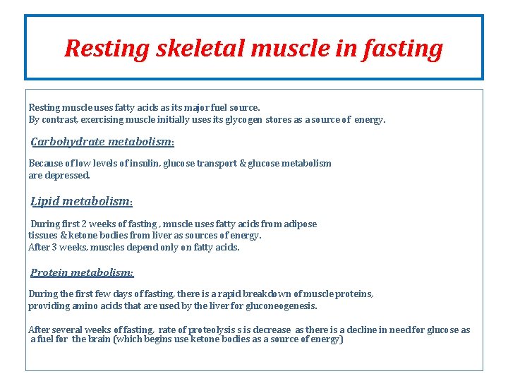
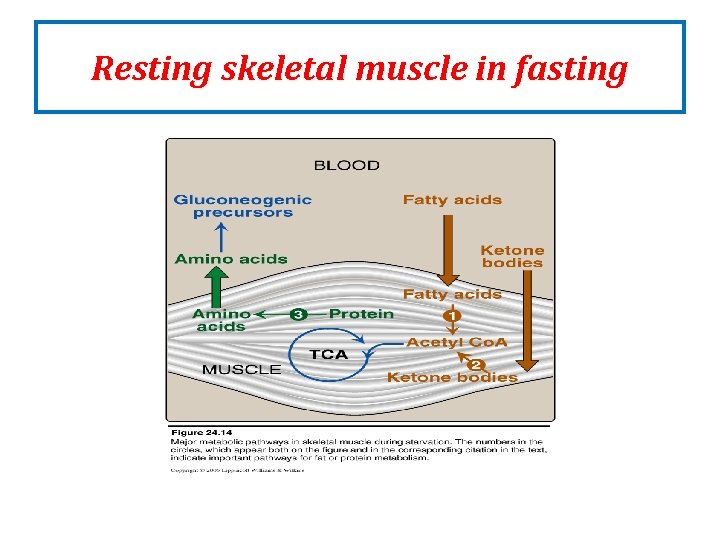
Resting skeletal muscle in fasting Resting muscle uses fatty acids as its major fuel source. By contrast, exercising muscle initially uses its glycogen stores as a source of energy. Carbohydrate metabolism: Because of low levels of insulin, glucose transport & glucose metabolism are depressed. Lipid metabolism: During first 2 weeks of fasting , muscle uses fatty acids from adipose tissues & ketone bodies from liver as sources of energy. After 3 weeks, muscles depend only on fatty acids. Protein metabolism: During the first few days of fasting, there is a rapid breakdown of muscle proteins, providing amino acids that are used by the liver for gluconeogenesis. After several weeks of fasting, rate of proteolysis s is decrease as there is a decline in need for glucose as a fuel for the brain (which begins use ketone bodies as a source of energy)
Resting skeletal muscle in fasting
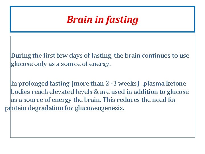
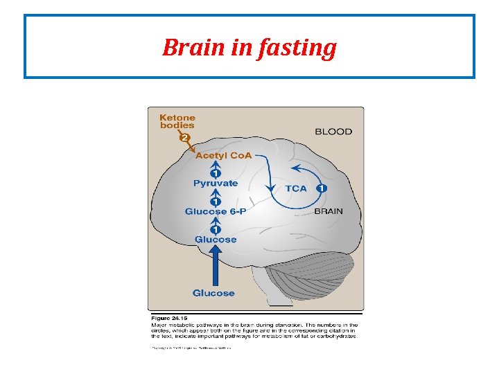
Brain in fasting During the first few days of fasting, the brain continues to use glucose only as a source of energy. In prolonged fasting (more than 2 -3 weeks) , plasma ketone bodies reach elevated levels & are used in addition to glucose as a source of energy the brain. This reduces the need for protein degradation for gluconeogenesis.
Brain in fasting
Organ map during the fasting state showing intertissue relationship
Glucose homeostasis A process that • Controls glucose metabolism and • Maintains blood glucose level in the body Glucose is a major source of body’s energy The liver plays a key role in maintaining blood glucose level Blood glucose level is tightly controlled because the brain constantly needs glucose Severe hypoglycemia can cause coma and death Chronic hyperglycemia results in glycation of proteins, endothelial dysfunction and diabetes
Sources of glucose Dietary sources: e. g. Starch provides glucose directly Hexose intake are converted to glucose in the liver ( fructose ) Metabolic sources (via gluconeogenesis): Glycerol, glucogenic amino acids, lactate
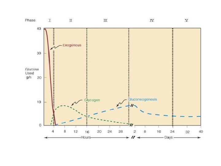
Phases of glucose homeostasis Five phases: Phase I (Well-fed state) Phase II (Glycogenolysis) Phase III (Gluconeogenesis) Phase IV (Glucose, ketone bodies (KB) oxidation) Phase V (Fatty acid (FA), KB oxidation)
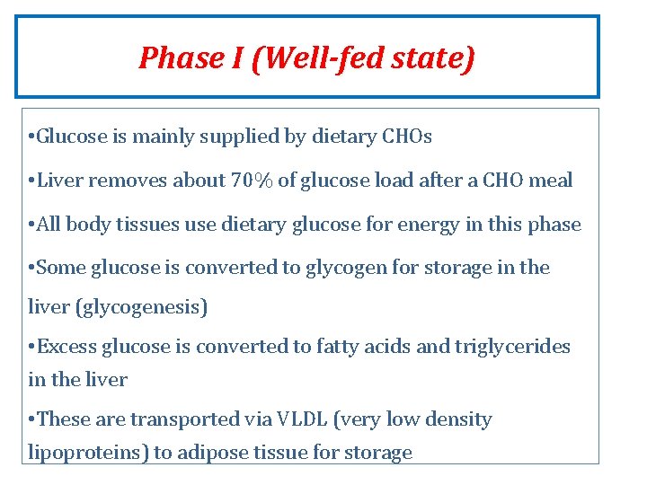
Phase I (Well-fed state) • Glucose is mainly supplied by dietary CHOs • Liver removes about 70% of glucose load after a CHO meal • All body tissues use dietary glucose for energy in this phase • Some glucose is converted to glycogen for storage in the liver (glycogenesis) • Excess glucose is converted to fatty acids and triglycerides in the liver • These are transported via VLDL (very low density lipoproteins) to adipose tissue for storage
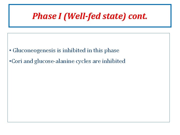
Phase I (Well-fed state) cont. • Gluconeogenesis is inhibited in this phase • Cori and glucose-alanine cycles are inhibited
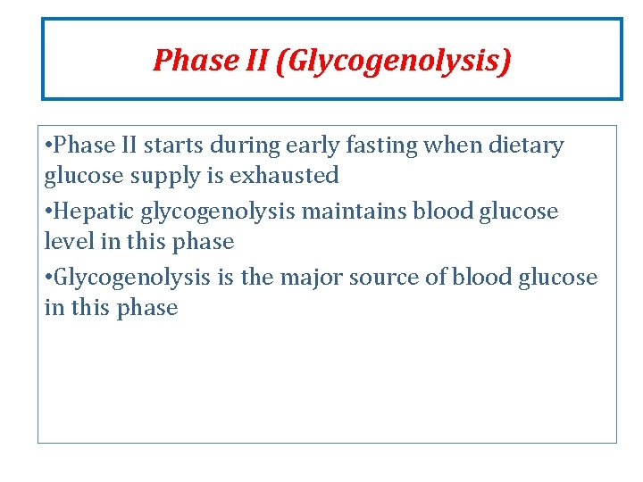
Phase II (Glycogenolysis) • Phase II starts during early fasting when dietary glucose supply is exhausted • Hepatic glycogenolysis maintains blood glucose level in this phase • Glycogenolysis is the major source of blood glucose in this phase
Phase III (Gluconeogenesis) Phase III starts when glycogen stores in liver are exhausted (within 18 hours) Duration of phase III depends on Feeding status Hepatic glycogen stores Physical activity Hepatic gluconeogenesis from lactate, pyruvate, glycerol and alanine maintains blood glucose level Gluconeogenesis is the major source of blood glucose in this phase
Phase IV (Glucose and KB oxidation) Several days of fasting leads to phase IV Gluconeogenesis starts to decrease KB accumulation increases which enter the brain for energy production Brain uses both glucose and KB for energy
Phase V (FA and KB oxidation) Prolonged fasting leads to phase V Less dependence on gluconeogenesis All body tissues use FA and KB oxidation for energy production Gluconeogenesis somewhat maintains blood glucose level in this phase
Phase V (FA and KB oxidation) • High KB conc. and glucose levels inhibit proteolysis in muscle (conservation of muscle) • When all fat and KBs are used up • Body uses muscle protein to maintain blood glucose level
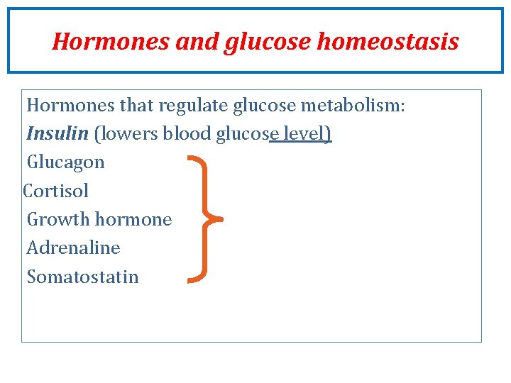
Hormones and glucose homeostasis Hormones that regulate glucose metabolism: Insulin (lowers blood glucose level) Glucagon Cortisol Growth hormone Adrenaline Somatostatin
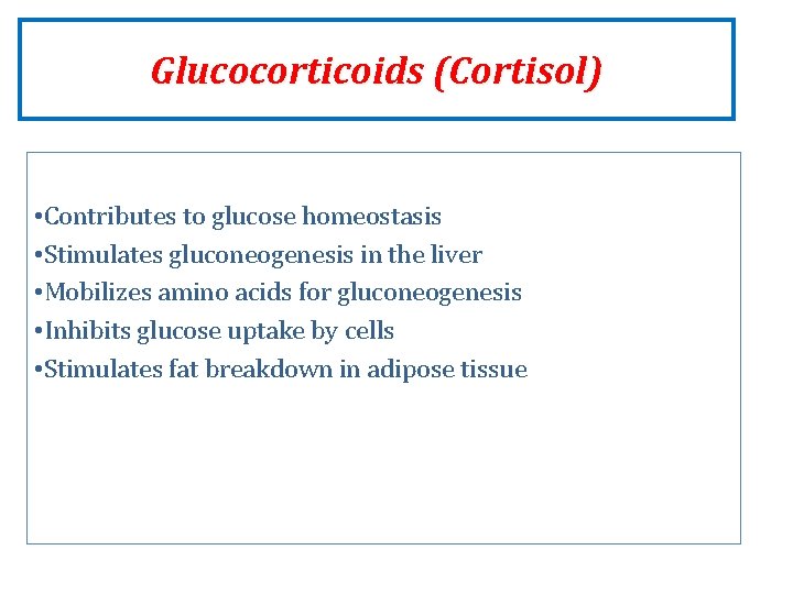
Glucocorticoids (Cortisol) • Contributes to glucose homeostasis • Stimulates gluconeogenesis in the liver • Mobilizes amino acids for gluconeogenesis • Inhibits glucose uptake by cells • Stimulates fat breakdown in adipose tissue
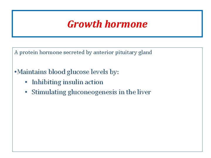
Growth hormone A protein hormone secreted by anterior pituitary gland • Maintains blood glucose levels by: • Inhibiting insulin action • Stimulating gluconeogenesis in the liver
Epinephrine A catecholamine hormone secreted by adrenal gland • Stimulates lipolysis in adipose tissue when glucose blood levels fall • Promotes glycogenolysis in skeletal muscle
Somatostatin A peptide hormone secreted by δ-cells of pancreatic islets, stomach and intestine • An inhibitory hormone • Inhibits secretion of both insulin and glucagon • Affects glucose homeostasis indirectly
References Lippincott’s Illustrated Biochemistry Textbook of biochemistry with clinical correlation by Devlin