
February EMS Training AV Blocks Pacing Used with
February EMS Training: AV Blocks & Pacing Used with permission of Silver Cross EMS System
Goals • Review different heart blocks found when completing an EKG • Identify how to differentiate between different heart blocks • Review transcutaneous pacing equipment and how to pace a patient • Identify any other relevant treatments for patients with a heart block 2
Cardiac Rhythm of the Month • AV Blocks 3
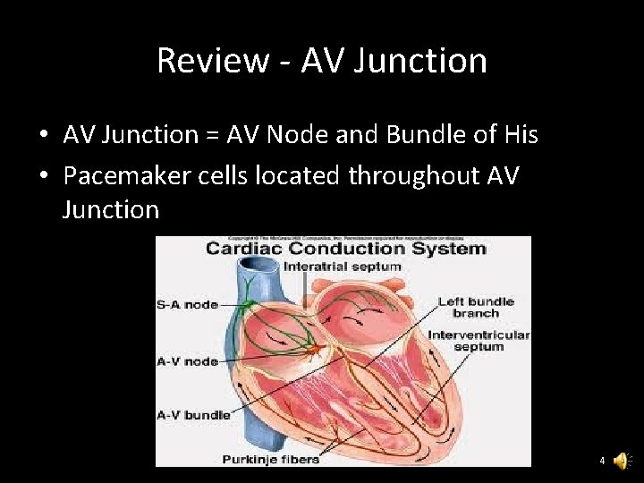
Review - AV Junction • AV Junction = AV Node and Bundle of His • Pacemaker cells located throughout AV Junction 4
Review - Functions of AV Node • Backup pacemaker for SA Node • Creates delay between atrial and ventricular depolarizations • Physiologic block for rapid supraventricular rhythms 5
Degrees of AV Blocks • First Degree - Delay in conduction • Second Degree - Some impulses blocked • Third Degree - All impulses blocked 6
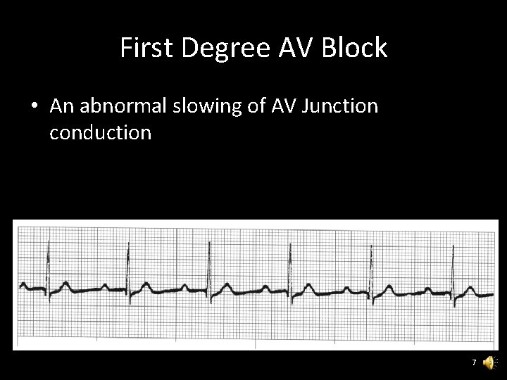
First Degree AV Block • An abnormal slowing of AV Junction conduction 7
First Degree AV Block ECG Criteria • Rate - Dependent on underlying rhythm – Interpretation must include underlying rhythm • Rhythm - Dependent on underlying rhythm • P-Waves - Normal morphology with one PWave for each QRS • PRI - >. 20 seconds and constant • QRS - Dependent on underlying rhythm 8
First Degree AV Block Clinical Significance • Not usually detrimental and often resolves when ischemia corrected • Must consider entire patient 9
Second Degree AV Blocks • Type I – Also called “Wenckebach” – Also called Mobitz I • Type II – Also called Mobitz II 10
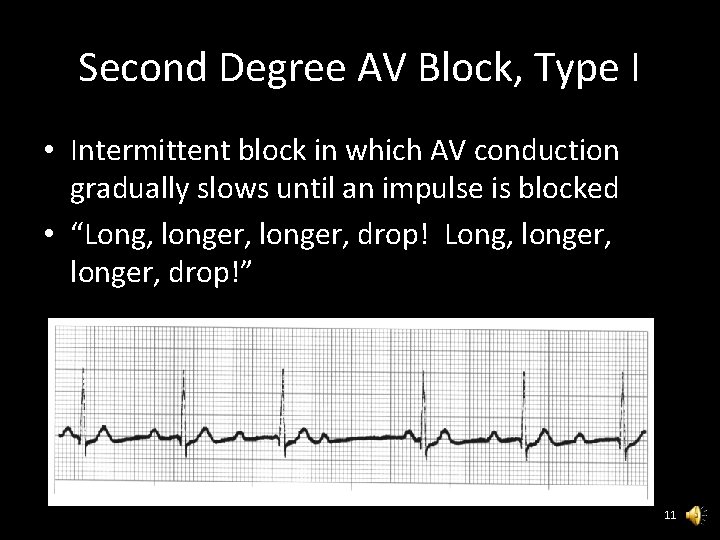
Second Degree AV Block, Type I • Intermittent block in which AV conduction gradually slows until an impulse is blocked • “Long, longer, drop!” 11
Second Degree AV Block, Type I ECG Criteria Rate - Atrial rate unaffected but ventricular rate is less than atrial rate Rhythm - Atrial rhythm usually regular. Ventricular rhythm is irregular with more PWaves than QRS Complexes. P-Waves - Unaffected with more P-Waves than QRS Complexes PRI - Progressively increases for consecutively conducted P-Waves until QRS Complex is dropped QRS - Unaffected 12
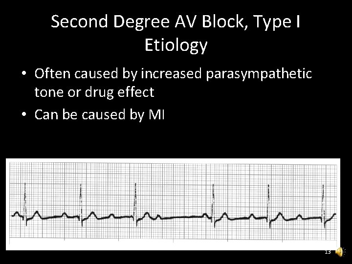
Second Degree AV Block, Type I Etiology • Often caused by increased parasympathetic tone or drug effect • Can be caused by MI 13
Second Degree AV Block, Type I Clinical Significance • Usually transient with good prognosis • Can reduce cardiac output due to bradycardia 14
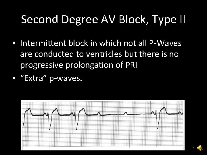
Second Degree AV Block, Type II • Intermittent block in which not all P-Waves are conducted to ventricles but there is no progressive prolongation of PRI • “Extra” p-waves. 15
Second Degree AV Block, Type II Etiology • Usually due to MI or other organic heart disease • Rarely the result of increased parasympathetic tone or drug effect 16
Second Degree AV Block, Type II Clinical Significance • Poorer prognosis than Type I • Usually requires pacemaker • Frequently develops into Complete Block 17
Second Degree AV Block, Type II ECG Criteria Rate - Atrial rate is unaffected but ventricular rate is less than atrial Rhythm - Atrial rhythm regular, Ventricular irregular with more P-waves than QRS Complexes P-Waves - Normal morphology with more PWaves than QRS Complexes PRI - Constant for consecutively conducted PWaves QRS - Usually wide but may be narrow if block is at His level or above 18
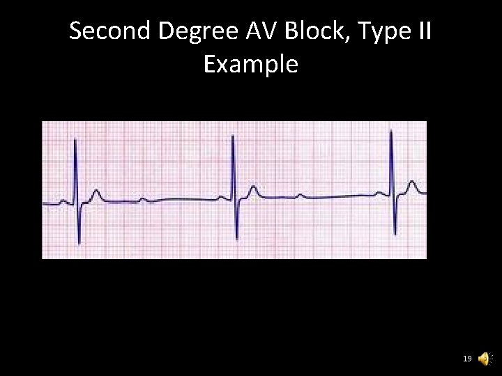
Second Degree AV Block, Type II Example 19
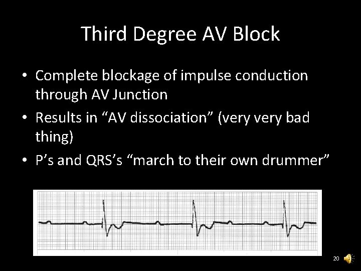
Third Degree AV Block • Complete blockage of impulse conduction through AV Junction • Results in “AV dissociation” (very bad thing) • P’s and QRS’s “march to their own drummer” 20
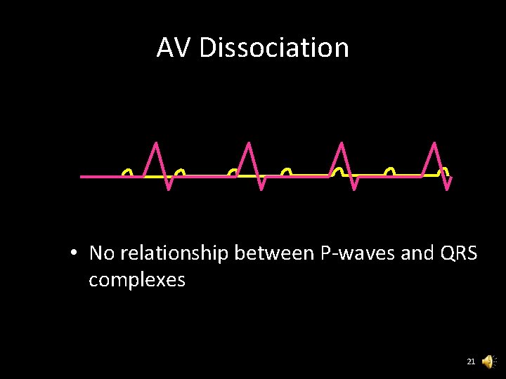
AV Dissociation • No relationship between P-waves and QRS complexes 21
Third Degree AV Block Etiology • MI • Increased parasympathetic tone • Drug toxicity 22
Third Degree AV Block ECG Criteria Rate - Atrial > 60, Ventricular based on escape Rhythm - Atrial and ventricular regular P-Waves - Normal PRI - No association between P-Waves and QRS complexes (P’s and QRS’s are divorced and do their own thing) • QRS - Narrow if intranodal, Wide if infranodal • • 23
Transcutaneous Pacing (TCP) • Non-invasive electrical therapy for symptomatic bradycardias/complete heart blocks • Fast to set up • Reasonably reliable 24
TCP Equipment • Give the patient Versed if they are awake, per SMO • Set milliamps (adjustable 0 -200 m. A typical) – Start low if they are awake, and high if they are out. • Set rate to 70. • Similar controls across brands • Be familiar with your equipment! 25
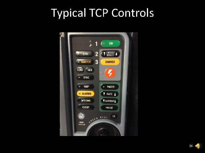
Typical TCP Controls 26
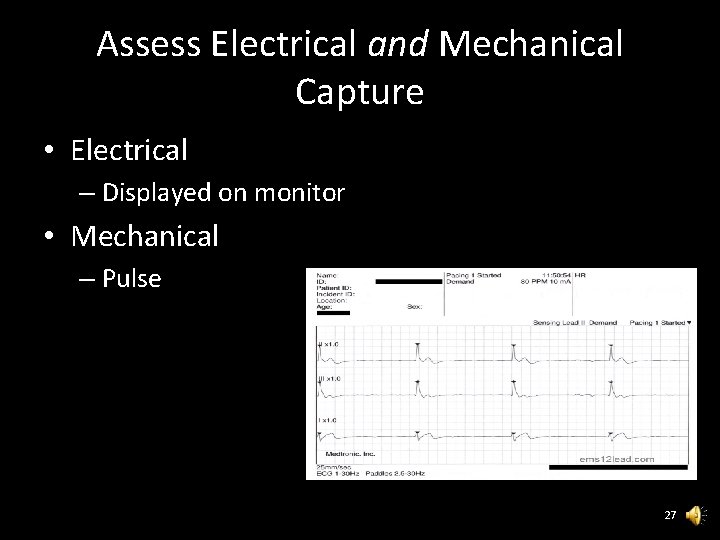
Assess Electrical and Mechanical Capture • Electrical – Displayed on monitor • Mechanical – Pulse 27
- Slides: 27