Fascicular blocks in the Left Ventricle Left interruption


Fascicular blocks in the Left Ventricle Left, interruption of the LAD results in an initial inferior (1) followed by a dominant superior (2) direction of activation. Right, interruption of the LPD results in an initial superior (1) followed by a dominant inferior (2) direction of activation
 - Degree Atrioventricular Block • During 1 st degree AV block,")
First (1 st) - Degree Atrioventricular Block • During 1 st degree AV block, every atrial impulse is conducted to the ventricles and a regular ventricular rate is produced, but the PR interval exceeds 0. 20 second in adults. • PR intervals as long as 1. 0 second have been noted and can at times exceed the P-P interval, a phenomenon known as skipped P waves. • Clinically important PR interval prolongation can result from a conduction delay in the AV node (A-H interval), in the His-Purkinje system (H-V interval), or at both sites.
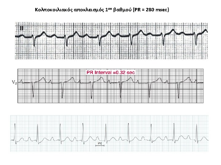

Marked 1 st degree heart block (PR interval > 300 ms, P waves are buried in the preceding T wave)
 - Degree Atrioventricular Block • Electrocardiographically, typical type I second-degree AV")
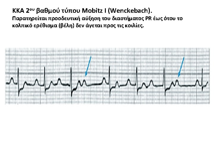
Second (2 nd) - Degree Atrioventricular Block • Electrocardiographically, typical type I second-degree AV block is characterized by progressive PR prolongation culminating in a non-conducted P wave, whereas in type II second-degree AV block, the PR interval remains constant before the blocked P wave. • In both cases the AV block is intermittent and generally repetitive and can block several P waves in a row. • Frequently, the eponyms Mobitz type I and Mobitz type II are applied to the two types of block, whereas Wenckebach block refers to type I block only.

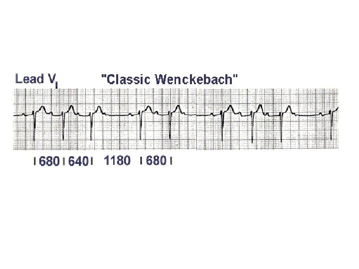
Mobitz type I 2 nd-degree atrioventricular block The PR interval progressively lengthens with each beat until the atrial impulse is not conducted and the QRS complex is dropped (Wenckebach phenomenon); AV nodal conduction resumes with the next beat, and the sequence is repeated.

Causes of Mobitz II • • Anterior MI (due to septal infarction with necrosis of the bundle branches). Idiopathic fibrosis of the conducting system (Lenegre’s or Lev’s disease). Cardiac surgery (especially surgery occurring close to the septum, e. g. mitral valve repair) Inflammatory conditions (rheumatic fever, myocarditis, Lyme disease). Autoimmune (SLE, systemic sclerosis). Infiltrative myocardial disease (amyloidosis, haemochromatosis, sarcoidosis). Hyperkalaemia. Drugs: beta-blockers, calcium channel blockers, digoxin, amiodarone. Clinical Significance • • Mobitz II is much more likely than Mobitz I to be associated with hemodynamic compromise, severe bradycardia and progression to 3 rd degree heart block. Onset of hemodynamic instability may be sudden and unexpected, causing syncope (Stokes-Adams attacks) or sudden cardiac death. The risk of asystole is around 35% per year. Mobitz II mandates immediate admission for cardiac monitoring, backup temporary pacing and ultimately insertion of a permanent pacemaker.

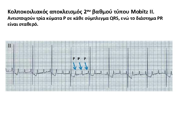
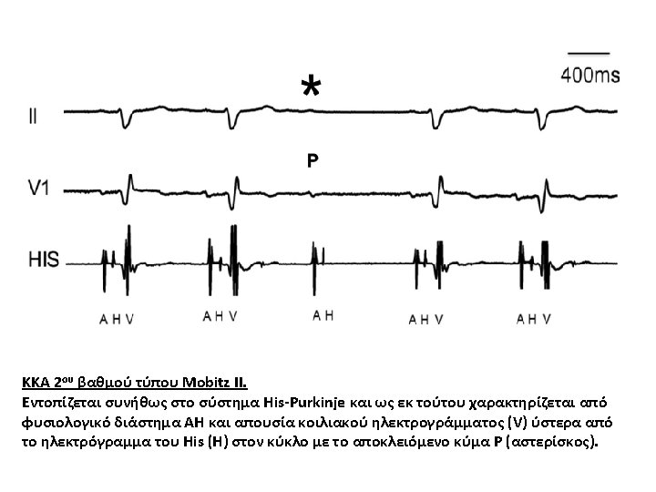
Mobitz type II 2 nd-degree atrioventricular block The PR interval remains constant. Beats are intermittently nonconducted, and QRS complexes dropped, usually in a repeating cycle of every 3 rd (3: 1 block) or 4 th (4: 1 block) P wave.
 • • Intermittent non-conducted")
Arrows indicate “dropped” QRS complexes (i. e. non-conducted P waves) • • Intermittent non-conducted P waves without progressive prolongation of the PR interval (compare this to Mobitz I). The PR interval in the conducted beats remains constant. The P waves ‘march through’ at a constant rate. The RR interval surrounding the dropped beat(s) is an exact multiple of the preceding RR interval (e. g. double the preceding RR interval for a single dropped beat, treble for two dropped beats, etc).
. In high-grade 2 nd-degree AV block, every 2 nd")
Second-degree atrioventricular block (high grade). In high-grade 2 nd-degree AV block, every 2 nd (or more) P wave is blocked • High-grade AV block (4: 1 conduction ratio). • Atrial rate is approximately 140 bpm. • Ventricular rate is approximately 35 bpm.

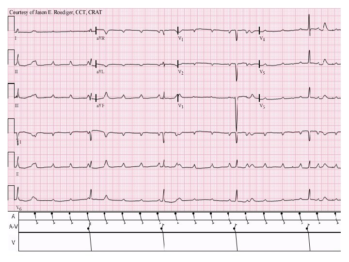
Sinus rhythm at 95 b/min with 2: 1 AV block
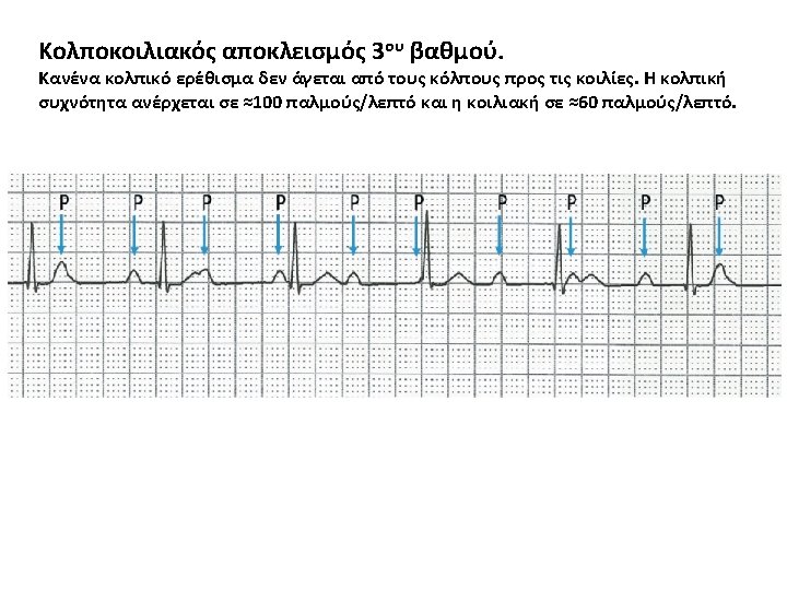

• • • The atrial rate is approximately 100 bpm. The ventricular rate is approximately 40 bpm. The two rates are independent; there is no evidence that any of the atrial impulses are conducted to the ventricles.

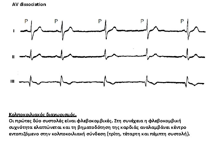
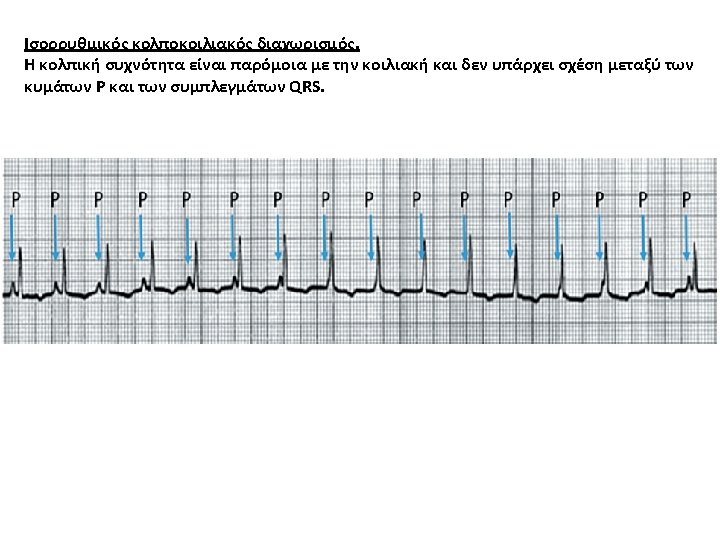
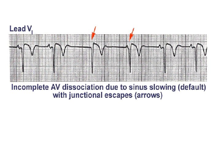
AV dissociation
- Slides: 34