Facial Bone Anatomy Positioning RTEC 233 Anterior Aspect

Facial Bone Anatomy & Positioning RTEC 233

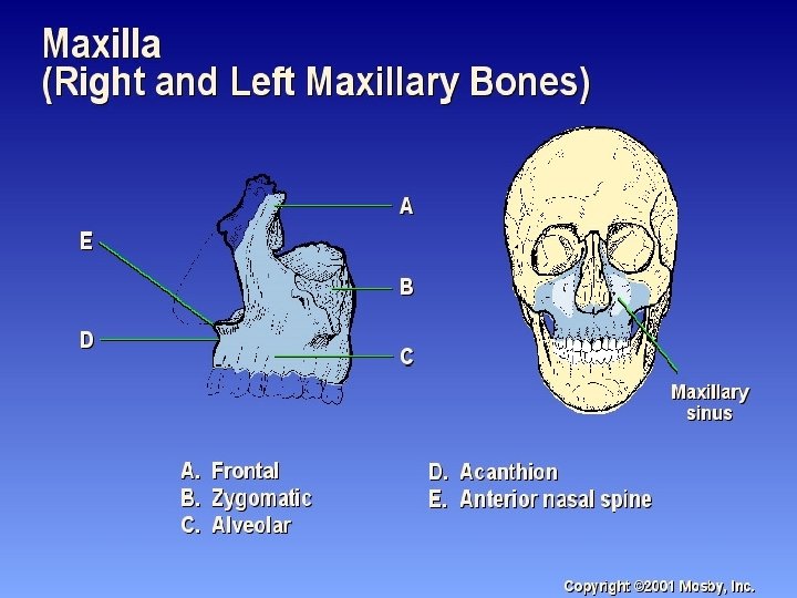
Anterior Aspect of Facial Bones n 2 Maxillae n 2 Zygomatic bones n 2 Lacrimal bones n 2 Nasal bones n 2 Inferior nasal conchae n 2 Palatine bones (not visualized n 1 vomer n 1 mandible

Palatine Bones n L-shaped bones n Horizontal portion forms posterior hard palate n Vertical portion extends between 1 maxillae and 1 pterygoid plate of sphenoid bone Articulates with 2 cranial bones and 4 facial bones n

Zygomatic Bones n n n Forms cheeks Forms lower outer margin of orbits Articulates with 3 cranial bones n n Frontal Sphenoid Temporal Articulates with maxillae

Inferior Nasal Cochae n The only pair of conchae that are separate facial bones n Articulates with 1 cranial bone and 3 facial bones n Covered with mucous membranes to warm, moisten and cleanse inhaled air

Lacrimal Bones n About the size & shape of a fingernail n Lacrimal foramen for tear duct n Lie anteriorly on the medial side of orbit n Can be seen on PA and lateral projections n Articulates with 2 cranial bones and 2 facial bones

n Nasal Bones Fused and form Nasal bridge of nose n Vary in size considerably n The point of junction with the frontal bone is the nasion n Articulates with 2 cranial and 2 facial bones

Vomer n Forms inferosuperior part of nasal septum n Deviated nasal septum n Depressions for blood vessels n Articulates with 2 cranial bones & 4 facial bones

Mandible n Only movable bone in the skull n Densest & largest facial bone n 2 bones at birth n Contains mental foramina

Pathologic Indications for Facial Radiography n Fractures n n Blowout Tripod Le. Fort Coutrecoup n Foreign Body n Osteomyelitis n Neoplasms n Secondary Osteomyelitis n TMJ Syndrome

Tri-pod Fracture

Blow out fracture

Le. Fort Fractures

FIG 3 - Le. Fort lines used for classifying fractures of the middle third of the face. Hodgkinson, D W et al. BMJ 1994; 308: 46 -50 Copyright © 1994 BMJ Publishing Group Ltd.

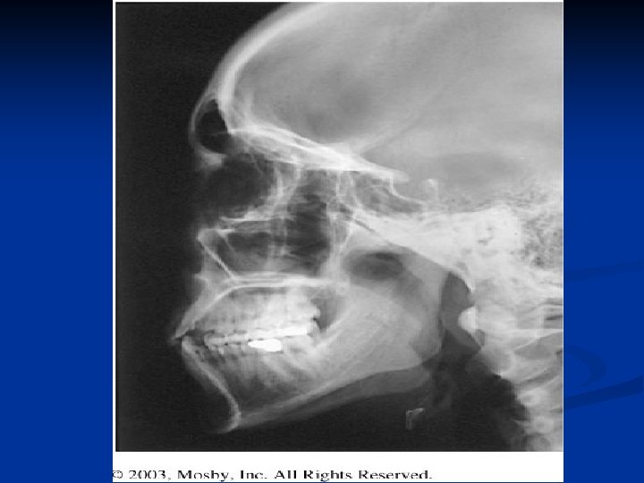
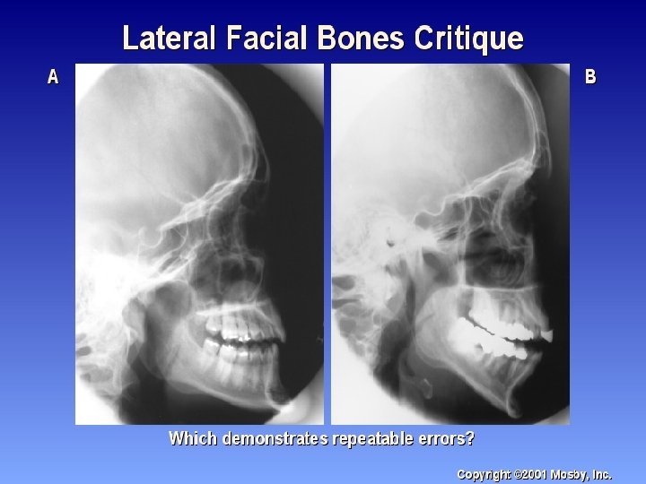
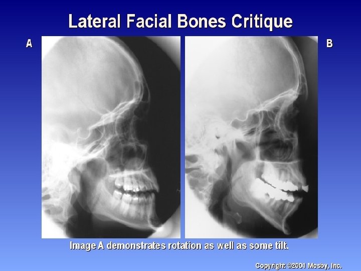
Positioning: Lateral Facial bones n Semiprone or seated n MSP parallel n IPL perpendicular n Suspend respiration n CR is perp and enters lateral zygomatic bone ½ way between outer canthus and EAM.

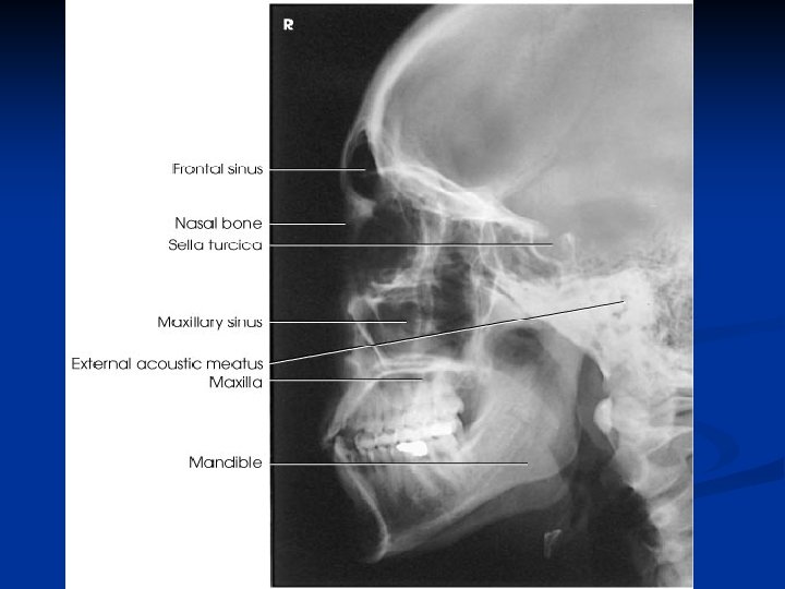
Lateral Facial Radiograph n All facial bones in with zygomatic bone in center n n Almost SI mandibular rami n SI orbital roofs (no tilt) n No rotation of sella turcica

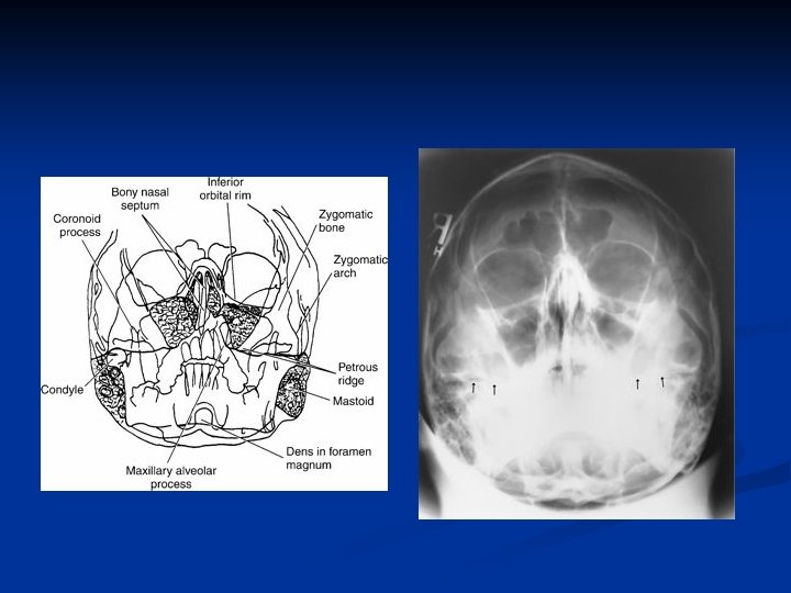
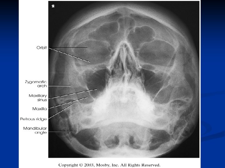
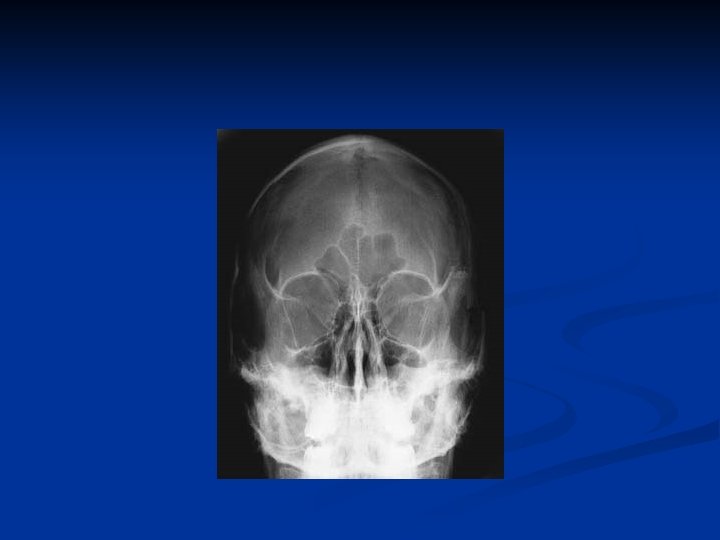
Anatomy Identity
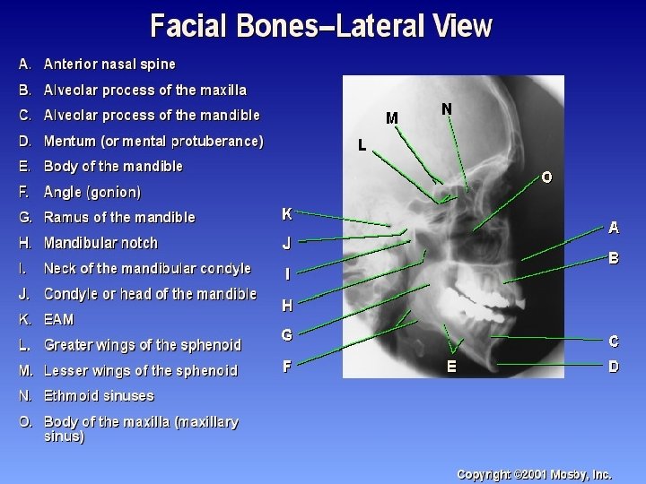
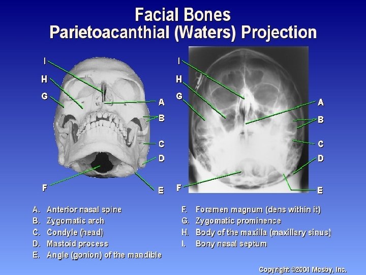
 Anterior nasal spine F) Alveolar process")
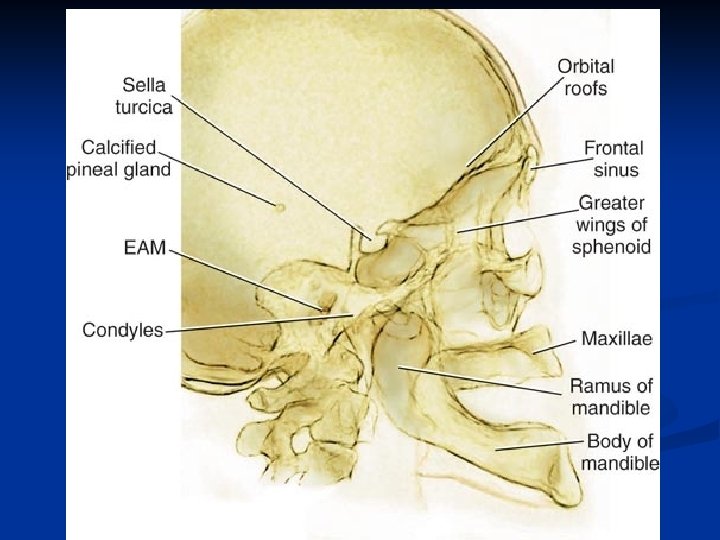
Radiograph Anatomy n n n n n E) Anterior nasal spine F) Alveolar process of maxilla G) Alveolar process of mandible H) Mentum J) Body of mandible K) Angle of mandib le L) Ramus of mandible M) Coronoid process O) Neck of mandibular condyle n n n n P) Condyle or neck of mandible Q) EAM R) Temporalmandibular fossa of temporal bone S) Greater wings of sphenoid T) Lesser wings of sphenoid U) Ethmoid sinuses between orbits V) Body of maxilla containing maxillary sinuses

Anatomy Identity
 zygomatic arch B) RT zygomatic")
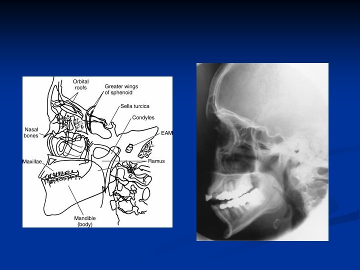
Lateral Skull Anatomy n n n n n A) zygomatic arch B) RT zygomatic bone C) RT nasal bone D) Frontal process of maxilla E) Anterior nasal spine F) Alveolar process of maxilla G) Alveolar process of mandible H) Mentum I) Mental foramen n n n n J) Body of mandible K) Angle (gonion) L) Ramus of mandible M) Coronoid process N) Mandibular notch O) Neck of mandibular condyle P) Condyle or head of mandible Q) EAM

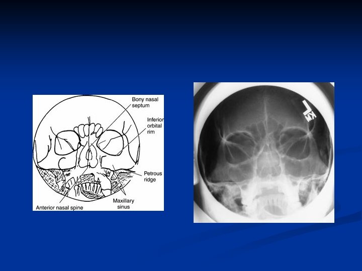
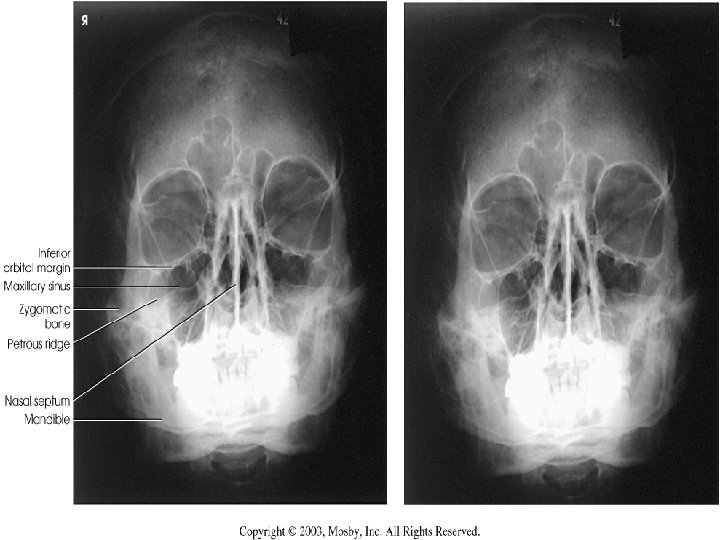
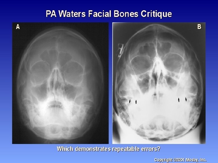
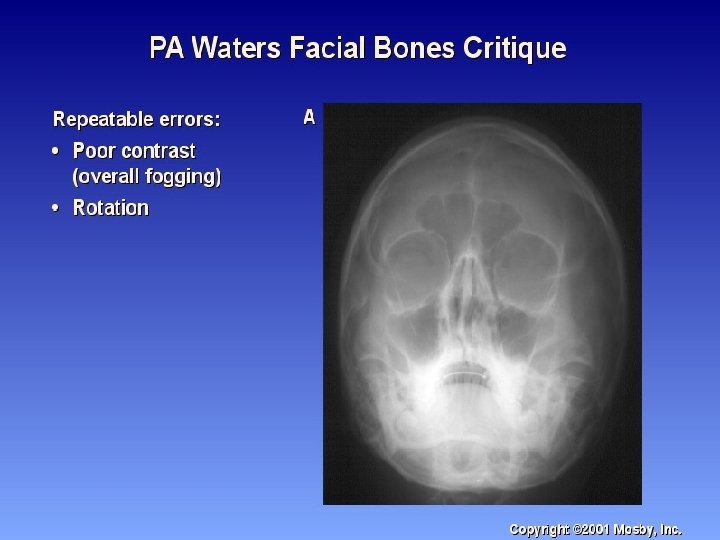
Positioning: Waters n Prone or seated upright n Chin on bucky -OML 37 angle with plane of cassette n MML & MSP perp n Nose 3/4 inch from IR n Suspend respiration n CR perpendicular to exit acanthion

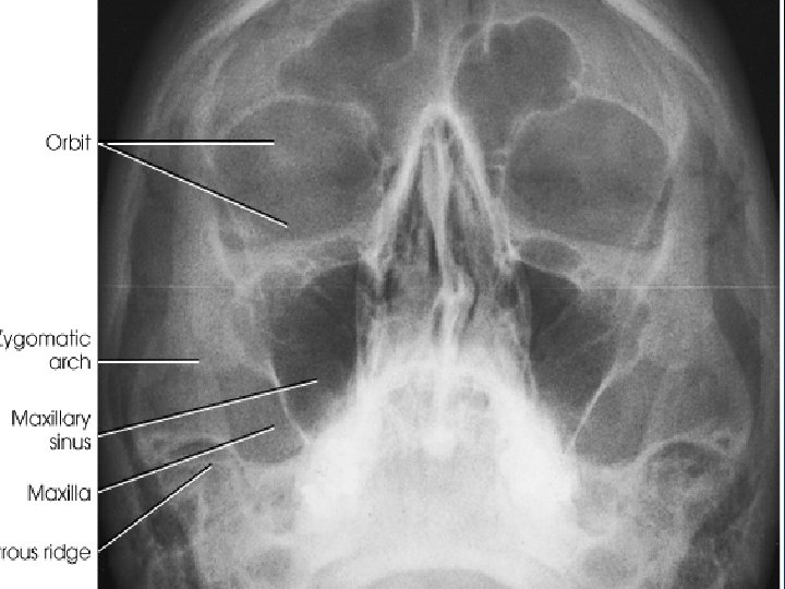
Waters Radiograph n Distance from lateral border of skull and orbit equal on each side n Petrous ridges projected immediately below maxillary sinuses

Trauma

Reverse Waters n Supine n Extend neck so OML is 37 degree with plane of IR n MML and MSP perp n Suspend respiration n CR perpendicular and enters acanthion

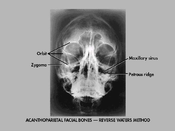
Reverse Waters Radiograph n Distance from lateral border of skull and orbit equal on each side n Petrous ridges projected immediately below maxillary sinuses

Modified Waters n OML 55 degree angle from plane of IR n MSP perp n CR perpendicular and exits acanthion

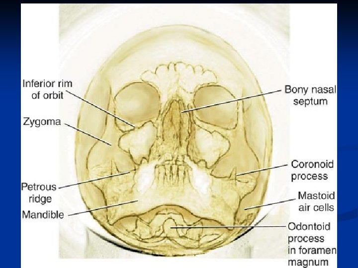
Modified Waters Radiograph n Petrous ridges projected immediately below the inferior border of the orbits n Equal distance from lateral orbit to lateral skull on both sides

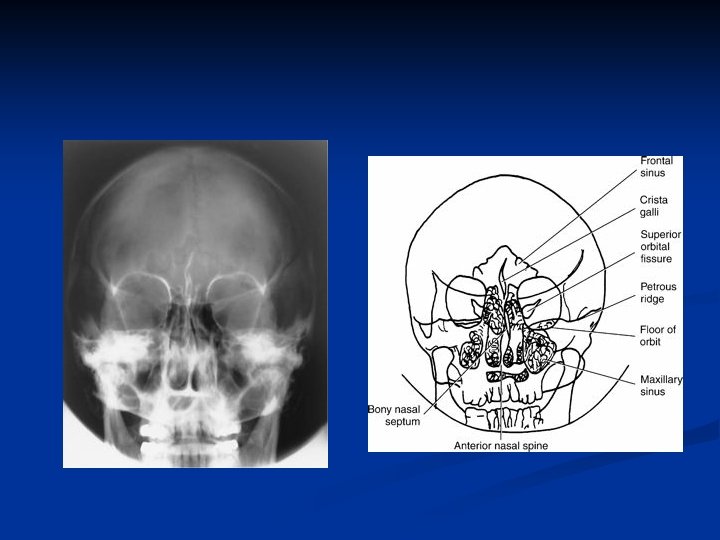
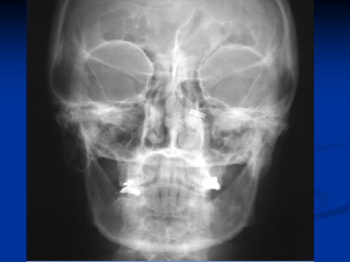
PA Axial - Caldwell n Prone or seated upright n Forehead & nose against grid device n OML perpendicular n CR 15 caudal to exit nasion n Suspend respiration

PA Axial- Caldwell Radiograph n Equal distance from lat skull to lat orbit n Symmetric petrous ridges in lower 1/3 orbit n Penetration of frontal bone without excessive density at lateral borders of skull.

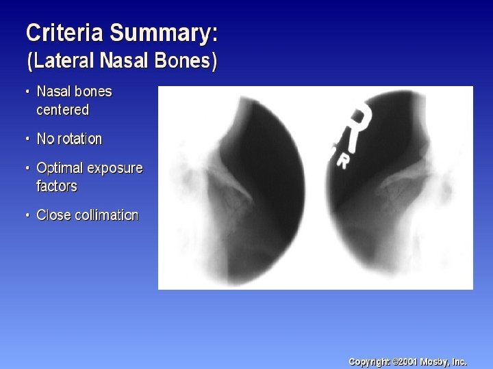
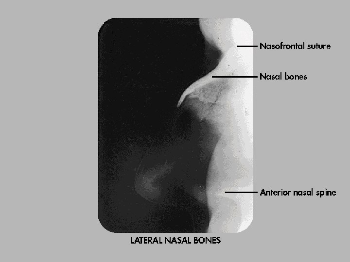
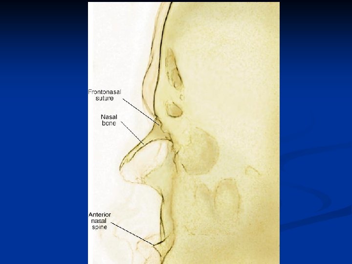
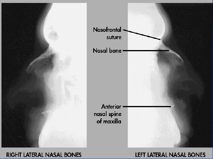
Lateral Nasal Bones n Semiprone n MSP & IOML parallel n IPL perpendicular n CR perpendicular to the bridge of nose at a point 1” distal to the nasion

Lateral Nasal bones Radiograph n No rotation of nasal bone and soft tissue n Anterior nasal spine and frontonasal suture evident n Close collimation
- Slides: 57