Face Scalp and Cervical Plexus Lecture Objectives Review

Face, Scalp, and Cervical Plexus

Lecture Objectives • Review the general anatomical features of the face and scalp. • Describe blood supply, innervation, and lymphatic drainage of the face and scalp. • Make a list of contributing roots to cervical plexus. • Discuss the general arrangement. • Describe the location of this plexus. • Make a list of the outcoming nerves. • Follow the branches to their target organs. • Make a list of the cutaneous nerves. • Follow the cutaneous branches to their destinations.
 • Subcutaneous layer (Layer 2) – Superficial fat")
Facial Layers • Skin (Layer 1) • Subcutaneous layer (Layer 2) – Superficial fat compartments • Superficial musculo‐aponeurotic system (SMAS) (Layer 3) • Retaining ligaments and deep compartments (Layer 4) – Deep fat compartments • Deep fascia &/or periosteum (Layer 5)
")
Layers of the Scalp • Layers of scalp – Skin – CT (subcutaneous layer) • Rich in BVs, lymphatics and nerves – Aponeurosis of occipitofrontalis muscle – Loose CT • Allows movement of above layers – Periosteum • Muscles of the External Ear – Auricularis anterior, posterior & superior

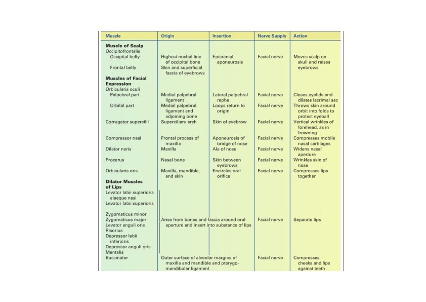
Muscles of Facial Expression • Arise from fascia or skull bones & insert onto skin • Encircle eyes, nose & mouth • Express emotions • Facial Nerve (VII) • Bell’s palsy = facial paralysis

Facial Muscles

Facial Muscles Action

Facial Muscles Innervation

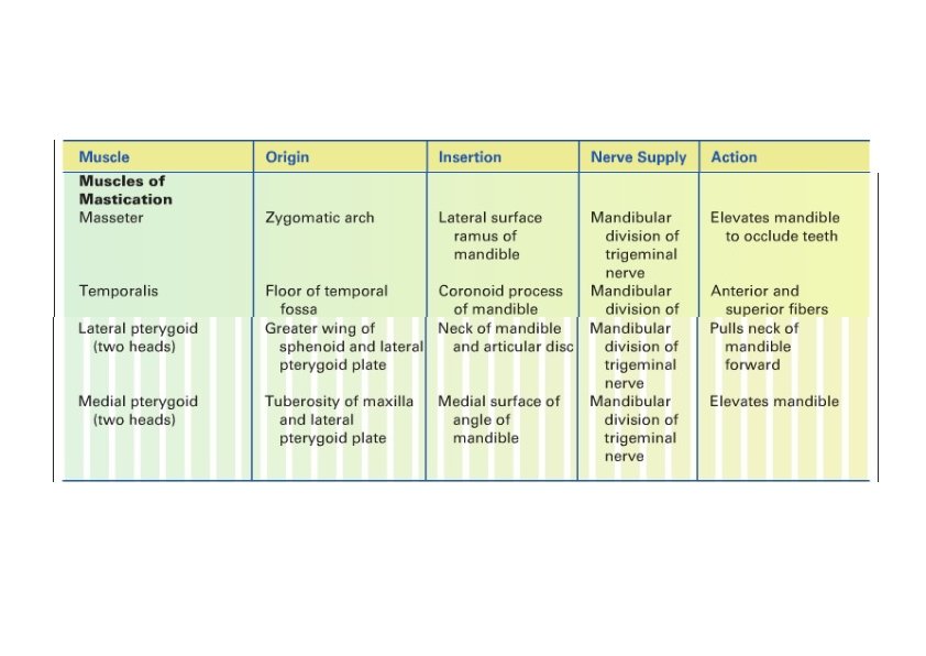
Muscles of Mastication • Masseter, temporalis & pterygoids • Arise from skull & insert on mandible • Cranial nerve V (trigeminal nerve) – mandibular division • Protracts, elevates or retracts mandible

Muscles of Mastication

Lateral Pterygoid

Cutaneous Nerves of Face and Scalp

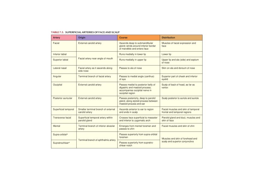
Arterial Supply for Face and Scalp

Arterial Supply for Face and Scalp

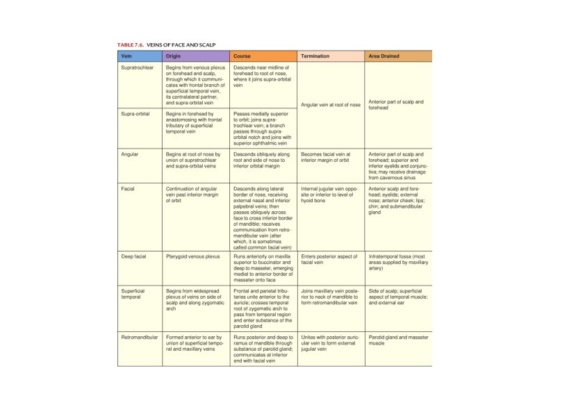
Venous Supply for Face and Scalp

Neck

Hyoid Bone • Position – C 3 – Between mandible & thyroid cartilage • Shape – U • Stylohyoid ligament • Parts – Body – Lesser horn – Greater horn

Neck Muscles • Superficial mm. of the side of the neck – Platysma ‐ VII – SCM ‐ XI • Suprahyoid mm. – Stylohyoid ‐ VII – Digastric • Posterior belly ‐ VII • Anterior belly ‐ V – Mylohyoid ‐ V – Geniohyoid – C 1 • Infrahyoid mm. – – Omohyoid ‐ AC Sternothyroid ‐ AC Thyrohyoid – C 1
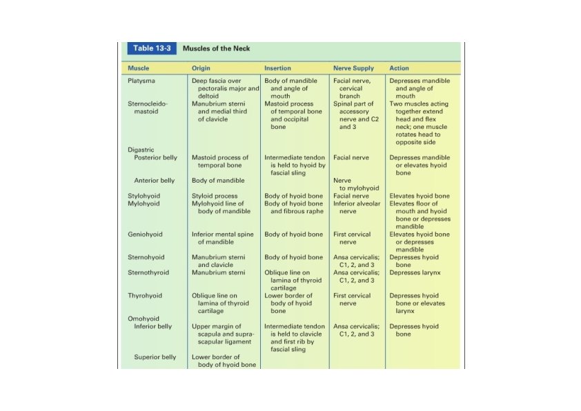

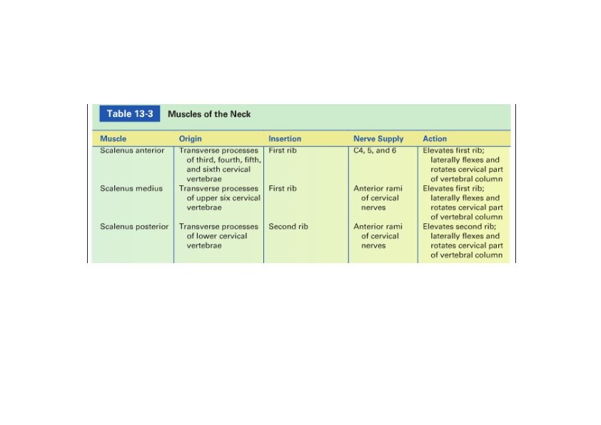
• Anterior and lateral vertebral mm. – Scalenus anterior • Relations – Scalenus medius – Scalenus posterior Neck Muscles
 •")
Cervical Plexus • Ventral rami of spinal nerves (C 1 to C 5) • Supplies parts of head, neck & shoulders

Cervical Plexus • Relations – Anterior to levator scapulae m. and middle scalene m. – Posterior to the sternocleidomastoid m. – Subcutaneous branches emerge behind the lateral border of the sternocleidomastoid m.

Cervical Plexus: Relations
 Greater")
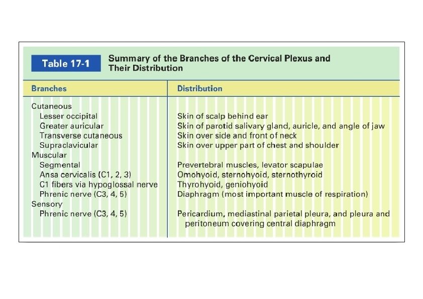
Cervical Plexus: Branches • Cutaneous branches – – Lesser occipital n. (C 2) Greater auricular n. (C 2‐C 3) Transverse cervical n. (C 2‐C 3) Supraclavicular nn. (C 3‐C 4) • Muscular branches – Ansa cervicalis (Infrahyoid mm. ) • Descending branch from hypoglossal n. (C 1) • Descending cervical n. (C 2‐C 3) – Phrenic n. (C 1‐C 3) • Diaphragm
- Slides: 30