FACE By Dr Leena N Associate Professor Department

FACE By Dr. Leena. N Associate Professor Department of Anatomy

FACE Extents • Superiorly from the adolescent position of hairline, • Inferiorly to the chin and the base of the mandible, and • Each side to the auricle

Skin Is very vascular. Is rich in sebaceous and sweat glands. Is very elastic and thick because the facial muscles are inserted into it.

FACIAL MUSCLES Embryologically, they develop from the mesoderm of the second branchial arch, and are, therefore, supplied by the facial nerve. .

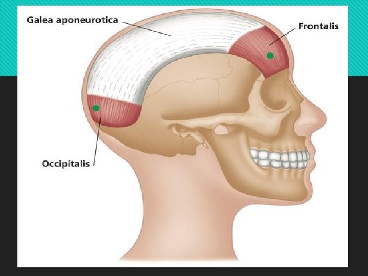
I. Muscle of the Scalp Occipitofrontalis II. Muscles of the Auricle—Vestigeal III. Muscles of the Eyelids/Orbital Openings 1. Orbicularis oculi 2. Corrugator 3. Levator palpebrae superioris IV. Muscles of the Nose 1. Procerus 2. Compressor naris 3. Dilator naris 4. Depressor septi
 3. Levator")
V. Muscles around the Mouth 1. Orbicularis oris 2. Buccinator (Latin cheek) 3. Levator labii superioris alaeque nasi 4. Zygomaticus major 5. Levator labii superioris 6. Levator anguli oris 7. Zygomaticus minor 8. Depressor anguli oris 9. Depressor labii inferioris 10. Mentalis (Latin chin) 11. Risorius (Latin laughter) VI. Muscles of the Neck Platysma
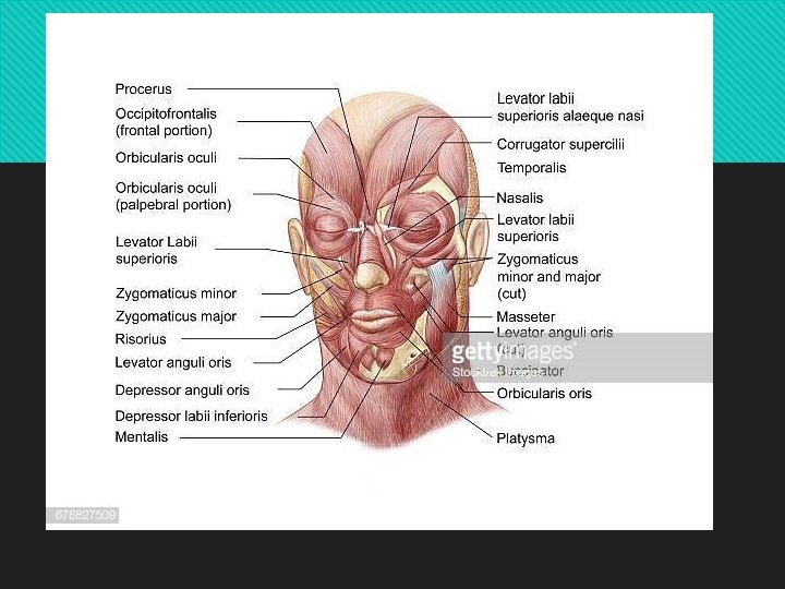

The facial muscles
Orbital Medial part of medial palpebral ligament. b)Palpabral Lateral")
Muscle Origin Orbiculari s occuli a)Orbital Medial part of medial palpebral ligament. b)Palpabral Lateral part of medial palpebral ligament. c)Lacrimal part Lacrimal fascia Insertion Nerve supply Concentric rings returns to the point of origin. Lateral palpebral raphae. Pass in front of eyelids to lateral palpebral Action Forcible closure of eyelids. Facial nerve Closes lids gently. Dilates lacrimal sac.
Intrinsic part-deep stratum, very thin b)Extrinsic part –two strata Origin Insertion")
Muscle Orbicularis oris a)Intrinsic part-deep stratum, very thin b)Extrinsic part –two strata Origin Insertion Superior incisivus Angle of mouth from Maxilla, Inferior incisivus from Mandible. Lips and angle of Thickest middle mouth stratum from Buccinator &thick superficial stratum from dilators of mouth Nerve supply Facial nerve Action Closes lips &protrude lips
Upper fibres from")
Muscle Origin Insertio Nerve n supply Buccinat or– muscle of cheek a)Upper fibres from maxilla opposite molar teeth Straight to upper lip Facial nerve b)Lower fibres from mandible opposite molar teeth Straight to lower lip c)Middle fibres from pterygomandibul ar raphae Middle fibres deccusat e Action Whistling muscle. Flatte ns cheek against gums &teeth, prevents accumulation of food in vestibule.

Some common facial expressions

ACTIONS OF THE MUSCLES A few of the common facial expressions and the muscles producing them are 1. Smiling and laughing: Zygomaticus major. 2. Sadness: Levator labii superioris and levator anguli oris. 3. Grief: Depressor anguli oris. 4. Anger: Dilator naris and depressor septi. 5. Dislike: Corrugator supercilii and procerus. 6. Horror, terror and fright: Platysma 7. Surprise: Frontalis, 8. Doubt: Mentalis, 8. Grinning: Risorius 9. Contempt: Zygomaticus minor. 10. Closing the mouth: Orbicularis oris 11. Whistling: Buccinator, and orbicularis oris.

NERVE SUPPLY OF FACE Motor Nerve Supply The facial nerve is the motor nerve of the face. Its five terminal branches supply the various facial muscles as follows. • Temporal—frontalis, auricular muscles, orbicularis oculi. • Zygomatic—orbicularis oculi (lower eyelid part). • Buccal—muscles of the cheek and upper lip. • Marginal mandibular—muscles of lower lip. • Cervical—platysma.

Terminal branches of the facial nerve

CLINICAL ANATOMY Infranuclear lesion of right facial nerve or Bell’s palsy

Supranuclear lesion of left facial nerve

nerves of Face of Sensory Mainly by the branches Ophthalmic (Supratrochlear, Supraorbital, palpebral branch of lacrimal, infratrochlear, external nasal) Maxillary(infraorbital, zygomaticofacial, zygomaticote mporal) & Mandibular (auriculotemporal, buccal &mental) divisions of Trigeminal nerve. Also branches from Cervical plexus. Anterior div of great auricular nerve and upper div of transverse cutaneous nerve of neck
 Supratrochlear, (2)")
Sensory Nerve Supply The sensory nerves of the face and neck. (1) Supratrochlear, (2) Supraorbital, (3) palpebral branch of lacrimal, (4) infratrochlear, (5) external nasal, (6) infraorbital, (7) zygomaticofacial, (8) zygomaticotemporal, (9) auricu-lotemporal, (10) buccal, (11) mental, (12) great auricular, (13) transverse cutaneous nerve of neck, (14) lesser occipital, and (15) supraclavicular

ARTERIES OF THE FACE The face is richly vascular. It is supplied by: 1. The facial artery, 2. The transverse facial artery, and 3. Arteries that accompany the cutaneous nerves.

Arteries of the face

VEINS OF THE FACE: These accompany the arteries Deep Connections: A communication between the supraorbital and superior ophthalmic veins. Another connection with the pterygoid plexus in.

VENOUS DRAINAGE The veins of the scalp, face and their deep connections with the cavernous sinus and the pterygoid plexus of veins
. Spread of infection from")
DANGEROUS AREA OF FACE Dangerous area of the face (stippled). Spread of infection from this area can cause thrombosis of the cavernous sinus
 drains")
LYMPHATIC DRAINAGE OF THE FACE The lymphatic territories of the face. Area (a) drains into the preauricular nodes, area (b) drains into the submandibular nodes, and area (c) drains into the submental nodes
- Slides: 26