Face and Neck Regions Chapter 1 Dental Embryology

Face and Neck Regions Chapter 1 Dental Embryology, Histology, and Anatomy
 A B C D")
Regions of the face (Fig. 1 -1) A B C D
 A B Reg. A Region B Region")
Regions of the face (Fig. 1 -1) A B Reg. A Region B Region C Region D C D
 A. Zygomatic B. Buccal C. Oral D.")
Regions of the face (Fig. 1 -1) A. Zygomatic B. Buccal C. Oral D. Mental
 E.")
Regions of the face (Fig. 1 -2) E.
 E. Sternocleidomastoid muscle")
Regions of the face (Fig. 1 -2) E. Sternocleidomastoid muscle
 A F. G")
Regions of the Face (Figure 1 -2) A F. G
 A F Zygomatic arch G Parotid salivary")
Regions of the Face (Figure 1 -2) A F Zygomatic arch G Parotid salivary gland
 H. I. J.")
Frontal view of face (Fig. 1 -3) H. I. J.
 H. I. J.")
Frontal view of face (Fig. 1 -3) H. I. J.
 H. Zygomatic arch I. Temporomandibular joint J.")
Frontal view of face (Fig. 1 -3) H. Zygomatic arch I. Temporomandibular joint J. Buccal region
 L M K")
Nose (Fig. 1 -4) L M K
 L Nasal septum M Ala K Nares (nostrils)")
Nose (Fig. 1 -4) L Nasal septum M Ala K Nares (nostrils)
 NQ NR NN NO NP NS _duct NT")
Buccal Region (Fig. 1 -5) NQ NR NN NO NP NS _duct NT
 Q R S N O P T")
Buccal Region (Fig. 1 -5) Q R S N O P T
 W U X Y V")
Frontal view of lips (Fig. 1 -6) W U X Y V
 V Philtrum W Vermilion U Tubercle X")
Frontal view of lips (Fig. 1 -6) V Philtrum W Vermilion U Tubercle X commissure Y zone Y border
 Z 2 1 3")
Mandible (Fig. 1 -8) Z 2 1 3
 Z. Coronoid process 2. Mental Foramen 1. Condyle 3. Ramus")
Mandible (Fig. 1 -8) Z. Coronoid process 2. Mental Foramen 1. Condyle 3. Ramus
 5. 4. 6.")
Neck region (Fig. 1 -11) 5. 4. 6.
 4. Sternocleidomastoid 5. Hyoid 6. Thyroid")
Neck region (Fig. 1 -11) 4. Sternocleidomastoid 5. Hyoid 6. Thyroid


Lips Darby and Walsh, Dental Hygiene Theory and Practice 2 nd Edition, Fig 12 -6.

Oral Cavity and Pharynx Chapter 2 Dental Embryology, Histology, and Anatomy
 upper C D A towards cheek")
Divisions of the Oral Cavity (Fig. 2 -1) upper C D A towards cheek B towards lip E F towards tongue lower
 C Maxillary D Palatal A Buccal")
Divisions of the Oral Cavity (Fig. 2 -1) C Maxillary D Palatal A Buccal B Facial / labial E Lingual F Mandibular
 J K G H I L mucosa M")
Vestibule (Fig. 2 -2) J K G H I L mucosa M
 J vestibule G Parotid papilla H Buccal mucosa I Labial")
Vestibule (Fig. 2 -2) J vestibule G Parotid papilla H Buccal mucosa I Labial mucosa K Alveolar mucosa L Muccobuccal fold M Vestibule

Describe appearance. n Smokeless Tobacco Keratosis Newland, Meiller, Wynn, and Crossley; Oral Soft Tissue Diseases, Lexi-Comp, Inc. , 2001, p. 27
 Oral cavity proper u (inside")
Divisions of the Oral Cavity n n Vestibule (entranceway) Oral cavity proper u (inside the teeth) n Surfaces: u Facial /labial u Buccal u Palatal u Lingual
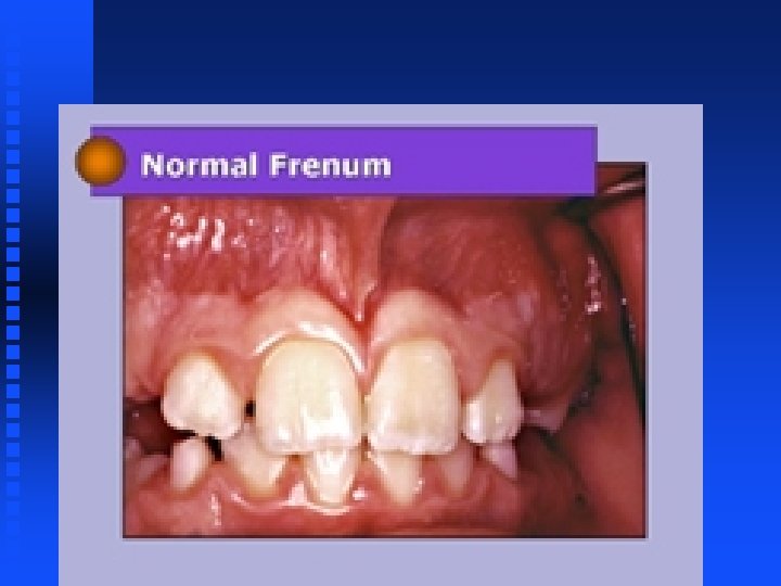
 N. Labial frenum u Labial mucosa u Buccal")
Oral Vestibules n Oral mucosa (membrane) N. Labial frenum u Labial mucosa u Buccal mucosa n Alveolar mucosa N. Labial frenum n Labial frenum

PRIMARY DENTAL ARCHES

GINGIVAL AREA

GINGIVAL AREA
 n n Ectopic sebaceous glands found in the buccal mucosa, labial")
Fordyce Granules (spots) n n Ectopic sebaceous glands found in the buccal mucosa, labial mucosa, or vermillion zone Multiple, small, white to yellow nodules Newland, Meiller, Wynn, and Crossley; Oral Soft Tissue Diseases, Lexi-Comp, Inc. , 2001, p. 17
 Sebaceous fat glands")
Clinical considerations wit h Oral Mucosa (Fordyce Granules with Linea Alba) Sebaceous fat glands Daniel and Harfst, Mosby’s Dental Hygiene, Concepts, Cases and Competencies, 2008, p. 298.
")
Abnormal condition of the buccal mucosa: Linea Alba (Frictional Keratosis)
")
Alveolar processes and permanent teeth (Fig. 2 -4)

Alveolar processes and permanent teeth Maxillary Mandibular
 What does the periodontal ligament (PDL) attach?")
Tooth tissues (Fig. 2 -5) What does the periodontal ligament (PDL) attach?
 (Fig. 2 -6) Alveolus (tooth socket) n Portions of gingiva n")
Alveolar Processes (Bones) (Fig. 2 -6) Alveolus (tooth socket) n Portions of gingiva n

Mandibular Tori Darby and Walsh, Dental Hygiene Theory and Practice 2 nd Edition, Fig 12 -27.
 O P Q")
Gingiva and Landmarks (Fig. 2 -9) O P Q
 O Alveolar m P Mucogingi juncion Q Attached")
Gingiva and Landmarks (Fig. 2 -9) O Alveolar m P Mucogingi juncion Q Attached gingiva

Vestibule
 R S T (inside)")
Gingiva and landmarks (Fig. 2 -10) R S T (inside)
 R Marginal gingiva S Interdental gingiva/papilla T Sulcus")
Gingiva and landmarks (Fig. 2 -10) R Marginal gingiva S Interdental gingiva/papilla T Sulcus
 Z 1 U V 2 3")
Oral cavity proper and boundaries (Fig. 2 -11) Z 1 U V 2 3 4 5 W X Y 6
 Z Fauces U Hard palate V")
Oral cavity proper and boundaries (Fig. 2 -11) Z Fauces U Hard palate V Soft palate 1 Max. tuberosity 2 3 4 5 Pterygomandibular fold Posterior faucial pillar Palatine tonsil Anterior faucial pillar W Uvula X Post. wall of pharynx Y Dorsal surface of tongue 6 Retromolar pad
 arch Uvula Palatine tonsil Palatopharyngeal (post.")
Posterior Oral Cavity Soft Palate Palatoglossal (ant. ) arch Uvula Palatine tonsil Palatopharyngeal (post. ) arch Posterior wall of the pharynx Daniel and Harfst, Mosby’s Dental Hygiene, Concepts, Cases and Competencies, 2002, p. 261.

Landmarks in the Oral Cavity - Dental Arches 77 8 9 Darby and Walsh, Dental Hygiene Theory and Practice 2 nd Edition, Fig 12 -21.

Landmarks in the Oral Cavity - Dental Arches 7 Max. tuberosity 8 Palatine tonsil 9 Retromolar pad Darby and Walsh, Dental Hygiene Theory and Practice 2 nd Edition, Fig 12 -21.

Gingival Area

GINGIVAL AREA

GINGIVAL AREA Attached gingiva Mucogingival junction Alveolar mucosa

GINGIVAL AREA Free gingival margin Interdental papilla

Atypical condition: Exostosis
 10 13 14 11 12")
Palate and landmarks (Fig. 2 -12) 10 13 14 11 12
 10 Incisive papilla 13 Palatine rugae 14 Median")
Palate and landmarks (Fig. 2 -12) 10 Incisive papilla 13 Palatine rugae 14 Median palatine raphe 11 Hard palate 12 Soft palate
")
Palatal torus (Fig. 2 -13)

Palate Darby and Walsh, Dental Hygiene Theory and Practice 2 nd Edition, Fig 12 -6.

Palate Incisive papilla Palatine rugae Lingual gingiva Palatine raphe Palatal glands Palatine fovea Daniel and Harfst, Mosby’s Dental Hygiene, Concepts, Cases and Competencies, 2002, p. 261.

Describe appearance. n A Nicotine stomatitis Identify A and B B Newland, Meiller, Wynn, and Crossley; Oral Soft Tissue Diseases, Lexi-Comp, Inc. , 2001, p. 28

Normal Anatomy A A Palatine raphe B Palatine fovea B Newland, Meiller, Wynn, and Crossley; Oral Soft Tissue Diseases, Lexi-Comp, Inc. , 2001, p. 28
 18 15 19 16 20 21 17")
Dorsal view of tongue (Fig. 2 -14) 18 15 19 16 20 21 17 22
 18 Palatine tonsil 15 Lingual 19 Foramen")
Dorsal view of tongue (Fig. 2 -14) 18 Palatine tonsil 15 Lingual 19 Foramen cecum 16 Circumvalle 20 Sulcus terminalis 21 Median lingual sulcus 17 Filiform 22 Fungiform
 23 24 25 26")
Lateral view of the tongue (Fig. 2 -15) 23 24 25 26
 23 Dorsal 24 Lateral 25 Ventral")
Lateral view of the tongue (Fig. 2 -15) 23 Dorsal 24 Lateral 25 Ventral 26 Foliate
 27 28")
Ventral surface of the tongue (Fig. 2 -16) 27 28
 27 veins 28 Plica fimbriateae")
Ventral surface of the tongue (Fig. 2 -16) 27 veins 28 Plica fimbriateae

Condition of tongue: Lingual Varicosities Circumvallate papilla n Filiform papilla n Foliate papilla n Fungiform papilla n Ventral aspect

TONGUE Identify the following papilla a b c d c

TONGUE Identify the following papilla a b c d 7 - 14 c filiform (hairlike) fungiform (red) foliate (lateral) circumvallate (7 -14) lateral hairlike red, mushroom-like

Floor of the Mouth 30 29 31 Lingual gingiva Daniel and Harfst, Mosby’s Dental Hygiene, Concepts, Cases and Competencies, 2002, p. 259.

Floor of the Mouth 29 Sublingual caruncle, opening to Wharton’s ducts connecting to submandibular and sublingual glands Lingual gingiva 30 Lingual frenum 31 Sublingual fold, Opening to Ducts of Ravinus or Bartholin’s duct connecting to sublingual salivary gland Daniel and Harfst, Mosby’s Dental Hygiene, Concepts, Cases and Competencies, 2002, p. 259.

Ventral Surface of Tongue Darby and Walsh, Dental Hygiene Theory and Practice 2 nd Edition, Fig 12 -26.

Divisions of the Pharynx

- Slides: 79