
Eye Structure and Refraction Prof K Sivapalan Structure
- Slides: 15
Eye- Structure and Refraction Prof. K. Sivapalan
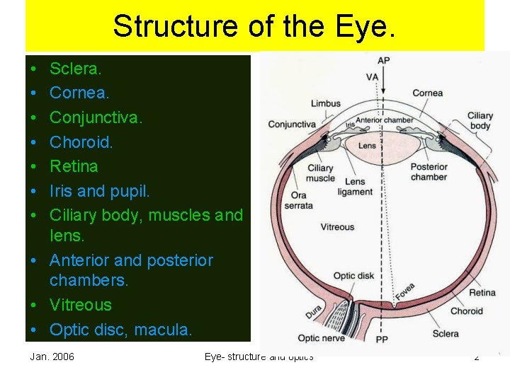
Structure of the Eye. • • Sclera. Cornea. Conjunctiva. Choroid. Retina Iris and pupil. Ciliary body, muscles and lens. • Anterior and posterior chambers. • Vitreous • Optic disc, macula. Jan. 2006 Eye- structure and optics 2
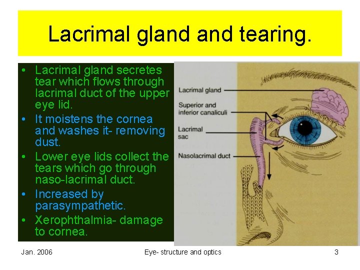
Lacrimal gland tearing. • Lacrimal gland secretes tear which flows through lacrimal duct of the upper eye lid. • It moistens the cornea and washes it- removing dust. • Lower eye lids collect the tears which go through naso-lacrimal duct. • Increased by parasympathetic. • Xerophthalmia- damage to cornea. Jan. 2006 Eye- structure and optics 3
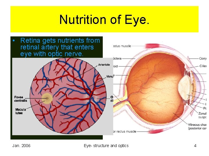
Nutrition of Eye. • Retina gets nutrients from retinal artery that enters eye with optic nerve. • Fovea has no vessols. Nurished by diffusion from choroid. • Other structures get the nutrients from vessels in the choroid. • There are no vessels in the anterior chamber, lens, posterior chamber and vitreous. Jan. 2006 Eye- structure and optics 4
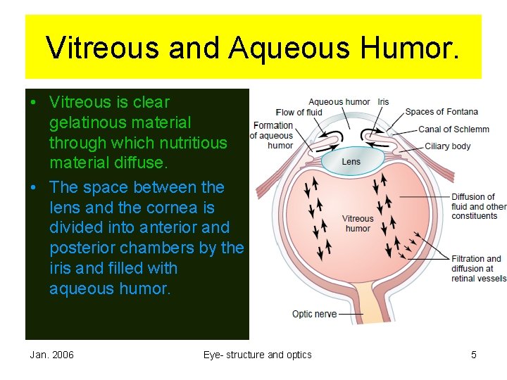
Vitreous and Aqueous Humor. • Vitreous is clear gelatinous material through which nutritious material diffuse. • The space between the lens and the cornea is divided into anterior and posterior chambers by the iris and filled with aqueous humor. Jan. 2006 Eye- structure and optics 5
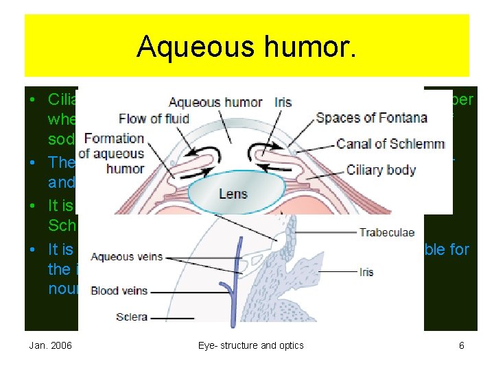
Aqueous humor. • Ciliary body has ciliary processes in the posterior chamber where aqueous humor is secreted by active transport of sodium and other nutrients. • The fluid passes through the pupil into anterior chamber and into the angle between the cornea and the iris. • It is absorbed through the trabeculae into the Chanal of Schlemm and into veins. • It is formed at a rate of 2. 5 μL / min. and is responsible for the intra-ocular pressure of 15 [10 -20] mm Hg and nourishes the cornea and lens. Jan. 2006 Eye- structure and optics 6
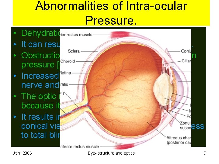
Abnormalities of Intra-ocular Pressure. • Dehydration reduces intra-ocular pressure. • It can result in abnormal curvatures in cornea. • Obstruction to absorption results in increase in pressure [Glaucoma] • Increased pressure compresses retina, optic nerve and retinal vessels. • The optic nerve is affected at the optic disc because it is the weak point in the sclera. • It results in loss of vision in peripheral fieldconical vision at early stages. This can progress to total blindness. Jan. 2006 Eye- structure and optics 7
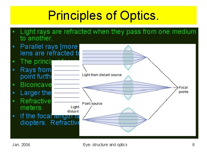
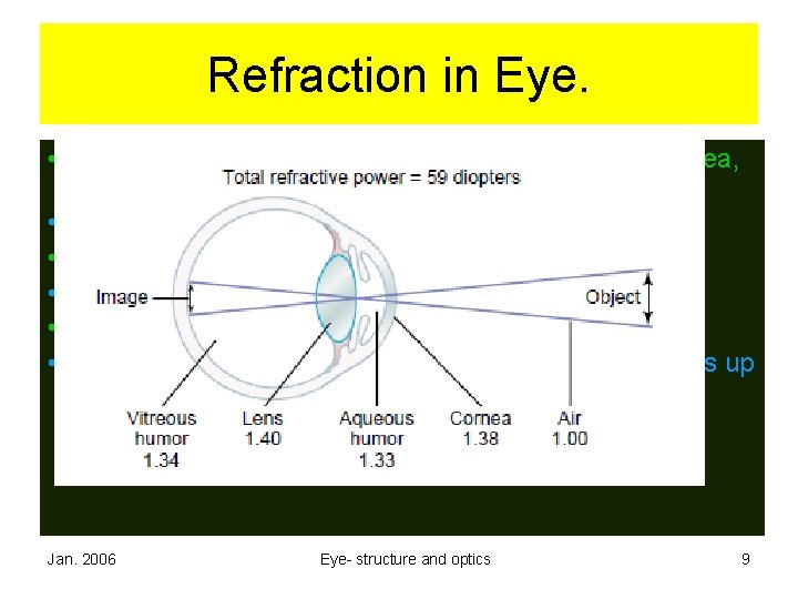
Principles of Optics. • Light rays are refracted when they pass from one medium to another. • Parallel rays [more than 6 meters] striking on biconvex lens are refracted to principal focus. • The principal focus is in the principal axis. • Rays from closer objects are diverging and focus on a point further than principal focus. • Biconcave lens causes the rays to diverge. • Larger the curvature greater the refractive power. • Refractive power is the reciprocal of focal length in meters. • If the focal length is 25 cm, refractive power is 1/0. 25= 4 diopters. Refractive power of human eye = 60 diopters Jan. 2006 Eye- structure and optics 8
Refraction in Eye. • When light rays from a distant object falls on the cornea, it is refracted: • On entry into cornea, • On entry into aqueous humor, • On entry into lens, and • On entry into vitreous humor. • Finally an image is formed on the retina. The image is up side down. Jan. 2006 Eye- structure and optics 9
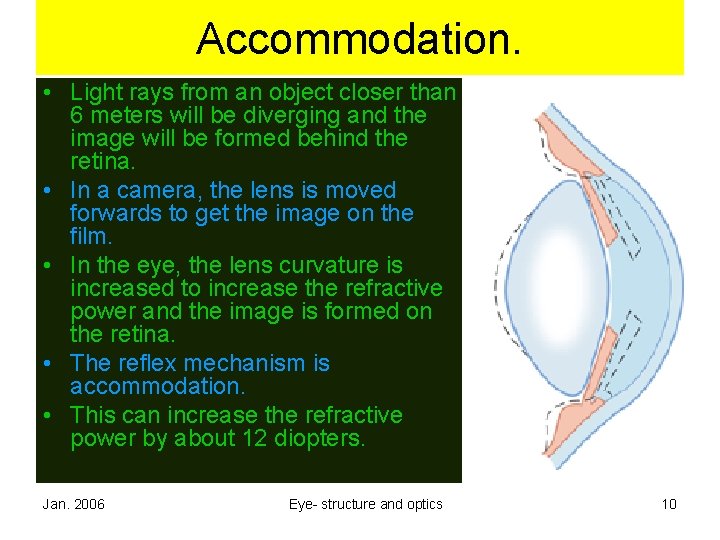
Accommodation. • Light rays from an object closer than 6 meters will be diverging and the image will be formed behind the retina. • In a camera, the lens is moved forwards to get the image on the film. • In the eye, the lens curvature is increased to increase the refractive power and the image is formed on the retina. • The reflex mechanism is accommodation. • This can increase the refractive power by about 12 diopters. Jan. 2006 Eye- structure and optics 10
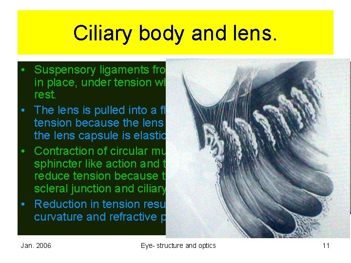
Ciliary body and lens. • Suspensory ligaments from the ciliary body keep lens in place, under tension when the ciliary muscles are at rest. • The lens is pulled into a flattened shape by the tension because the lens substance is malleable and the lens capsule is elastic. • Contraction of circular muscles reduce tension by sphincter like action and the meridional muscles reduce tension because they are attached to corneoscleral junction and ciliary body. • Reduction in tension results in increase in the curvature and refractive power of the lens. Jan. 2006 Eye- structure and optics 11
Near Point. • Light rays from very near to eye cannot be focused on the retina. • The nearest point from which an object can be focused on the retina is the ‘near point’. • Near point receds through out the life, 9 cm at 10 years, 10. 5 at 20 years and 83 cm at 60 years. • This is known as presbyopia and can be corrected by convex lens. Jan. 2006 Eye- structure and optics 12
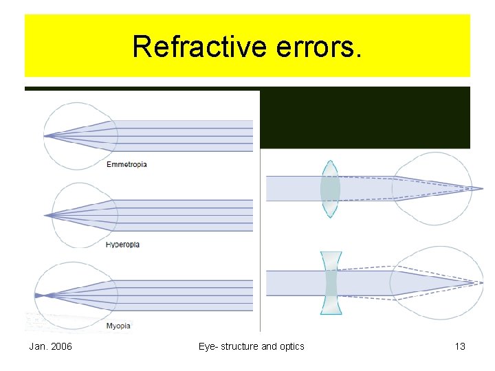
Refractive errors. Jan. 2006 Eye- structure and optics 13
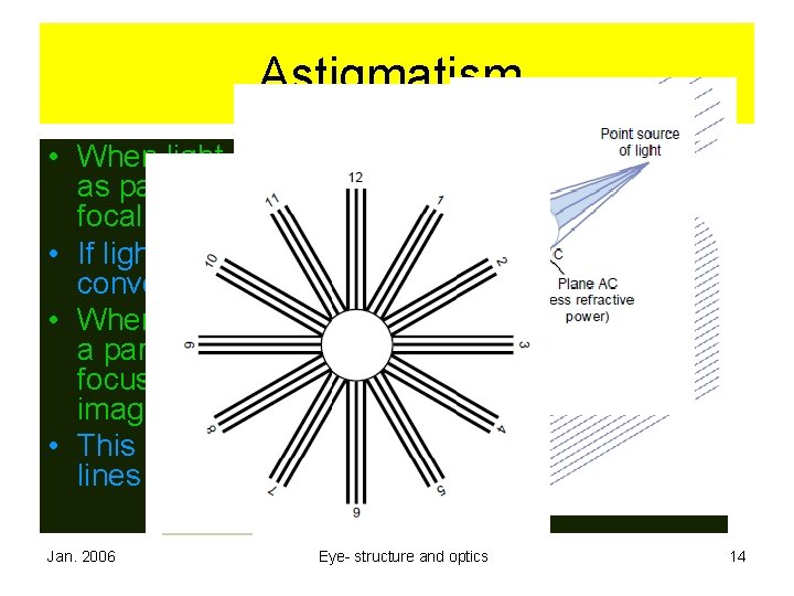
Astigmatism. • When light rays go through lens with surfaces as part of sphere, the rays converge at the focal point. • If light goes through cylindrical lens, the rays converge in a line parallel to the lens. • When the surface of the cornea happens to be a part of distorted sphere, rays in different axis focus at different points resulting in blurred image. • This can be identified by chart with radiating lines and corrected by cylindrical lens. Jan. 2006 Eye- structure and optics 14
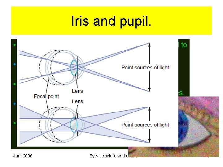
Iris and pupil. • Iris is a pigmented, opaque membrane attached to ciliary body. • Pupil is the opening at the centre if iris through which light rays reach the lens. • The diameter of the pupil can be altered from 8 mm to pinpoint by the circular and radial muscles. • The size of the pupil determines the amount of light reaching the retina. • Smaller diameter increases the depth of focus and reduces spherical aberration of the periphery of the lens. Jan. 2006 Eye- structure and optics 15